


Abstract
Background/Aim: The hepatoprotective role of various molecules in drug-induced hepatotoxicity arouses great interest. We investigated the effect of liposomal curcumin (LCC) on experimental acetaminophen (APAP)-induced hepatotoxicity. Materials and Methods: Rats were randomly allocated into 5 groups, and the effect of two LCC concentrations was studied: group 1 – 1 ml intraperitoneal (i.p.) saline, group 2 – APAP pretreatment, group 3 – APAP+silymarin (extract of the silybum marianum with anti-inflammatory, anti-oxidant, and anti-fibrotic properties), group 4 – APAP+LCC1, group 5 – APAP+LCC2. The biomarkers of oxidative stress (nitric oxide and malondialdehyde) and antioxidant status of plasma (thiols and catalase), TNF-α, MMP-2 and MMP-9 serum levels were evaluated. Results: An improvement in oxidative stress, antioxidant status, and TNF-α, MMP-2 and MMP-9 levels was obtained in groups pretreated with LCC compared to silymarin treatment, in a dose-dependent manner. Histopathological examination reinforced the results. Conclusion: Liposomal curcumin improves the oxidative stress/antioxidant balance and alleviates inflammation in experimental APAP-induced hepatotoxicity.
- Curcumin
- acetaminophen-induced hepatotoxicity
- tumor necrosis factor alpha
- oxidative stress
- matrix metalloproteinases
Acetaminophen, also known as paracetamol or N-acetyl-p-aminophenol (APAP), has anti-inflammatory, analgesic and antipyretic properties when used in its normal therapeutic doses (1, 2). It is widely available and can be used by all age groups. The tendency to use it in high doses for more rapid effects is the main concern, due to increased risk for acute liver failure that can be very severe, requiring liver transplantation (1). The liver is a target for the biotransformation of many drugs; therefore, liver tissue lesions are commonly associated with various medications (3). Acetaminophen-related liver toxicity is reported in many studies and is mainly associated with drug overdose, genetic factors, concurrent medication, concomitant alcohol consumption or nutritional status (4). Hepatic tissue lesions can be very severe, leading to acute liver failure (ALF) (4). ALF is related to cell death that has been documented to occur mainly by necrosis, but the apoptotic process can also have an important contribution (5). Inhibition of caspase activity (involved in the apoptotic process) proved to promote liver tissue regeneration and recovery (6).
Mitochondrial dysfunction is one of the mechanisms involved in hepatic toxicity of APAP, consequently inducing hepatic cell death (7). Cell response to mitochondrial dysfunction consists of various deleterious consequences, including excessive production of reactive oxygen species (ROS) (8). Mitochondrial dysfunction has been proven to be related to a decrease in ATP production, an increase in ROS, and a decrease in the production of glutathione (GLUT) as a scavenger molecule for ROS (7). APAP hepatotoxicity is also related to increased production of reactive metabolite N-acetyl-p-benzoquinone (NAPQI) as a consequence of the metabolic activity of the cytochrome P450 system (9). The metabolite depletes GLUT from liver tissue, decreasing the cell's antioxidant defense system (9). GLUT production is important for the hepatic metabolism of APAP, acting as a scavenger for NAPQI protein (8). Changes in cellular enzymatic activity (involved in drug detoxification) can also cause cellular damage to liver tissue (10). Oxidative stress and inflammatory reactions were also incriminated in overdoses of APAP inducing renal toxicity (11). Depletion of the GLUT pool is another important mechanism that contributes to APAP hepatotoxicity (12). Hepatic injury is associated with increased synthesis of matrix metalloproteinases (MMPs), which are a group of proteolytic enzymes that can promote extracellular matrix (ECM) degradation, being a good indicator of liver damage in liver failure (13). MMPs can also act as regulators of inflammation and immunity by influencing cytokine and chemokine production (14). The normal liver expresses several MMPs, including MMP-2 and MMP-9, which have an important role in liver vascular homeostasis (15). Increased MMP-9 activity is associated with extensive leukocyte recruitment in severe liver injury (16). During their activation at the injury site, leukocytes can express MMP-9 (responsible for ECM degradation and for increased vascular permeability due to cleavage of endothelial junctional proteins) (17, 18).
Thus, the major issues in APAP-induced hepatotoxicity are to identify potential biomarkers to predict the severity of liver damage and to develop new therapies able to limit the extension of injury and promote liver regeneration. Natural products are attracting the interest of many researchers to study their effects on various disorders. Among the nutraceutical compounds commonly appreciated for their antioxidant and anti-inflammatory properties is curcumin, which can improve cell viability in various disorders by reducing cell apoptosis and necrosis (19). The antioxidant activity of curcumin is based on its scavenging properties for ROS (20, 21). Its anti-inflammatory effect was reported to be related to the down-regulation of nuclear factor-kB (NF-kB), cyclooxygenase 2 (COX2) and pro-inflammatory cytokines such as interleukin-1 and interleukin-6 (22). We previously demonstrated the beneficial effect of oral curcumin administration on hepatic function in fructose-induced metabolic syndrome and in myocardial ischemia (23-25). Despite the beneficial effect of curcumin, the low bioavailability of this natural compound presents interest for new formulations. Novel nanoformulations of curcumin are emerging and can enhance its systemic bioavailability and tissue distribution (26). Among curcumin nanoformulations, liposomal curcumin offers a better water solubility of curcumin, leading to 8- to 20-fold increased systemic exposure compared to the standard curcumin suspension formulation (27). Liposomal curcumin is a phospholipid bilayer vesicle that can carry curcumin; it is easy to prepare and safe to use (28-30).
The levels of the investigated markers of liver damage (ALT and AST) and inflammation (TNF-α) in each treatment group, expressed as mean and standard deviation.
Silymarin (SIL) is an active component of Silybum marianum extracts with anti-inflammatory, anti-oxidant, and anti-fibrotic properties. It contains a family of flavonolignans and a flavonoid (taxifolin), among which silybin accounts for 50% to 70% and is identified as the major biologically active component (31).
The aim of this study was to observe the beneficial effect of liposomal curcumin administration in two concentrations and to compare it to the effect of silymarin in APAP-induced hepatotoxicity in rats. Several biomarkers were assessed and compared: ALT and AST for hepatic function, oxidative stress parameters (NO and MDA), antioxidant parameters (thiols and catalase), inflammatory cytokine (TNF-alpha), MMP-2 and MMP-9 as biomarkers for ECM degradation.
Materials and Methods
Animals. Male albino rats (Wistar-Bratislava) were provided by the Animal Department of the Faculty of Medicine and Pharmacy, Cluj-Napoca. The animals weighing 200-250 mg were kept in polypropylene cages, each group in a separate cage, at a constant temperature (24±2°C) and 60±5% humidity, in a 12/12 h light-dark cycle. Unrestricted access to food (standard pellets from Cantacuzino Institute, Bucharest, Romania) and water was provided. Prior to blood sample collection, the animals were fasted overnight. The experimental protocol was approved by the Ethics Committee of “Iuliu Hatieganu” University of Pharmacy, Cluj-Napoca, Romania (No 16/20.05.2019), and complied with the rules of the European Convention for the Protection of Vertebrate Animals used for Experimental and other Scientific Purposes.

The levels of ALT between the APAP group and the control group were significantly different (p<0.01). The same results (p<0.01) were obtained from the comparison of the APAP group with APAP+S, APAP+LCC1 and APAP+LCC2 groups, and for the comparison of different pretreatments (APAP+S with APAP+LCC1 and APAP+LCC2). LCC1 and LCC2 pretreatments yielded also statistically significant differences with p<0.05. C: Control; APAP: acetaminophen administration; APAP+S: APAP and sylimarin administration as pretreatment; APAP+LCC1: APAP and pretreatment with 1 mg/100 g bw LCC; APAP+LCC2: APAP and pretreatment with 2 mg/100 g bw LCC.
Experimental design. In this study, five groups of 7 rats each were used as follows: Group 1 - control group - intraperitoneal (i.p.) administration of 1 ml saline solution 0.9%; Group 2 - i.p. administration of APAP [a single dose of 250 mg/kg body weight (bw)] after 16 h of fasting (32, 33); Group 3 - APAP+silymarin (silymarin (100 mg/kg bw) was administered i.p. once per day for 5 consecutive days before APAP administration) (34);
Group 4 - APAP+LCC1 - i.p. – 1 mg/100 g bw; Group 5 - APAP+LCC2 - i.p. – 2 mg/100 g bw.
LCC1 and LCC2 pretreatments were performed 30 min before APAP administration.
Substances. Liposomal curcumin was encapsulated in long-circulating liposomes at a concentration of 4.7 mg/ml, using the film hydration method with a lipid molar ratio of 9.5:0.5:1 (1,2-dipalmitoyl-sn-glycero-3-phosphocholine:1,2-distearoyl-sn-glycero-3-phosphoethanolamine-N-[amino(polyethylene glycol)-2000]-DPPC:PEG-2000-DSPE:CHO) as previously described (35, 36). The proposed formulation had appropriate quality attributes for i.p. administration, such as monodisperse size around 140 nm and zeta potential of about -50 mV. Silymarin and the substances used for biochemical determinations were purchased from Sigma-Aldrich (St. Louis, MO, USA). Quantitative assessment of TNF-α was performed using the ELISA technique according to the manufacturer's instructions (kit purchased from Signosis Inc., Santa Clara, CA, USA). For the quantitative determination of rat matrix metalloproteinases 2 and 9 (MMP-2, MMP-9), we used an ELISA kit according to the manufacturer's protocol (Elabscience Biotechnology Inc., Houston, TX, USA).
Blood collection and assessment of serum markers for oxidative stress/antioxidant status, transaminases, TNF-α, MMP-2 and MMP-9. Blood samples were collected at the end of the experiment (24 h) from the retro-orbital plexus of each animal, under ketamine anesthesia (5 mg/kg bw, i.p. route) (37), and biochemical measurements were used for serum aspartate aminotransferase (AST) and alanine aminotransferase (ALT). The oxidative stress parameters were measured according to Tsikas for indirect assessment of NOx and according to Janero for MDA determinations (38, 39). Antioxidant parameters, catalase and thiols, were assessed as previously described (40, 41). At the end of the experiment, the animals were euthanized [by ketamine overdose – intramuscular (i.m.) route]. Spectroscopic measurements were performed using a Jasco V-350 UV-VIS spectrophotometer (Jasko International Co, Ltd., Japan) for all biochemical analyses. TNF-α and MMP measurements were made using the ELISA method with an ELISA plate reader (DAS, Rome, Italy).
Histopathological analysis. The livers were collected and fixed in 10% formaldehyde solution. Subsequently, they were paraffin embedded and sectioned at 5 μm, stained with hematoxylin-eosin and examined for histological changes using a light microscope. The graded lesions were subjectively classified as absent, moderate and severe, according to the presence of lesions.
Statistical analysis. The results were expressed as mean±standard deviation for each group. Differences between groups were compared for the degree of oxidative/antioxidant parameters, ALT, AST levels, TNF-α, MMP-2, and MMP-9 levels, using Man-Whitney test. Statistix 10 software was used and differences were considered significant at p<0.05.

The levels of AST between the APAP group and the control group were significantly different (p<0.01). The same results (p<0.01) were obtained from the comparison of the APAP group with APAP+S, APAP+LCC1 and APAP+LCC2 groups, and for the comparison of different pretreatments (APAP+S with APAP+LCC1 and APAP+LCC2). LCC1 and LCC2 pretreatments yielded also statistically significant differences with p<0.05. C: Control; APAP: acetaminophen administration; APAP+S: APAP and sylimarin administration as pretreatment; APAP+LCC1: APAP and pretreatment with 1 mg/100 g bw LCC; APAP+LCC2: APAP and pretreatment with 2 mg/100 g bw LCC.
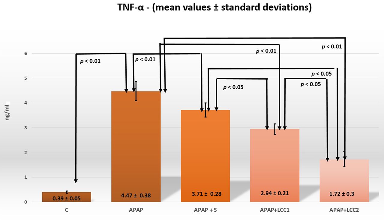
The levels of TNF-α were significantly different between the APAP group and the control group (p<0.01). Similar results (p<0.05) were obtained for the comparison between APAP+S, APAP+LCC1 and APAP+LCC2 groups. C: Control; APAP: acetaminophen administration; APAP+S: APAP and sylimarin administration as pretreatment; APAP+LCC1: APAP and pretreatment with 1 mg/100 g bw LCC; APAP+LCC2: APAP and pretreatment with 2 mg/100 g bw LCC.
Results
Treatment of rats with APA resulted in hepatoxicity as indicated by the increase, compared to control, in ALT and AST, markers of liver damage, and TNF-α, marker of inflammation (Table I, Figures 1, 2 and 3). In addition, after induction of hepatotoxicity, the oxidative stress parameters (NOx and MDA) were also significantly (Mann-Whitney test p<0.002) increased (Table I, Figures 4 and 5) as well as matrix metalloproteinases MMP-2 and MMP-9 (Table I, Figures 6 and 7). However, a significant decrease in the antioxidant capacity, quantified by thiols and catalase, was observed after APAP administration (Table I, Figures 8 and 9).

NOx value showed statistically significant differences in all the comparisons (p<0.001). C: Control; APAP: acetaminophen administration; APAP+S: APAP and sylimarin administration as pretreatment; APAP+LCC1: APAP and pretreatment with 1 mg/100 g bw LCC; APAP+LCC2: APAP and pretreatment with 2 mg/100 g bw LCC.

The levels of MDA were statistically significantly different at the level of p<0.01, when comparing C with APAP; APAP with APAP+S, APAP+LCC1 and APAP+LCC2, and at the level of p<0.05 when comparing APAP+S with APAP+LCC1 and APAP+LCC2; APAP+LCC1 and APAP+LCC2. C: Control; APAP: acetaminophen administration; APAP+S: APAP and sylimarin administration as pretreatment; APAP+LCC1: APAP and pretreatment with 1 mg/100 g bw LCC; APAP+LCC2: APAP and pretreatment with 2 mg/100 g bw LCC.
Effect of LCC1 and LCC2 pre-treatment on the levels of transaminases.
Pretreatment of rats with silymarin significantly (p<0.01) decreased ALT and AST levels following APAP administration (Figures 1 and 2). Furthermore, if the animals were pre-treated once with LCC, both ALT and AST levels were significantly (p<0.01) reduced compared with the APAP group (Figures 1 and 2). When comparing the hepatoprotective effect of the combination of silymarin with two liposomal curcumin concentrations, we observed an even more significant (p<0.01) decrease in AST and ALT levels compared to silymarin treatment alone, showing better improvement in hepatic cell function. Both doses of LCC were compared. The lowest values of AST and ALT were obtained with the high dose LCC2 (Figures 1 and 2, p<0.05).

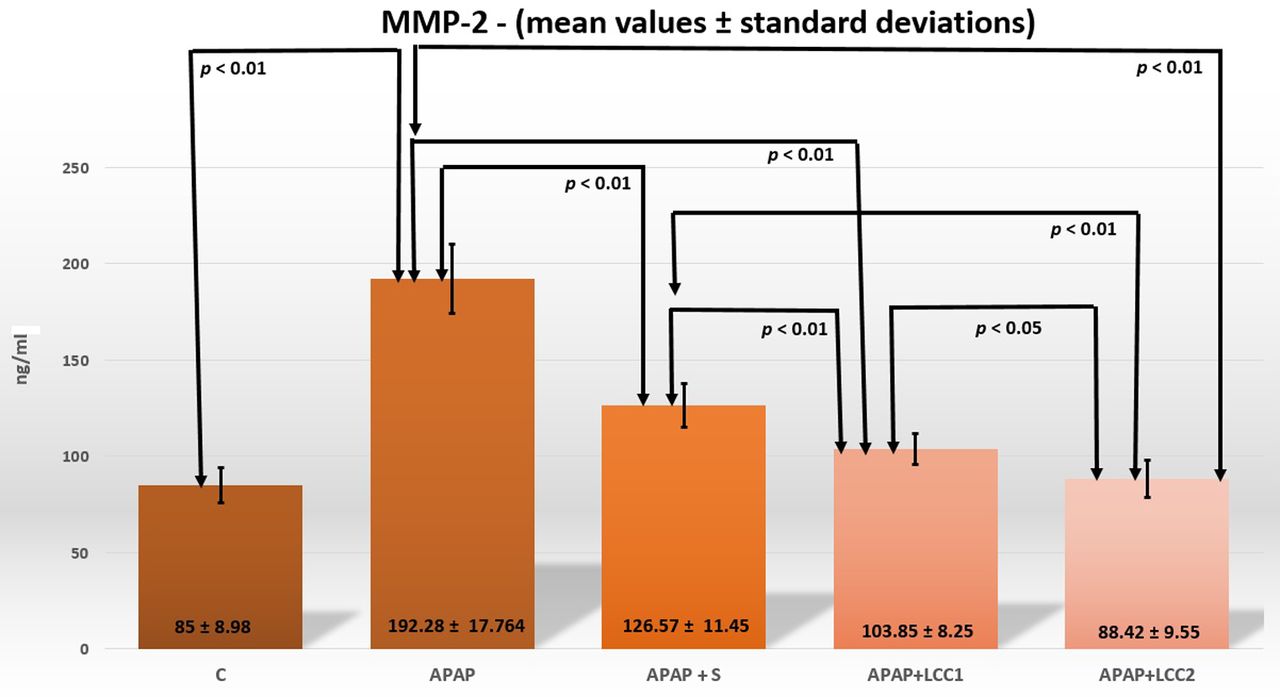
The levels of MMP-2 were significantly different between all compared groups (p<0.01) except between the LCC1 pretreatment group and the LCC2 pretreatment group p<0.05. C: Control; APAP: acetaminophen administration; APAP+S: APAP and sylimarin administration as pretreatment; APAP+LCC1: APAP and pretreatment with 1 mg/100 g bw LCC; APAP+LCC2: APAP and pretreatment with 2 mg/100 g bw LCC.

The levels of MMP-9 were statistically significantly different between all compared groups (p<0.01). C: Control; APAP: acetaminophen administration; APAP+S: APAP and sylimarin administration as pretreatment; APAP+LCC1: APAP and pretreatment with 1 mg/100 g bw LCC; APAP+LCC2: APAP and pretreatment with 2 mg/100 g bw LCC.
Effect of LCC1 and LCC2 pre-treatment on inflammation. TNF-α level was significantly (p<0.01) decreased is rats pretreated with the combination of silymarin with two doses of LCC (Figure 3), showing the anti-inflammatory effect of both substances during liver damage. Both doses of LCC were more efficient in decreasing inflammation than silymarin (p<0.05), however, the higher dose of LCC2 was more efficient than LCC1 in decreasing the levels of TNF-α (p<0.05) (Figure 3).

The levels of thiols, an antioxidant parameter, were statistically significant different at the level of p<0.01 when comparing C with APAP; APAP with APAP+S, APAP+LCC1 and APAP+LCC2, and at the level of p<0.05 when comparing APAP+S with APAP+LCC1 and APAP+LCC2; APAP+LCC1 and APAP+LCC2. C: Control; APAP: acetaminophen administration; APAP+S: APAP and sylimarin administration as pretreatment; APAP+LCC1: APAP and pretreatment with 1 mg/100 g bw LCC; APAP+LCC2: APAP and pretreatment with 2 mg/100 g bw LCC.
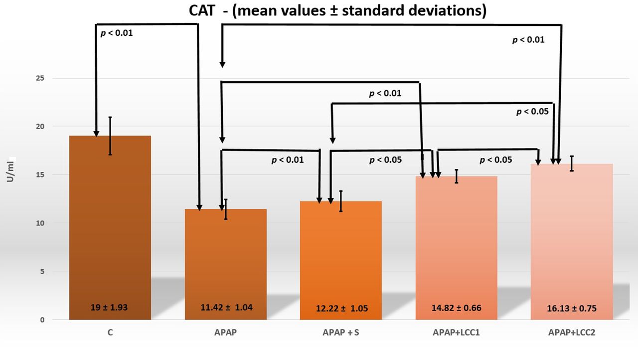
The levels of CAT, an antioxidant parameter, were statistically significant different at the levels of p<0.01 when comparing C with APAP; APAP with APAP+S, APAP+LCC1 and APAP+LCC2, and at the level of p<0.05 when comparing APAP+S with APAP+LCC1 and APAP+LCC2; APAP+LCC1 and APAP+LCC2. C: Control; APAP: acetaminophen administration; APAP+S: APAP and sylimarin administration as pretreatment; APAP+LCC1: APAP and pretreatment with 1 mg/100 g bw LCC; APAP+LCC2: APAP and pretreatment with 2 mg/100 g bw LCC.
Oxidative/antioxidant balance after LCC1 and LCC2 treatment. Oxidative stress markers induced by APAP administration were significantly decreased if the animals pre-treated with silymarin or LCC (Figures 4 and 5). The most significant decrease was observed for NOx, with a p<0.001 (Figure 4). Pre-treatment with LCC had a stronger effect than silymarin on reducing oxidative stress, as showed by the significant (p<0.05) decrease in MDA (Figure 5) and even more significant (p<0.001) decrease in NOx (Figure 4). The effect of LCC was dose-dependent and the higher dose had a stronger effect (p<0.05 for MDA and p<0.001 for NOx).

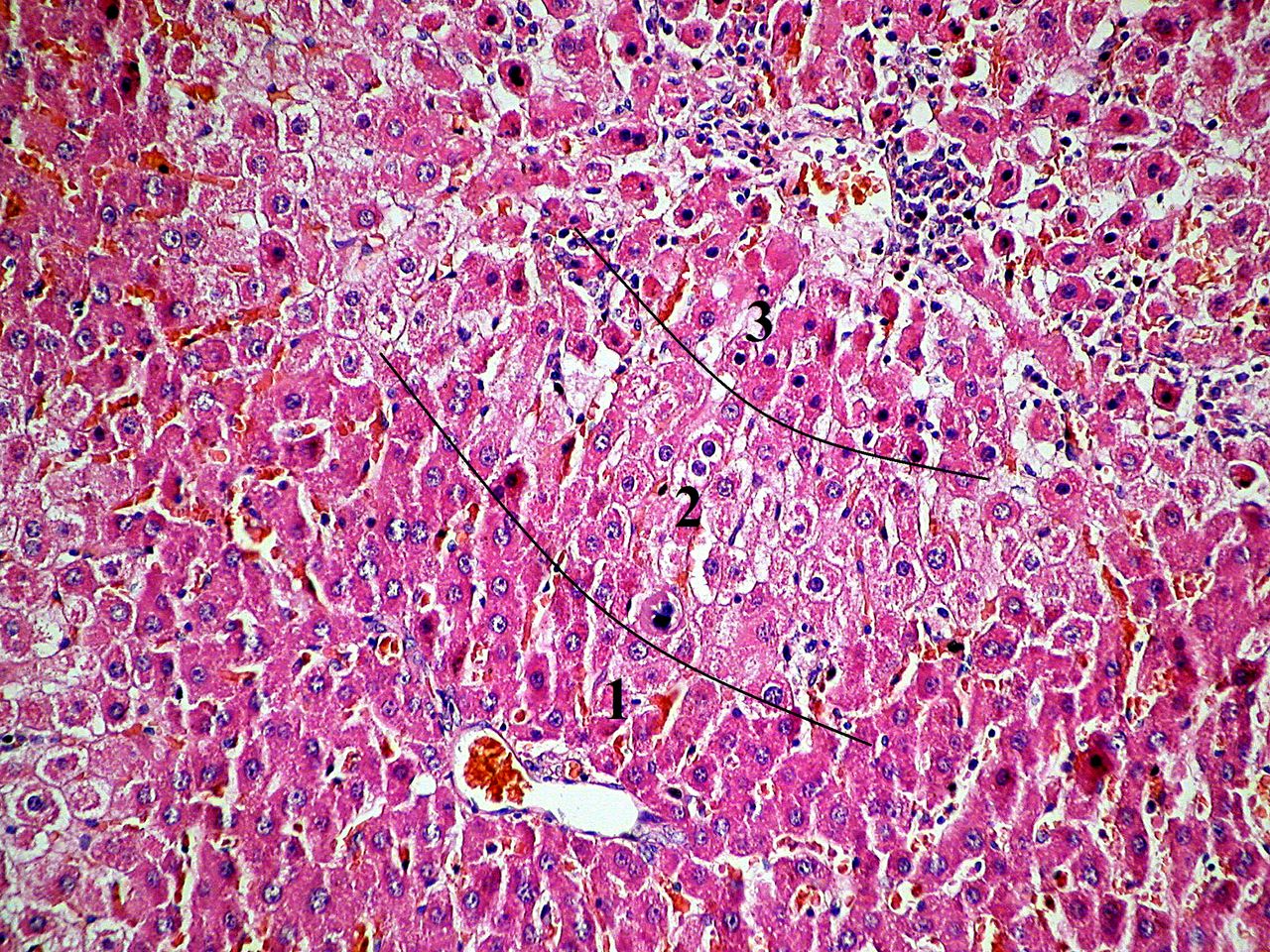
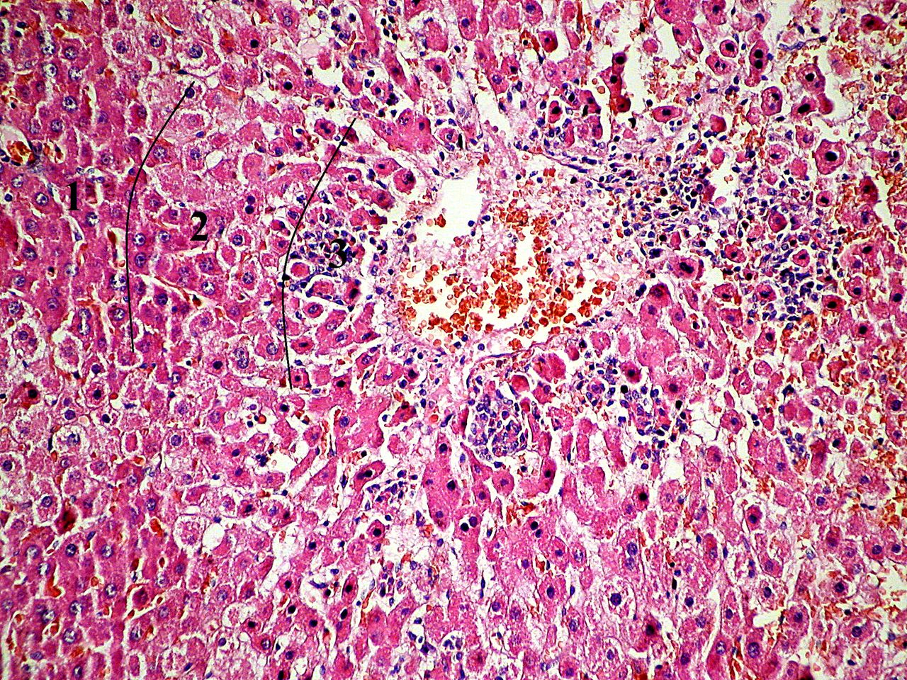
Liver group 2, hematoxylin-eosin staining, 40X ob; 1 – zone 1 of the hepatic acinus; 2 – zone 2 of the hepatic acinus; 3 – zone 3 of the hepatic acinus.
The antioxidant capacity was evaluated by measuring thiols and catalase. APAP administration resulted in reduced anti-oxidant capacity, which was reversed upon pre-treatment with silymarin or LCC; thus, significantly increased levels of thiols (p<0.01, Figure 8) and catalase (p<0.01, Figure 9) were observed in the treated groups. LCC pre-treatment reversed the antioxidant capacity to a higher level than silymarin (p<0.05 for thiols and catalase). The higher dose of LCC (LCC2) was the most effective in restoring antioxidant activity (p<0.05 for both thiols and catalase, when compared to LCC1).
Effect of LCC1 and LCC2 pre-treatment on MMP-2 and MMP-9. As shown above APAP treatment resulted in the activation of MMP-2 and MMP-9. This effect was significantly reduced (p<0.01) if the animals were pre-treated with silymarin or LCC (Figures 6 and 7). LCC had a significant (p<0.01) better effect in lowering both MMP-2 and MMP-9 levels than silymarin. The values of MMP-2 and MMP-9 reached those of the control group in the group treated with the highest dose of LCC (LCC2) (Figure 6 with p<0.05 for MMP-2 and Figure 7 with p<0.01 for MMP-9).
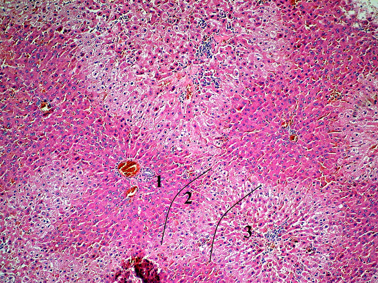
Histology assessment. In group 2 of animals, a large number of hepatocytes showed hydropic degeneration. Hepatocytes around the centrilobular venule (zone 3 of the hepatic acinus) were affected in high numbers. In zone 2 of the hepatic acinus, cells in various hydropic degeneration stages were found, among which cells with discrete signs of granular degeneration were interspersed (Figure 10). In group 3 of animals, lesions were less extensive; in zone 2 of the hepatic acinus, the number of hepatocytes with hydropic degeneration was reduced (Figure 11). In group 4 of rats, lesions had a predominantly granular appearance and mainly intersected hepatocytes in zone 2 of the hepatic acinus. In zone 3 of the hepatic acinus, hepatocytes had agranulovacuolar degeneration appearance (Figure 12). In group 5 of animals, the number of cells in zone 3 of the hepatic acinus with granulovacuolar degeneration appearance was further reduced compared to group 4. In zone 2 of the hepatic acinus, the majority of hepatocytes showed discrete granular degeneration and only in some places the degenerative appearance was more pronounced. In zone 1 of the hepatic acinus, degeneration seemed to be at an earlier stage compared to the other zones of the acinus (Figure 13).

Liver group 3, hematoxylin-eosin staining, 40X ob; 1 – zone 1 of the hepatic acinus; 2 – zone 2 of the hepatic acinus; 3 – zone 3 of the hepatic acinus.
Discussion
Liposomal curcumin effect on hepatic function and oxidative stress/antioxidant balance in APA-induced hepatotoxicity. Administration of an overdose of APAP resulted in destruction of hepatocytes and elevation of serum ALT and AST levels (Table I; Figures 1 and 2). Transaminase elevations are the most commonly used biomarkers for hepatocyte lesions. Hepatocyte destruction results from depletion of GLUT, a component of the liver antioxidant system, and from increased oxidative stress due to hepatic metabolism of APAP (42). Administration of APAP caused a dose-dependent depletion of hepatic GLUT (that plays a protective role against APAP overdose) (43). APAP is also oxidized by cytochrome P450 to a reactive metabolite, which can cause liver lesions (44). This mechanism leads to mitochondrial oxidative stress, increased mitochondrial membrane permeability and hepatic cell death (45). Mitochondrial oxidative stress is considered to be the main cellular dysfunction in APAP-induced liver injury (46). In our study, the levels of oxidative stress molecules (NOx and MDA) significantly increased after APAP administration (250 mg/kg) (Table II, Figures 4 and 5). Similar results were reported by other authors, thus, the suggested mechanism is APAP-induced activation of endothelial nitric oxide synthase (eNOS) and inducible nitric oxide synthase (iNOS) in hepatocytes (47). Other authors have reported that APAP can induce liver injury via an oxidative stress mechanism caused by increased MDA production (48). Besides GLUT, which has been proven to be depleted by APAP overdose, a decrease in other antioxidant molecules could change the cell oxidant/antioxidant balance. A significant reduction of thiol and catalase levels was obtained after APAP administration (Table II; Figures 8 and 9). Global protein sulfhydryl levels have been reported to decrease significantly starting one hour after APAP overdose, and slowly decreased after 24 h (49). This fact may provide a new insight into studying novel therapeutic molecules that can provide hepatoprotection; one of the mechanisms could be represented by improving the level of thiols. Improving catalase levels in experimental APAP-induced hepatotoxicity could also provide a new approach for hepatoprotection strategies (50). Therefore, modulation of oxidative stress/antioxidant balance could be the target of nutraceuticals used to provide a hepatoprotective effect. One of the most studied natural compounds for its antioxidative properties is curcumin. We have already demonstrated the beneficial effect of orally administered curcumin on hepatic function and on oxidative stress/antioxidant balance in experimental fructose-induced metabolic syndrome in rats (51). In this study, after APAP-induced hepatotoxicity challenge, the elevation in plasma ALT, AST, and oxidative stress molecules was ameliorated by pretreatment with nanoformulation of curcumin (lipososmal curcumin). We also obtained better results with LCC pretreatment compared to silymarin and a dose-dependent effect LCC, where a higher dose (LCC2) had higher efficiency (Table I and II; Figures 1, 2, 3, 4 and 5, 8, 9). The hepatoprotective effect of LCC could be linked to its already proven inhibitory effect on iNOS activity (52) and MDA production (51) as well as its ability to maintain the thiol pool (53) and to improve catalase production (54). In addition to mitochondrial oxidative stress, many other cellular processes, including inflammation, microcirculatory dysfunction and extracellular matrix degradation, have been shown to be involved in the pathogenesis of APAP-induced liver injury (46, 55).

Liver group 4, hematoxylin-eosin staining, 10X ob; 1 – zone 1 of the hepatic acinus; 2 – zone 2 of the hepatic acinus; 3 – zone 3 of the hepatic acinus.

Liver group 5, hematoxylin-eosin staining, 10X ob; 1 – zone 1 of the hepatic acinus; 2 – zone 2 of the hepatic acinus; 3 – zone 3 of the hepatic acinus.
Liposomal curcumin effect on pro-inflammatory cytokine TNF-α in APAP-induced hepatotoxicity. In APAP hepatotoxicity, increased oxidative stress results in DNA fragmentation followed by an inflammatory reaction and production of pro-inflammatory cytokines (5). One of the most representative pro-inflammatory cytokines associated with hepatic liver injury is TNF-α (56). TNF-α is produced in response to liver injury induced by APAP overdose (57). Activated liver macrophages (Kupffer cells) are the main cells responsible for TNF-α production that further mediates hepatocyte necrosis (58). Our study observations (Table I; Figure 3) are in agreement with other studies that have reported an increase in TNF-α in APAP-induced hepatotoxicity (57, 59, 60). A possible mechanism is the enhancement of an intense inflammatory reaction that includes TNF-α increase through oxidative stress (61). TNF-α and other pro-inflammatory cytokines can initiate the pathological changes following APAP overdose (62), which were also observed in our study (Table I, Figure 3). TNF-α is an important cytokine that can trigger the apoptosis cascade through activation of the caspase-dependent apoptosis mechanism (63). Continuous release of TNF-α during inflammation may rapidly lead to necroptosis and necrosis through an unknown mechanism (62). Multiple studies have demonstrated the role of curcumin in TNF-α modulation (63-65), but not as a liposomal formula. Our study demonstrated the hepatoprotective effect of both LCC concentrations in reducing the serum levels of TNF-α in APAP-induced hepatotoxicity (Table I, Figure 3). Both LCC concentrations were more efficient than silymarin pretreatment (Table I, Figure 3). TNF-α reduction was also demonstrated by oral curcumin administration or by other curcumin nanoformulations (66-68). The liposomal formulation of curcumin can enhance its anti-inflammatory properties through down-regulation of TNF-α due to its higher bioavailability, and a better distribution in therapeutic concentration at the injury site (69).
Levels of the investigated markers of oxidative stress and antioxidant parameters in each treatment group, expressed as mean and standard deviation.
Liposomal curcumin effect on MMP-2 and MMP-9 in APAP-induced hepatotoxicity. Significantly higher levels of MMP-2 and MMP-9 were observed following APAP administration (Table III; Figures 6 and 7). Both LCC1 and LCC2 reduced the MMP-2 and MMP-9 with LCC2 having the stronger effect, when compared with APAP+S group (Table III; Figures 6 and 7). Increased serum MMP levels have been shown to be associated with hepatocellular damage, contributing to hepatic microcirculation dysfunction and facilitating the arrival of leukocytes at the injury site (70). MMP-2/MMP-9 inhibition has been proven to reduce parenchymal and microvascular injury through minimizing endothelial injury (70). After liver contusion, some authors have reported an enhancement of MMP-2 at 6 h, with a peak at 24 h, then a gradual reduction with normalization of the levels after 7 days (71). ECM degradation by MMP is added to the hepatic microcirculation disturbances caused by NOx excess (72). Intracellular damage induced by APAP overdose is amplified by increased MMP and elicits a robust inflammatory response with the release of pro-inflammatory cytokines that will attract more neutrophils at the injury site (73). After the endothelial injury, accumulated neutrophils together with MMP will contribute to liver injury, but also to liver regeneration after the lesion stimulus ceases (73). It is important to mention that MMPs have an important physiological role in the liver. MMPs are produced by various cells such as Kupffer cells, hepatocytes, cholangiocytes and, in normal amounts, can control inflammation and fibrosis in hepatic tissue (74). Based on these findings and on our results, we consider that the excess of MMP-2 and MMP-9 contributes to the increase of oxidative stress and to the propagation of inflammation, amplifying the hepatic cytolysis process. Similarly to our results, other studies have reported a reduction in liver lesions by down-regulation of MMP production (75-78). Curcumin suppression of MMP expression has also been reported by other studies (79-80), but our findings proved that the liposomal formulation of curcumin can have beneficial effects in improving oxidative stress/antioxidant balance and inflammation in hepatotoxicity induced by APAP in rats. To our knowledge, this is the first report regarding the hepatoprotective effects of a liposomal formulation of curcumin.
Metalloproteinase levels in each treatment group.
Conclusion
Despite all studies performed so far, there are very few therapy choices for liver injuries. This study provides a new perspective for therapeutic strategies to alleviate hepatic lesions related to APAP overdose. Our results provide evidence that liposomal curcumin can exert a protective effect against APAP-induced hepatotoxicity by reducing NOx and MDA production, improving thiol and catalase levels, and reducing the serum concentration of TNF-α, MMP-2 and MMP-9. Therefore, liposomal curcumin formula may be a promising therapy for APAP-induced liver injury, with beneficial effects on associated oxidative stress and inflammation.
Acknowledgements
The Authors would like to thank Dr. Alina Porfire and Lucia Tefas for the liposomal curcumin preparation, Ana Uifalean for biochemical assessments, and Mirel Molnar for helping in rat confinement and for collecting the blood samples.
Footnotes
Authors' Contributions
Bulboacă Adriana Elena – study concept and design, analysis and interpretation of the data, writing the first draft of the manuscript; Dogaru Gabriela – study concept, organization of manuscript, review and critique; Gheban Dan – histological analysis; Boarescu Paul Mihai – organization of the manuscript, analysis and interpretation of the data; Rus Vasile – manuscript review and critique; Festila Dana – organization of the manuscript, analysis and interpretation of the data; Sitar-Taut Adela-Viviana – statistical analysis; Stănescu Ioana – organization of manuscript, review and critique.
This article is freely accessible online.
Conflicts of Interest
The Authors declare no conflicts of interest regarding this study.
- Received October 30, 2019.
- Revision received December 2, 2019.
- Accepted December 3, 2019.
- Copyright© 2020, International Institute of Anticancer Research (Dr. George J. Delinasios), All rights reserved


