


Abstract
Background: It was recently confirmed that ethacrynic acid (EA) inhibits Wnt/beta catenin signalling in myeloma. Materials and Methods: This study investigated the antitumor effect of EA in vitro and in vivo in a murine myeloma model. Results: EA demonstrated major apoptotic activity in different human and murine myeloma and lymphoma cell lines, as well as in human primary cells. In addition β-catenin expression was down-regulated when EA was added to lymphoma cells. In vivo, tumor growth, as well as overall survival, was significantly reduced in mice treated with EA as compared to untreated mice. Interestingly, in vitro, a significant additive effect was seen with the combination of lenalidomide plus EA as compared to single applications. Conclusion: These results reveal a significant selective induction of apoptosis by EA and suggest a significant in vivo effect against myeloma.
Major progress has been achieved in the treatment of multiple myeloma by the introduction of novel agents such as thalidomide, lenalidomide and bortezomib. Nevertheless, myeloma remains an incurable disease. In newly diagnosed patients, the combination of lenalidomide and dexamethasone has a response rate of 91%.
Several groups have shown that the Wnt/β-catenin pathway plays an important role in the regulation of cell proliferation, differentiation and apoptosis (1-3). Aberrant activation of the Wnt signaling pathway has major oncogenic effects (4-7). In the canonical Wnt pathway, the secreted Wnt proteins bind to a receptor complex, consisting of a member of the frizzled (Fzd) family, and the low-density lipoprotein receptor-related proteins (LRP) 5 or LRP6. Subsequently the cytoplasmic adaptor protein disheveled (Dvl) is phosphorylated and inhibits glycogen synthase kinase (GSK)-3β activity through its association with axin. Unphosphorylated β-catenin accumulates in the cytoplasm and translocates into the nucleus, where it interacts with T-cell (TCF) and lymphoid-enhancing (LEF) factors to activate transcription of Wnt target genes (4, 5, 8). In addition, it has been demonstrated that the Wnt pathway is activated in lymphoma. Therefore, the Wnt/β-catenin signaling molecules are attractive candidates for development of targeted approaches in lymphoma treatment.
It was recently confirmed that the diuretic agent ethacrynic acid (EA) and the antifungal agent ciclopirox olamine (cic) inhibit Wnt/beta catenin signaling (9). EA is already used clinically as a diuretic agent. Glutathione-S-transferase (GST), which is over expressed in human tumors, in form of GST-P binds glutathione (GSH) to electrophilic compounds leading to detoxification of the cell (10-13). GSH is a reducing agent and an antioxidant. The binding of EA to GSH can enhance the cytotoxicity of chemotherapeutic agents (14, 15).
For all of these reasons, we tested the effect of EA on myeloma cells in combination with the commonly used therapeutic drugs.
Materials and Methods
Cell lines and culture conditions. The lymphoma cell lines LAM-53, SU-DHL-4, Daudi and Raji, as well as the myeloma cell lines OPM-2, RPMI-8226 and U-266 (all obtained from DSMZ, Collection of Microorganisms and Cell Culture, Braunschweig, Germany) were cultured in RPMI-1640 medium consisting of 10% heat-inactivated fetal calf serum (FCS; Invitrogen, Karlsruhe, Germany), 2.5% 1 M HEPES, and 1× penicillin/streptomycin (all from PAA Laboratories GmbH, Cölbe, Germany). Cells were cultured at a density of 3.3×10⁵ cells/ml and incubated at 37°C with 5% CO2 and 95% humidity.
MPC11 (DMSZ) is a murine plasmocytoma cell line derived from the Balb/c strain expressing IgG2b. Cells were cultured in RPMI-1640 medium (PAA Laboratories GmbH) supplemented with 5% FCS, 2 mM glutamine (both from PAA, Cölbe, Germany), 100 U/ml penicillin/100 U/ml streptomycin (both from Seromed, Jülich, Germany) at 37°C in a humidified 5% CO2 atmosphere.
Human samples. Peripheral blood mononuclear cells (PBMCs) and peripheral blood lymphocytes (PBLs) were isolated from blood samples of healthy volunteers by Ficoll density gradient centrifugation (Lymphoprep, Nycomed, Oslo, Norway). In addition, bone marrow samples from patients with myeloma were obtained. Ethics approval had been obtained according to the guidelines of the host institution and all samples were taken after signed informed consent.
Drugs and chemical reagents. The following drugs were used in this study: ethacrynic acid (EA; Sigma-Aldrich GmbH, Seelze, Germany), doxorubicin (Cellpharm GmbH, Germany), rituximab (Roche Pharma AG, Grenzach Wyhlen, Germany), bortezomib (Janssen Cilag, Neuss, Germany), thalidomide (Grünenthal Pharma GmbH, Aachen, Germany), and lenalidomide (Celgene, Munich, Germany). All drugs were tested at different concentrations for 24-72 hours.
DiOC6 and propidium iodide (PI)-staining. A total of 1×10⁵ Cells were cultured in 3 ml medium in 6-well plates. EA was dissolved in dimethyl sulfoxide (DMSO), and added at an optimized concentration in the range of 20-100 μM alone or in combination with the therapeutic agents at different concentrations for three days. The apoptosis assay was performed with 3’,3-dihexyloxacarbocyanine iodide (DiOC6) detecting mitochondrial membrane potential in viable cells, and propidium iodide (PI) which binds to DNA in necrotic cells, measured by a fluorescence-activated cell sorter (FACS).
For FACS analysis, 500 μl staining solution containing 80 nM DiOC6 in FACS buffer, consisting of deficient RPMI medium with 0.5% bovine serum albumin (BSA), was mixed with equal volumes of the cell sample in a glass tube and incubated at 37°C for 15 min. After a washing step with PBS/1% BSA, the cells were re-suspended in 500 μl PBS/1% BSA. After addition of 5 μl PI solution (100 μg/ml), the cells were analyzed by FACS. Using this assay, viable cells reveal high fluorescence intensity for DiOC6 and a low expression for PI. On the contrary, apoptotic cells show a low expression for DiOC6 and also a low expression for PI (2). Finally, necrotic cells show a low expression for DiOC6 and a high expression for PI.
A mean IC50 value in myeloma cells was determined using the mean of the IC50 results determined in OPM-2, U-266 and RPMI-8226 cells.
Isolation of PBMCs. PBMCs were isolated from blood of healthy donors by Ficoll-Hypaque density gradient centrifugation. Blood from buffy coats was mixed 1:2 with PBS/1% BSA (both PAA, Cölbe, Germany) and used for a ficoll gradient (LymphoPrep). After the centrifugation at 800×g for 30 min, the leukocyte layer was removed and transferred to new tubes. Subsequently, these cells were washed three times with PBS/1% BSA and re-suspended in fresh medium, consisting of RPMI (PAA, Cölbe, Germany) with 10% FCS (Invitrogen), 2.5% HEPES buffer solution (PAA, Cölbe, Germany), and 1% penicillin/streptomycin (PAA, Cölbe, Germany).
Bone marrow samples. Single-cell suspensions were generated from patient bone marrow samples and the cells were ficolled. Cells were incubated with or without EA at an optimized concentration in the range of 20-100 μM for three days and then measured for viability.
Western blot. The effect of EA on the Wnt/beta catenin pathway was analyzed by Western blot. Western blot was performed as described elsewhere (16).
Animal studies. All animal experiments were performed at least in duplicate with groups of six BALB/c mice (6 weeks old, 25 g, female, Charles River, Sulzfeld, Germany). MCP11 murine myeloma cells (5×10⁵) were injected subcutaneously into each Balb/c mouse. Mice were treated orally with 450 μg/day EA and 750 μg lenalidomide/day, respectively for a time frame of 60 days. Overall survival and tumor growth were measured. Tumor volume was calculated as follows: volume=length×width2×0.52. Animals were sacrificed when the tumor volume reached 2000 mm3.
Statistical analysis. For statistical analysis, the results comprising the relative viability were expressed as the mean±standard deviation (SD). Different sample sizes (n) were chosen for different cell lines. Student's t-test was used for statistical analysis. A p-value <0.05 was considered significant. Statistical survival analyses were performed with the software GraphPad InStat, Version 3.0.0 (GraphPad Software, San Diego, CA, USA). The Mann-Whitney test (non-paired, non-parametric) was also applied.
Results
Effect of EA on Wnt/beta catenin signaling. In our recent research we used a 96-well plate-based TOPflash reporter system to screen the Gen-plus drug library (Microsource) that contained 960 compounds. The screen identified EA and cic as Wnt/beta catenin inhibitors (16). Since the canonical Wnt signaling pathway is activated in lymphoma and myeloma cells, we further investigated if EA could induce apoptosis and reduce the viability of lymphoma and myeloma cell lines, respectively.
Titration of EA. As a first step, we determined the optimal concentration of EA in various lymphoma and myeloma cell lines. Subsequently, we determined the optimal concentration of EA in PBMCs derived from healthy individuals in order to check for toxicity. Determination of the optimal concentration of EA revealed that 30 μM EA was the most effective concentration to initialize cell death in both lymphoma and myeloma cell lines, without significantly reducing the viability of normal PBMCs (data not shown). The effect of DMSO as a toxic solvent was only observed in the myeloma cell line OPM-2 (Figure 1C). As a control, PBMCs were investigated by FACS analysis. A toxic effect on the cells induced a shift from viable, DiOC6-positive cells to apoptotic, DiOC6-negative cells.

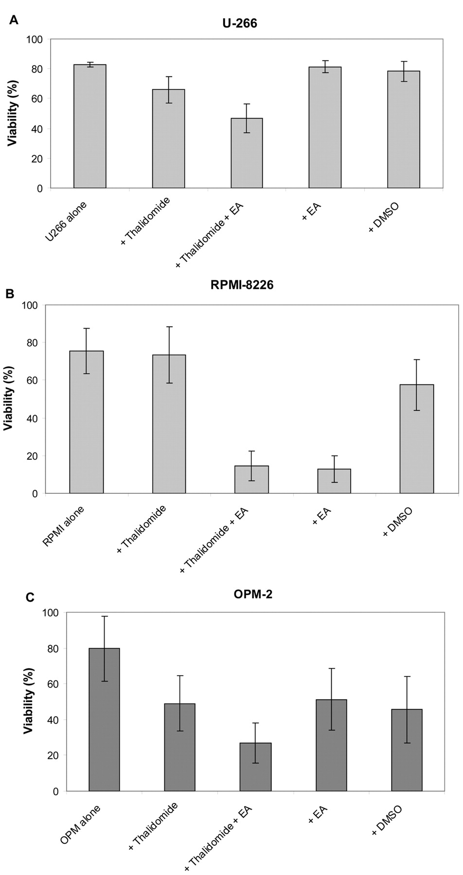
Effect of thalidomide in combination with EA on viability of U-266 (A), RPMI-8226 (B) and OPM-2 (C) myeloma cells. Cells (1×10⁵) were cultured with different compounds for three days. The concentration of thalidomide was 160 μM for U266, 20 μM for RPMI, and 40 μM for OPM cells. EA was used at a concentration of 20 μM. Cell viability was then measured by DiOC6 staining in flow cytometry. Results represent data from three, two and five separate experiments, respectively. U-266 n=3, RPM-8226 n=2, OPM n=5 experiments. Data are shown as the mean±SD.

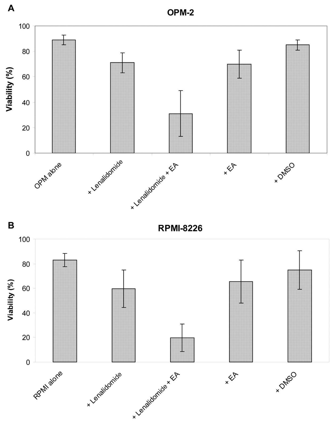
Effect of lenalidomide in combination with EA on viability of OPM-2 (A) and RPMI-8226 (B) myeloma cells. Cells were cultured with the different compounds for three days. The concentration of lenalidomide was 12 μM for OPM-2 and 80 μM for RPMI-8226 cells. EA was used at a concentration of 20 μM. Cell viability was then measured by DiOC6 staining in flow cytometry. Results represent data from OPM n=8 and RPMI n=5 separate experiments, respectively. Data are shown as mean±SD.
Effect of EA on viability of cell lines and PBMCs. Viability decreased slowly over time. After 72 h, the relative viability for the lymphoma cell lines SU-DHL-4, LAM-53 and Raji in the presence of EA (30 μM) was 97.6±1.1%, 91.0±4.9% and 78.9±2.1%, respectively (17). These values were similar to values of the control of PBMCs at 89.5±3.1%.
Effect of DMSO on viability of cell lines. In contrast to PBMC and lymphoma cell lines, the myeloma cell line OPM-2 was highly sensitive towards DMSO and showed a decrease of relative viability to 66.8±2.3%.
Effect of doxorubicin in combination with EA on viability of lymphoma cell lines. In general, lymphoma patients are treated with doxorubicin and rituximab besides other drugs. Therefore, we tested these drugs in lymphoma cells in combination with EA.
For Daudi and Raji cells, the addition of doxorubicin led to a significant decrease in viability. However, the combination of EA plus doxorubicin did not further decrease viability of Raji cells. Therefore, we excluded there being a synergistic effect (data not shown).
Effect of rituximab in combination with EA on viability of lymphoma cell lines. For Daudi and Raji cells, the addition of rituximab led to significantly reduced viability of these cells. However, the combination of EA plus rituximab did not further reduce viability of Daudi and Raji cells. Like with doxorubicin, we excluded there being a synergistic effect of this combination (data not shown).

Effect of EA on the Wnt/beta catenin pathway in lymphoma cells. A: Flow cytometric results. B: Effect of EA on the Wnt/β-catenin pathway by Western blot.

Western Blot analysis for the detection of β-catenin in myeloma cell lines. A: Under normal conditions. The myeloma cell lines RPMI-8226, KMS-18 and U-266 were analyzed according for their β-catenin expression. B: Under treatment with EA at 90 μM for 24 h of KMS-18 and RPMI-8226 cells. Cells were treated with EA for 24 h, cells were lysed and then Western blot analysis was performed for β-catenin. β-Actin immunoblotting served as the loading control.
Effect of bortezomib in combination with EA on viability of myeloma cell line OPM-2. Treatment of myeloma has changed in recent times. More and more patients are treated with novel drugs, such as bortezomib, lenalidomide and thalidomide. Therefore, we tested these drugs in OPM-2 myeloma cells in combination with EA.
In OPM-2 cells, the addition of bortezomib led to a significant decrease in viability of OPM-2 myeloma cells. However, the combination of EA plus bortezomib did not further decrease viability of OPM cells. A synergistic effect of this combination was also excluded (data not shown).
Effect of thalidomide, lenalidomide and EA on viability of myeloma and lymphoma cell lines. Thalidomide and EA significantly (p<0.05) reduced the viability of myeloma cell lines in vitro (Table I). From several myeloma cell lines, a mean IC50 value in myeloma cells was determined: EA (30 μM) was significantly more effective than lenalidomide (>200 μM) and thalidomide (>251 μM; Table I).
In contrast, the effect on normal PBMCs and PBLs was marginal with EA and lenalidomide. Here, no effect was found for thalidomide (Table I).
Only EA significantly (p<0.05) reduced the viability of lymphoma cell lines in vitro (Table I). In contrast, lenalidomide and thalidomide showed no effect (Table I).
Effect of thalidomide in combination with EA on viability of myeloma cell lines. For OPM-2, U-266 and RPMI-8226 cells, the addition of thalidomide led to a significant decrease in viability of OPM-2, U-266 and RPMI-8226 myeloma cells. Interestingly, the combination of EA plus thalidomide led to a further decrease of viability of OPM-2, U-266 and RPMI-8226 cells; suggesting a synergistic effect for this combination (Figure 1).
Effect of lenalidomide in combination with EA on viability of myeloma cell lines. For OPM-2 and RPMI-8226 cells, the addition of lenalidomide led to a significant decrease in viability of OPM-2 and RPMI-8226 myeloma cells. Similarly to thalidomide, the addition of lenalidomide to EA led to further reduced viability of both OPM-2 and RPMI-8226 cells. These results indicate a synergistic effect for this combination (Figure 2).
Effect of EA on the Wnt/beta catenin pathway in lymphoma and myeloma cells. As a next step, we analyzed the effect of EA on the Wnt/beta catenin pathway. The myeloma cell line OPM-2 did not express as much β-catenin compared to lymphoma cell lines. However, β-catenin was down-regulated when EA was added to lymphoma cells (Figure 3).
The effect of EA on β-catenin expression in myeloma cells was tested. KMS18 and RPMI cells were found to be β-catenin-positive in contrast to U-266 cells, which where negative (Figure 4A). When exposed to EA, β-catenin expression increased in KMS-18 cells and slightly increased in RPMI-8226 cells (Figure 4B).

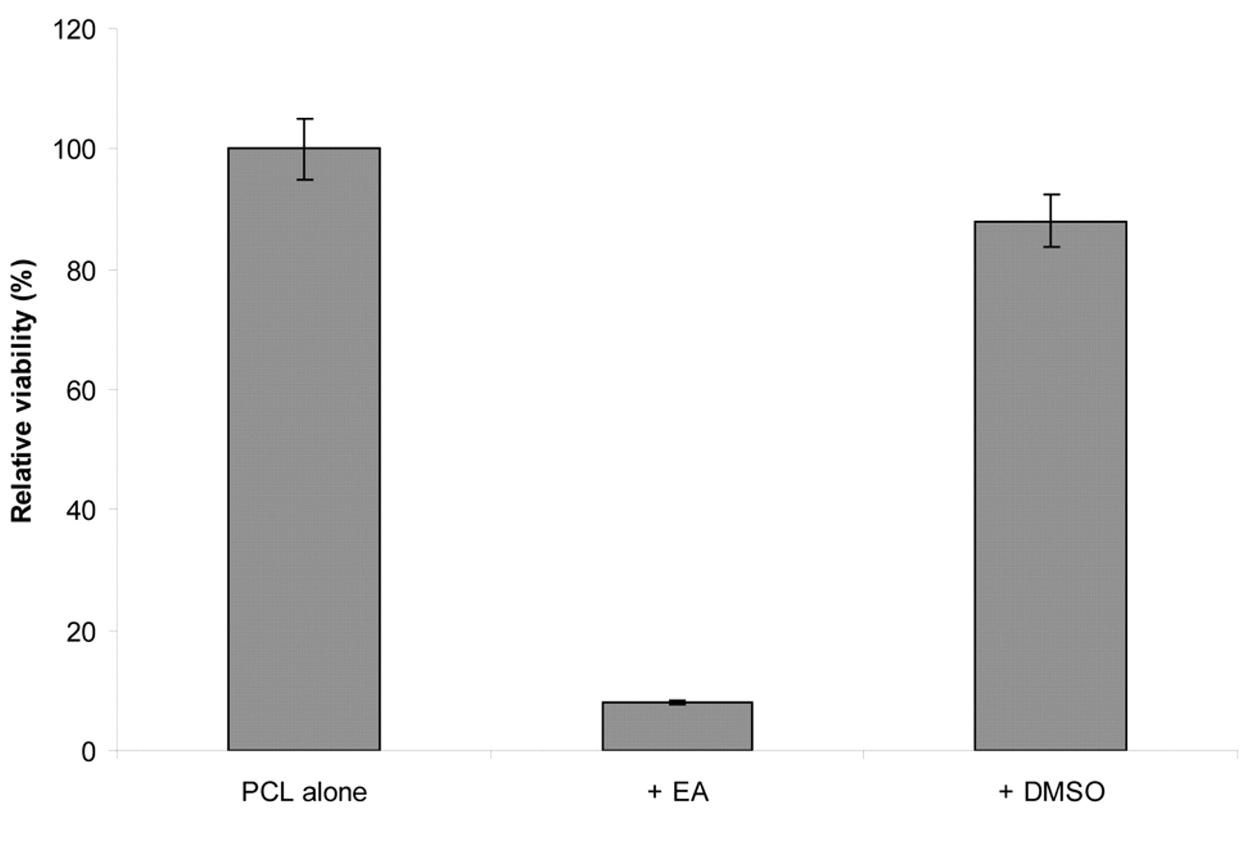
Effect of EA on viability of primary lymphoma cells. Primary lymphoma cells (PCL) derived from blood from patients with chronic lymphocytic lymphoma were incubated for three days with 100 μM EA. Cell viability was then measured by DiOC6 staining in flow cytometry. Results represent data from two separate experiments. Data are shown as the mean.
Effect of EA on viability of primary myeloma cells. Preliminary data suggest that EA inhibits the growth of primary myeloma cells derived of bone marrow from patients with multiple myeloma (Figure 5).
Effect of lenalidomide, EA and of the combination of lenalidomide and EA in vivo. The effect of lenalidomide, EA and of the combination of lenalidomide and EA was tested in vivo. BALB/c mice were injected with 5×10⁵ MPC11 myeloma cells subcutaneously. Tumor growth as well as overall survival were significantly reduced (p<0.001, respectively) in mice treated with EA or EA plus lenalidomide as compared to mice treated with lenalidomide alone (Figure 6).
Discussion
The Wnt signaling pathway has been shown to play a critical role in the early phases of B lymphocyte development. Multiple myeloma (MM) cells, but not cells from healthy donors and patients with monoclonal gammopathy of undetermined significance or other plasma cell dyscrasias involving bone marrow express the Wnt-signaling antagonist DKK1 (18). It has been reported that secretion of DKK1, a stress responsive gene, by MM cells likely contributes to the formation of osteolytic lesions in this disease by inhibiting Wnt signaling, the latter being essential for osteoblast differentiation and survival. Changes of DKK1 expression in MM cells can be traced through disturbances in the JNK signaling cascade which is differentially modulated by oxidative stress and interactions between MM cells with osteoclasts in vitro. Despite its role as a tumor suppressor and mediator of apoptosis in other cell types, including osteoblasts, the data indicate that DKK1 does not mediate apoptotic signaling, is not activated by TP53, and its forced overexpression does not inhibit cell growth or sensitize MM cells towards apoptosis, following treatment with either thalidomide or lenalidomide. Therefore, specific strategies may be beneficial to modulate persistent activation of the JNK pathway in preventing disease progression and treating myeloma-associated bone disease by inhibiting DKK1 expression (18).
The effect of ethacrynic acid (EA) on myeloma (OPM-2, U-266, RPMI-8226, KMS-18 and MPC11) and lymphoma (OCI-Ly8 LAM53, SU-DHL-4 and Raji) cell lines was assayed. Alternatively, thalidomide and lenalidomide were used for comparison. Peripheral blood lymphocytes (PBLs) derived from healthy individuals were used as normal controls. A total of 1×10⁵ cells were cultured with each compound using different concentrations for three days. Then cell viability was measured by DiOC6 staining by flow cytometry. Results represent data from two to four separate experiments each. ND: Not done.
Many hypotheses have been proposed to explain the molecular mechanism of thalidomide teratogenicity, in particular regarding limb defects. Most in vivo experimental evidence has been provided for a model suggesting the generation of oxidative stress by thalidomide with subsequent down-regulation of both Wnt and Akt survival pathways. In addition, transcription factors Tbx5 and Sall4 are involved in thalidomide-induced molecular pathology (19).
Since thalidomide down-regulates the Wnt pathway and is used for treatment of myeloma patients, testing the combination of thalidomide and Wnt inhibitors is of particular interest. We used EA, a drug that has recently been shown to induce apoptosis and down-regulate β-catenin expression in lymphoma cells. Most interestingly, we demonstrated a synergistic effect of the combination of thalidomide and EA for myeloma cells.
In addition to thalidomide, patients with myeloma are also being treated more frequently with the novel agents bortezomib and lenalidomide. Therefore, we tested these drugs in myeloma cells in combination with the Wnt inhibitor EA. Our research demonstrates a synergistic effect of the combination of lenalidomide/thalidomide and EA for myeloma cells. Recently, similar results were obtained with other Wnt inhibitors (20, 21). However, we failed to demonstrate such an effect for the combination of bortezomib and the Wnt inhibitors. This might be explained by the molecular relationship of thalidomide and lenalidomide, in contrast to bortezomib which belongs to a completely different class of drugs. In the lymphoma cell lines SU-DHL4 and LAM-53, the Wnt inhibitor EA induced apoptosis through down-regulation of β-catenin, an important molecule within the Wnt pathway.
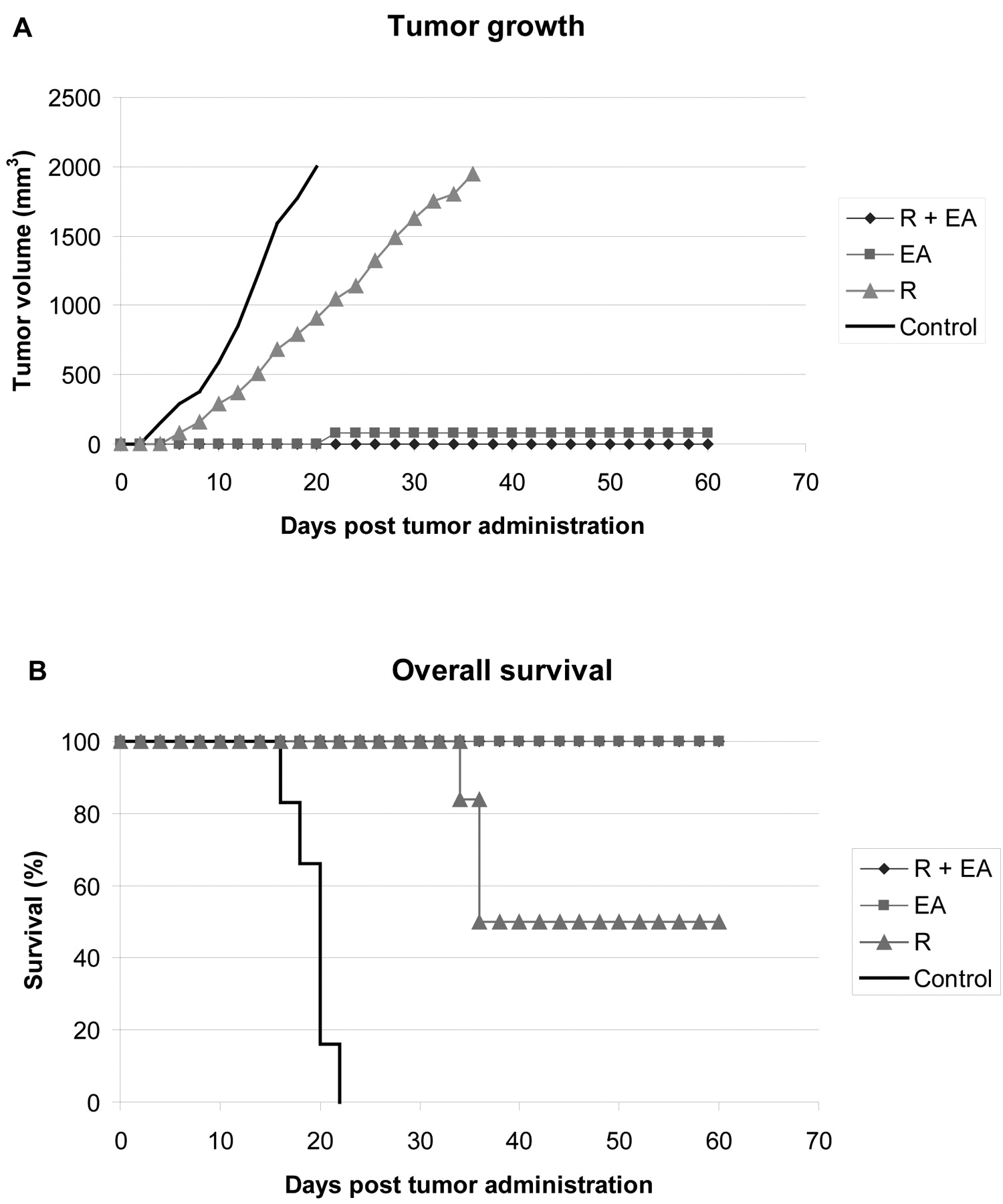
The effect of lenalidomide (R) (750 μM/mouse/day), EA (450 μM/mouse/day) and of their combination was tested in vivo. BALB/c mice were injected with 5×10⁵ MPC11 myeloma cells subcutaneously. Tumor growth (A) and overall survival (B) were measured. Six animals were assayed per group.
Recently, we also showed that EA is efficacious in primary cultures derived from patients with chronic lymphocytic leukemia (CLL) (16). EA was identified as a Wnt inhibitor using a cell-based Wnt reporter assay. In vitro assays further confirmed the inhibitory effect of EA on Wnt/β-catenin signaling. Cell viability assays showed that EA selectively induced cell death in primary CLL cells. Exposure of CLL cells to EA reduced the expression of Wnt/β-catenin target genes, including LEF-1, cyclin D1 and fibronectin. Immune co-precipitation experiments demonstrated that EA was able to directly bind to LEF-1 protein and destabilize the LEF-1/β-catenin complex. N-Acetyl-L-cysteine (NAC), which can react with the α,β-unsaturated ketone in EA, but not other antioxidants, prevented the inhibition of Wnt/β-catenin activation by EA and its ability to induce apoptosis in CLL cells.
Our results are in accordance with a recent report of Sukhdeo et al. demonstrating that the canonical Wnt signaling pathway is activated in MM through constitutively active β-catenin (22). Interestingly, we demonstrated a synergistic effect of the combination of thalidomide as well as lenalidomide and Wnt inhibitors in myeloma cells. This observation might lead to novel treatment options being developed for patients with MM.
Acknowledgements
We kindly acknowledge the support of T. Kipps, La Jolla, U.S.A. and P. Brossart, Bonn, Germany, the excellent technical expertise of Petra Alpmann and Sabine Blaum-Feder and Axel Glasmacher, Bonn for his support. This study was kindly supported in part by the Tumorinitiative von Haller e.V., Bonn and by the Deutsche Krebshilfe, Bonn.
- Received December 28, 2010.
- Revision received March 2, 2011.
- Accepted March 3, 2011.
- Copyright © 2011 International Institute of Anticancer Research (Dr. John G. Delinassios), All rights reserved
References
In this issue


Jump to section
Related Articles
Cited By...
- Optimization of 3-Cyano-7-cyclopropylamino-pyrazolo[1,5-a]pyrimidines Toward the Development of an In Vivo Chemical Probe for CSNK2A
- Optimization of 3-Cyano-7-cyclopropylamino-pyrazolo[1,5-a]pyrimidines Toward the Development of an In Vivo Chemical Probe for CSNK2A
- In Vitro Apoptosis Induction by Fenofibrate in Lymphoma and Multiple Myeloma
- Griseofulvin Efficiently Induces Apoptosis in In Vitro Treatment of Lymphoma and Multiple Myeloma
- Glutathione-S-transferases and Chemotherapy Resistance of Hodgkin's Lymphoma Cell Lines
- Clofibrate Demonstrates Efficacy in In Vitro Treatment of Lymphoma and Multiple Myeloma
- In Vitro Efficacy of Naftifine Against Lymphoma and Multiple Myeloma
- Flunarizine Exhibits In Vitro Efficacy Against Lymphoma and Multiple Myeloma Cells
- In Vitro Efficacy of Cinnarizine Against Lymphoma and Multiple Myeloma
- Targeting the Wnt/Beta-Catenin Pathway in Renal Cell Carcinoma
- Targeting the Wnt/Beta-Catenin Pathway in Multiple Myeloma
- Targeting Renal Cancer with a Combination of WNT Inhibitors and a Bi-Functional Peptide