Article Text

Abstract
Objective Several epidemiological studies have shown that regular exercise can prevent the onset of colon cancer, although the underlying mechanism is unclear. Myokines are secreted skeletal muscle proteins responsible for some exercise-induced health benefits including metabolic improvement and anti-inflammatory effects in organs. The purpose of this study was to identify new myokines that contribute to the prevention of colon tumorigenesis.
Methods To identify novel secreted muscle-derived proteins, DNA microarrays were used to compare the transcriptome of muscle tissue in sedentary and exercised young and old mice. The level of circulating secreted protein acidic and rich in cysteine (SPARC) was measured in mice and humans that performed a single bout of exercise. The effect of SPARC on colon tumorigenesis was examined using SPARC-null mice. The secretion and function of SPARC was examined in culture experiments.
Results A single bout of exercise increased the expression and secretion of SPARC in skeletal muscle in both mice and humans. In addition, in an azoxymethane-induced colon cancer mouse model, regular low-intensity exercise significantly reduced the formation of aberrant crypt foci in wild-type mice but not in SPARC-null mice. Furthermore, regular exercise enhanced apoptosis in colon mucosal cells and increased the cleaved forms of caspase-3 and caspase-8 in wild-type mice but not in SPARC-null mice. Culture experiments showed that SPARC secretion from myocytes was induced by cyclic stretch and inhibited proliferation with apoptotic effect of colon cancer cells.
Conclusions These findings suggest that exercise stimulates SPARC secretion from muscle tissues and that SPARC inhibits colon tumorigenesis by increasing apoptosis.
- Skeletal muscle
- colon cancer
- apoptosis
- colorectal physiology
- colorectal cancer
- oxidative stress
- inflammatory bowel disease
- inflammatory mediators
- immunology
- ibd models
- gastric inflammation
- amino acids
- antioxidants
- cell biology
- ageing
- amino acids
- ibd
- mucosal repair
- oxidative stress
Statistics from Altmetric.com
- Skeletal muscle
- colon cancer
- apoptosis
- colorectal physiology
- colorectal cancer
- oxidative stress
- inflammatory bowel disease
- inflammatory mediators
- immunology
- ibd models
- gastric inflammation
- amino acids
- antioxidants
- cell biology
- ageing
- amino acids
- ibd
- mucosal repair
- oxidative stress
Significance of this study
What is already known about this subject?
-
Epidemiological studies have shown that regular exercise can prevent the onset of colon cancer, although the underlying mechanism is unclear.
-
We recently reported that regular exercise prevents the formation of aberrant crypt foci, which are the precursor lesions of colon adenocarcinoma, on the mucosal surface of the colon in a colon cancer mouse model.
-
Myokines, which are secreted by muscle cells, are increased in response to exercise and can regulate the functions of other organs, thereby promoting additional benefits of exercise.
What are the new findings?
-
We identified a new myokine, secreted protein acidic and rich in cysteine (SPARC), which is increased in skeletal muscle and secreted into the circulation in response to exercise in both mice and humans.
-
Surprisingly, in a colon cancer mouse model, regular exercise did not reduce the formation of aberrant crypt foci in SPARC-null mice.
-
In addition, regular exercise enhanced apoptosis in colon mucosal cells and increased the cleaved forms of caspase-3 and -8 in wild-type mice but not in SPARC-null mice.
How might it impact clinical practice in the foreseeable future?
-
Our findings have important clinical implications in that administration of SPARC may be useful for the treatment and prevention of colon cancer and may contribute to establishing an evidence-based exercise prescription for prevention of colon carcinogenesis.
Introduction
Adequate exercise has numerous health benefits. For example, it prevents various diseases such as type 2 diabetes, cardiovascular disease and carcinogenesis. In addition, regular exercise improves the prognosis of existing diseases including diabetes, ischaemic heart disease, heart failure and chronic obstructive pulmonary disease. Several mechanisms underlie the benefits of regular exercise. For example, exercise improves nutrient metabolism in skeletal muscle by regulating the expression and activity of key metabolic enzymes as well as improving arterial stiffness and microcirculation via the production of vasodilators and regulation of the autonomic nervous system. In addition, growing evidence indicates that myokines, which are secreted by muscle cells, are increased in response to exercise and can regulate the functions of other organs, thereby promoting additional benefits of exercise.1 ,2
Several myokines, such as interleukin (IL)-6, IL-15 and brain-derived neurotrophic factor, are known to mediate exercise-induced metabolic changes and anti-inflammatory effects in an endocrine, autocrine or paracrine manner.3–8 However, many different types of studies have suggested that other proteins secreted from muscle have not yet been identified. For example, a bioinformatics study reported that the secretome of human muscle cells includes more than 300 proteins.9 In addition, an in vitro study demonstrated that myocytes secrete many proteins into the medium during differentiation.10 ,11 Furthermore, transcriptome and proteome studies of human and rodent muscle tissue have shown that the expression of many genes and proteins increases in response to exercise.12–15
Several epidemiological studies have shown that regular exercise can prevent the onset of colon cancer,16–19 although the underlying mechanism is unclear. We recently reported that regular exercise prevents the formation of aberrant crypt foci (ACF), which are the precursor lesions of colon adenocarcinoma, on the mucosal surface of the mouse colon.20 We also showed that the endogenous defence system, such as antioxidant and chaperone proteins, was unchanged,20 which suggested that the antitumorigenesis effect of regular exercise is affected by the levels of circulating factors rather than endogenous proteins in the colon. Here we report a novel myokine, secreted protein acidic and rich in cysteine (SPARC), which inhibits the initiation of colon tumorigenesis via physical exercise by increasing apoptosis.
Materials and methods
Animals
ICR mice were used for the identification of SPARC. SPARC-null (B6;129S-Sparctm1Hwe/J) and wild-type mice were used in azoxymethane (AOM)-induced colon tumorigenesis experiments. All animal studies were performed in accordance with the guidelines of the Japanese Council on Animal Care and approved by the Committee for Animal Research of Kyoto Prefectural University of Medicine.
Cell culture and cyclic stretching
C2C12 myocytes and colon-26 carcinoma cells were used in all experiments. In C2C12 cells, an 8% uniaxial sinusoidal stretch (ie, 108% of the original length) was applied at 1 Hz for 60 min in the presence of 100 μM cycloheximide and 5 μg/ml actinomycin D or dimethyl sulfoxide as the control.
Human studies
Ten and nine healthy young men volunteered to participate in experiments 1 and 2, respectively. In experiment 1, all participants performed a single bout of steady-state cycling exercise at 70% maximal oxygen uptake (Vo 2max) for 30 min. Blood samples were collected from the antecubital vein pre- and post-exercise (0, 3, 6, and 24 h later). In experiment 2, all participants exercised at 70% Vo 2max for 30 min three times per week for 4 weeks. Subsequently, Vo 2max was measured again. A single bout of exercise was performed before and after the 4 weeks of physical training for 60 min at 70% Vo 2max and blood samples were collected in the resting state and immediately after the exercise. All subjects provided written informed consent before inclusion in the study. The experimental protocol was approved by the Review Board on Human Experiments, Kyoto Prefectural University.
Statistics
Analysis of variance or the Student t test was used to determine statistically significant differences between groups. If analysis of variance indicated statistical significance, a post hoc test was used to determine the significance of the differences between the mean values. In all analyses p<0.05 indicated statistical significance.
Detailed methods are provided in the online supplement.
Results
Identification of SPARC
To identify novel secreted muscle-derived proteins we used DNA microarrays to compare the transcriptome of muscle tissue in sedentary and exercised young and old mice. A total of 381 genes in gastrocnemius muscle were upregulated in mice that exercised for 4 weeks compared with sedentary mice (supplementary figure 1A online) and 100 genes were downregulated in 24-month-old sedentary mice compared with 3-month-old sedentary mice. Among these genes there were 24 common genes (supplementary table 1 online), including the secretory protein SPARC. The regular exercise- and age-related changes in the expression of SPARC mRNA and protein were confirmed using reverse transcription PCR (RT-PCR) and immunoblotting analysis, respectively (supplementary figure 1B,C online). The band position of SPARC was confirmed by both molecular weight and recombinant mouse SPARC in the immunoblotting analysis (supplementary figure 2 online). In addition, muscle tissue from exercised mice secreted more SPARC into ex vivo incubation media than that from sedentary mice (supplementary figure 1D online). However, the plasma level of SPARC in the sedentary and exercise groups was not significantly different, although it tended to be higher in the exercised mice (supplementary figure 1E online). This finding suggested that the increase of SPARC in muscle tissues due to regular exercise does not contribute substantially to the circulating concentration at rest.
A single bout of exercise increases the secretion of SPARC in mice
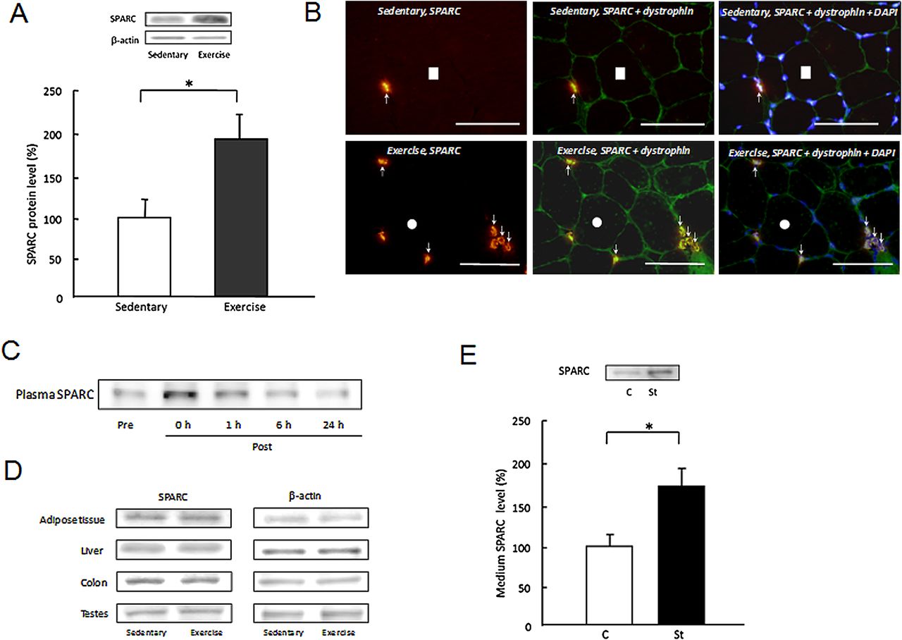
Since myokines are secreted into the blood from muscles in response to a single bout of exercise,4 ,5 ,7 we examined the level of secreted SPARC in mice that performed a single bout of exercise. The level of SPARC protein in gastrocnemius muscle was significantly increased, as shown by immunoblotting (figure 1A) and immunohistochemistry (figure 1B, supplementary figure 3 online). This increase in muscle SPARC was found in the entire muscle section in exercised muscle and specifically pronounced around the plasma membrane. A time-course analysis of the plasma levels of SPARC showed that it was increased immediately after a single bout of exercise, and then gradually returned to the pre-exercise level (figure 1C). The expression of SPARC in adipose tissue, liver, colon and testes, which are rich in SPARC, was not changed by a single bout of exercise (figure 1D). Furthermore, 60 min cyclic stretching of C2C12 myotubes stimulated SPARC secretion into the extracellular medium (figure 1E). These findings suggest that a single bout of exercise accelerates SPARC secretion from contracting muscle into the blood.

Secretion of secreted protein acidic and rich in cysteine (SPARC) in response to a single bout of exercise in mice. (A, B) Changes in SPARC protein expression in skeletal muscle immediately after a single bout of exercise using immunoblotting (A) and immunohistochemical analysis (B). Each cryosection was immunohistochemically stained for SPARC, dystrophin or 4′-6-diamidino-2-phenylindole (DAPI). White circles and squares indicate identical fibres in different immunoimages. White arrows denote identical positions of SPARC immunoreactivity. Scale bars 50 μm. (C) Time course analysis of plasma levels of SPARC in response to a single bout of exercise. (D) SPARC protein expression in adipose tissue, liver, colon and testes in sedentary mice and immediately after mice performed a single bout of exercise. (E) Relative amount of SPARC in the extracellular medium after cyclic stretching (8%, 1 Hz, 60 min) of C2C12 mouse myotubes. The amount of SPARC in the control medium is defined as 100%. C, unstretched control cells; St, stretched cells. Results are shown as mean±SE (n=6–8). *p<0.05.
Secretion of SPARC requires protein translation
We next investigated the underlying mechanism of SPARC secretion in response to muscle contraction using C2C12 cells. Cyclic stretching increased SPARC in both cells and medium, suggesting that mechanical contraction accelerates the secretion of SPARC from muscle cells into the extracellular media (figure 2A,B, supplementary figure 4 online). However, cycloheximide, a protein translation inhibitor, inhibited these elevations (figure 2A,B, supplementary figure 4B online). In contrast, actinomycin D, a transcription inhibitor, had no effect on SPARC elevations (figure 2A,B, supplementary figure 4A online). Mechanical stretching did not increase the SPARC mRNA expression level in C2C12 cells (figure 2C). These in vitro results suggested that mechanical stretching of muscle cells induces SPARC secretion by stimulating protein translation. Furthermore, mechanical stretching did not affect the level of lactate dehydrogenase (LDH) in the extracellular medium (figure 2D), which suggested that cells that had been injured by stretching did not release intracellular SPARC into the extracellular medium.
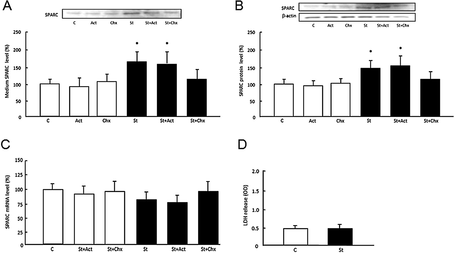
Secretion of secreted protein acidic and rich in cysteine (SPARC) from mechanically stretched C2C12 mouse myotubes requires protein translation. (A) The amount of SPARC in the extracellular medium after cyclic stretching (8%, 1 Hz, 60 min) of C2C12 myotubes in the presence or absence of 5 μg/ml actinomycin D (Act) or 100 μM cycloheximide (Chx) for 60 min before stretching. Changes in (B) SPARC protein and (C) mRNA in C2C12 cells after stretching. (D) Level of lactate dehydrogenase (LDH) in the extracellular medium after stretching. C, unstretched control cells; St, stretched cells. Results are shown as mean±SE (n=4–6). *p<0.05 vs unstretched control cells.
Exercise increases serum SPARC levels in humans
In experiment 1, serum SPARC was transiently increased in young healthy men immediately after a single bout of cycling at 70% Vo 2max for 30 min (figure 3A), which is consistent with the results of the mouse experiments. However, the serum SPARC level gradually decreased until it returned to the baseline level 6 h after exercise. In experiment 2, 4 weeks of training at 70% Vo 2max significantly promoted the exercise-induced increase in the serum level of SPARC (figure 3B). In contrast, the level of serum SPARC in the resting state did not change between baseline and post-training. Furthermore, the platelet count, which may be a source of serum SPARC, was not significantly affected by a single bout of exercise, and resting state and post-exercise change ratios of SPARC concentration and platelet counts were not correlated (R2=0.041) (supplementary figure 5 online), which suggested that exercise did not alter the secretion of SPARC from platelets into serum.

Elevation of serum secreted protein acidic and rich in cysteine (SPARC) in response to a single bout of exercise in humans. (A) Time course of serum SPARC after steady-state cycling at 70% maximal oxygen uptake (Vo 2max) for 30 min (n=10). *p<0.05 vs resting state (Rest). (B) Changes in serum SPARC after steady-state cycling at the intensity of 70% Vo 2max for 60 min in baseline and post-training (n=9). *p<0.05, **p<0.01 difference between resting state (Rest) and immediately after exercise (Post-Ex). Results are shown as mean±SE.
Exercise-induced expression and secretion of SPARC inhibits colon tumorigenesis
Since we previously demonstrated that regular exercise suppresses the formation of ACF in colon20 and several other studies21–23 have suggested that SPARC can function as a tumour suppressor, we compared the number of ACF in wild-type and SPARC-null mice (supplementary figure 6A online). In wild-type mice, regular low-intensity exercise significantly reduced the number of ACF and aberrant crypts (AC) in the colons of AOM-treated mice compared with sedentary mice (figure 4A). In contrast, more ACF and AC formed in AOM-treated SPARC-null mice than in wild-type mice, and exercise did not have an inhibitory effect (figure 4A).
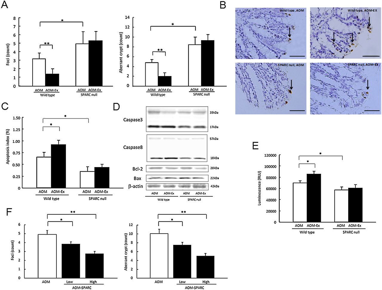
Effect of regular exercise on azoxymethane (AOM)-induced colonic tumorigenesis in wild-type and secreted protein acidic and rich in cysteine (SPARC)-null mice. (A) Number of aberrant crypt foci (ACF) (left) and aberrant crypts (AC) (right) on the mucosal surface of the colon were counted under a light microscope. (B, C) Terminal deoxyribonucleotidyl transferase (TdT) dUTP nick end labeling (TUNEL) assay of apoptotic colon mucosal cells in wild-type and SPARC-null mice. Histological results (B) and percentage of apoptotic cells (C). Arrows denote the TUNEL-positive cells. Scale bars 50 μm. The apoptosis index is the percentage of TdT-labelled cells. The mean number of TUNEL-positive cells in a sample was calculated from eight different fields. (D) Immunoblots of apoptotic proteins in the colon mucosa of wild-type and SPARC-null mice with or without exercise. (E) Caspase 3/7 activity in colonic mucosa of these mice, as measured by a luminescent assay. (F) Number of ACF (left) and AC (right) on the mucosal surface of the colon counted in AOM-treated wild-type mice injected with saline or SPARC at doses of 3 μg/kg (low) and 15 μg/kg (high). Results are shown as mean±SE (n=10–12). AOM, AOM-treated sedentary mice; AOM-Ex, AOM-treated exercised mice; RLU, relative light unit. *p<0.05; **p<0.01.
In AOM-treated wild-type mice, 6 weeks of regular exercise also significantly increased SPARC mRNA expression in muscle but not in colon (supplementary figure 6B online), which is consistent with the above results. In addition, the levels of SPARC in muscle and plasma increased immediately after a single bout of exercise compared with the sedentary state in regularly exercised mice (supplementary figure 6C,D online). However, the level of urinary corticosterone, a psychological stress marker, was unchanged between sedentary and regularly exercised mice (supplementary figure 6E online). In both wild-type and SPARC-null mice, regular exercise reduced the epididymal adipose tissue weight while body weight and muscle tissue weight were not changed (supplementary table 2 online).
In addition, we examined the effect of exogenous SPARC on ACF formation in the colon by injection of recombinant SPARC in AOM-treated mice. Injection of low-dose SPARC, which is equivalent to the elevation of response to exercise, suppressed AC and ACF formation; furthermore, formation of more AC and ACF was suppressed in mice who received a high dose (ie, five times the low dose) injection of SPARC (figure 4F).
Effect of SPARC on apoptosis in the colon
The terminal deoxyribonucleotidyl transferase dUTP nick end labelling assay showed that regular exercise increased the number of apoptotic colon cells in wild-type mice; however, sedentary and exercised SPARC-null mice did not differ (figure 4B,C). Furthermore, the levels of cleaved caspase-3 (17 kDa) and caspase-8 (18 kDa) were higher in wild-type mice than in SPARC-null mice, and regular exercise further increased the levels of these apoptosis markers in wild-type mice but not in SPARC-null mice (figure 4D). Similarly, regular exercise increased the activity of caspase 3/7 in wild-type mice but not in SPARC-null mice (figure 4E). However, regular exercise did not affect the levels of B cell lymphoma 2 (Bcl-2) or Bcl-2-associated X protein (Bax) in either wild-type or SPARC-null mice. These findings suggested that SPARC mediates exercise-induced colon tumorigenesis via caspase-3- and caspase-8-dependent apoptosis.
In addition, we examined the effect of exogenous SPARC on colon tumour using colon-26 cells. The treatment of SPARC inhibited proliferation of the colon cancer cells in a dose-dependent manner (figure 5A). In contrast, culture medium obtained from muscle cells treated with short interfering RNA directed against SPARC demonstrated accelerated cell proliferation compared with medium obtained from normal muscle cells (figure 5C). Apoptosis of cells was increased by the addition of SPARC in a dose-dependent manner (figure 5B). These in vitro results supported the hypothesis that SPARC prevents proliferation of colon tumour cells via an apoptotic effect.

Effect of secreted protein acid and rich in cystein (SPARC) on proliferation in colon carcinoma cells. (A) Changes of cell counts in response to SPARC. Mouse recombinant SPARC was incubated for 48 h at concentrations of 0, 0.2, 0.5, 1, 2, and 5 μg/ml. *p<0.05 versus 0 μg/ml. (B) Changes in apoptotic cell counts in response to SPARC. Recombinant SPARC was incubated for 12 h at concentration of 0 (control), 1, 2, and 5 μg/ml, respectively. (C) Changes of cell counts in response to culture medium obtained from muscle cells which treated RNA interference of SPARC. Control, free oligonucleotide; Negative control, random oligonucleotides. *p<0.05; **p<0.01. Results are shown as mean ± standard error (n=3–6).
Discussion
This study aimed to identify novel secretory proteins from muscle. Skeletal muscle has various physiological functions, including nutrient metabolism and power output, which are improved by regular exercise but impaired by inactivity and ageing. We hypothesised that secreted myokines are associated with muscle function, and then used microarray analysis to identify novel secretory proteins in skeletal muscle using mouse models of exercise and ageing. This analysis revealed 24 genes that are upregulated by regular exercise and downregulated by ageing. One gene that encoded the secretory protein, SPARC, was included among these genes. SPARC is a secreted matricellular glycoprotein that is involved in the development, remodelling and tissue repair by modulating cell-cell and cell-matrix interactions,21 ,22 as well as other functions such as antitumorigenesis.23 We therefore chose to focus on SPARC in this study rather than the other 23 genes.
This study is the first to show that SPARC, a novel myokine that is secreted by skeletal muscle contraction, is involved in the exercise-mediated suppression of colon tumorigenesis. Our results revealed that a single bout of exercise rapidly increased circulation and muscle levels of SPARC, and cyclic stretching increased secretion of SPARC from C2C12 cells into the medium, which suggested that contracting muscle cells secrete SPARC into the extracellular milieu. This exercise-induced increase in SPARC appeared to be muscle-specific because no increase was observed in other organs where SPARC is abundant. Moreover, the lack of an effect of a single bout of exercise on the platelet count and the absence of a correlation between changes in platelet count and serum SPARC in humans suggested that exercise did not increase the secretion of SPARC from platelets. The increase in SPARC in response to exercise is therefore most likely due to increased secretion from skeletal muscle.
Cyclic stretching increased the amount of SPARC protein in both muscle cells and extracellular medium, but not the mRNA level. In addition, cycloheximide but not actinomycin D inhibited the secretion of SPARC. These results suggested that secretion of SPARC requires protein translation in muscle cells. Muscle contraction has been shown to rapidly activate some protein translation pathways,24 which may be associated with the increase in SPARC. On the other hand, regular exercise increased SPARC expression in mouse and human muscle tissue in the resting state but did not affect the circulating level of SPARC. However, a single bout of exercise promoted the increase in SPARC secretion after training compared with before training. Therefore, regular exercise can enhance the secretory capacity of SPARC in response to muscle contraction by increasing the amount of SPARC in muscle tissue in the resting state.
In an AOM-induced colon cancer model, regular low-intensity exercise suppressed the formation of ACF in the colons of wild-type mice. This result suggested that regular exercise prevents colon tumorigenesis, which is consistent with our previous study.20 However, in SPARC-null mice, exercise did not suppress the formation of ACF, which suggested that SPARC is involved in the exercise-mediated inhibition of colon tumorigenesis. Since circulating SPARC increases immediately after a single bout of exercise but not in the resting state, it is suggested that repetitive increases in SPARC induced by each bout of exercise during a 6-week regular exercise period led to the antitumorigenesis. These results are consistent with the findings of many previous studies demonstrating that SPARC is a tumour suppressor.23 ,25–27 For example, previous studies have shown that a lack of SPARC increases pancreatic and ovarian tumorigenesis in vivo.25 ,26 In addition, the addition of exogenous SPARC to cancer cell lines in vitro reduces cell proliferation.26 ,27 Furthermore, epigenetic silencing of the SPARC gene via hypermethylation of its promoter is frequent in colon cancers, and SPARC inactivation is related to rapid progression of colon cancer.27 ,28 Moreover, modulation of SPARC expression affects the sensitivity of colorectal tumours to radiation and chemotherapy.23 ,29 ,30 Interestingly, a clinical study showed that the 5-year survival of patients with tumours who expressed high levels of SPARC was significantly better than those with tumours that did not express SPARC.31 Our results also showed that the AOM-induced ACF in SPARC-null mice were markedly higher than in wild-type mice, which supports the hypothesis that SPARC is involved in colon tumorigenesis. In addition, a single bout of exercise increased the plasma levels of SPARC, even at low intensity. However, neither a single bout of exercise nor regular exercise changed the expression of SPARC in the colon. We also found that injection of recombinant SPARC at a dose which was equivalent to the increase in response to exercise suppressed ACF formation; however, the extent of suppression was less than that in mice treated with regular exercise. Furthermore, in a culture experiment the addition of recombinant SPARC to colon carcinoma cells inhibited cell proliferation. In contrast, addition of short interfering RNA-treated muscle medium accelerated the proliferation. These results suggested that secreted SPARC to some extent suppresses colon tumorigenesis.
One of the causes of ACF formation is dysregulation of apoptosis.32 Unlike wild-type mice, regular exercise did not increase apoptotic cells in SPARC-null mice. Similarly, regular exercise increased the cleaved form of caspase-3 in wild-type but not in SPARC-null mice. In addition, treatment of colon-26 cells with exogenous SPARC accelerated cell apoptosis. These observations suggested that exercise-induced SPARC suppresses the formation of ACF by promoting apoptosis, which is consistent with the tumour suppressor function of SPARC.23 ,25–27 In addition, we observed that regular exercise increased the cleaved form of caspase-8 in wild-type mice; however, exercise did not affect the levels of Bax and Bcl-2 in either type of mice. These results suggested that SPARC activates extrinsic apoptotic signalling which is triggered by external signals such as death receptor ligands, and results in the formation of the death-inducing signalling complex including caspase-8.33 This hypothesis is consistent with the findings of Tang and Tai34 who demonstrated that SPARC interacts with the N-terminal region of pro-caspase-8 and activates caspase-8 in colorectal cancer cells. Our results therefore suggest that the antitumorigenesis mechanism of SPARC involves stimulation of apoptosis via caspase-3 and caspase-8. However, further studies are needed to elucidate the underlying mechanism in more detail.
A few explanatory hypotheses have previously been proposed concerning the mechanism underlying the preventive effect of exercise on tumorigenesis. The anti-inflammatory effect associated with metabolic improvement has been the target of particular focus. Previous studies have demonstrated that long-term exercise decreased the circulating levels of inflammatory cytokines and reduced the concentration of C reactive protein.35 ,36 Conversely, exercise can increase the concentrations of anti-inflammatory factors such as IL-10 and IL-1 receptor antagonist in the blood.37–39 The anti-inflammatory effect of exercise may be represented by a reduction of adipose tissue mass with metabolic improvement. In this study, regular exercise led to a decrease in adipose tissue mass in mice, which may affect antitumorigenesis. Several studies have suggested that SPARC can regulate inflammatory and metabolic states in various experimental conditions.40–42 The detailed mechanism underlying the antitumorigenesis effect of SPARC associated with metabolic improvement and anti-inflammatory activity induced by exercise should be investigated in future studies.
In conclusion, our results showed that a single bout of exercise increased the secretion of SPARC, a novel myokine, into the blood in both mice and humans. In addition, in a colon cancer mouse model, regular exercise significantly reduced the formation of ACF in wild-type mice but not in SPARC-null mice. Regular exercise enhanced apoptosis in colon mucosal cells and increased the cleaved forms of caspase-3 and caspase-8 in wild-type mice but not in SPARC-null mice. These observations suggest that SPARC inhibits colon tumorigenesis via exercise by increasing apoptosis.
Acknowledgments
The authors thank the animal center staff of Kyoto Prefectural University of Medicine, the human study participants and all members of the Department of Gastroenterology of Kyoto Prefectural University of Medicine and Laboratory of Health Science of Kyoto Prefectural University.
References
Supplementary materials
Supplementary Data
This web only file has been produced by the BMJ Publishing Group from an electronic file supplied by the author(s) and has not been edited for content.
Files in this Data Supplement:
- Data Supplement 1 - Online Supplementary material
Footnotes
-
Funding This work was supported by Grants-in-Aid from the Japan Society for the Promotion of Science (23700776WA, 08101559YN and 21390184TY) from the Ministry of Education, Culture, Sports, Science and Technology of Japan and research grants from the Adaptable and Seamless Technology Transfer Program through Target Driven R&D from the Japan Science and Technology Agency, Kyoto Prefectural University Corporation, Nakatomi Foundation and Uehara Memorial Foundation.
-
Correction notice This article has been corrected since it was published Online First. The following sentence has been amended to read: SPARC demonstrated accelerated cell proliferation compared with medium obtained from normal muscle cells (figure 5C).
-
Competing interests None.
-
Patient consent Obtained.
-
Ethics approval The experimental protocol was approved by the Review Board on Human Experiments, Kyoto Prefectural University and complied with the Helsinki Declaration.
-
Provenance and peer review Not commissioned; externally peer reviewed.