


Abstract
Aim: We have previously reported that caffeine can enhance chemotherapy efficacy of bone and soft tissue sarcoma via cell-cycle perturbation. Valproic acid has histone deacetylase (HDAC) inhibitory activity. We have also reported the anti-tumor efficacy of combination treatment with caffeine and valproic acid against osteosarcoma primary tumors in a cell-line orthotopic mouse model. Materials and Methods: In this study, we performed combination treatment of caffeine and valproic acid on osteosarcoma cell lines in vitro and in spontaneous and experimental lung metastasis mouse models of osteosarcoma. Survival of 143B-RFP human osteosarcoma cells after exposure to caffeine and valproic acid for 72 hours was determined using the WST-8 assay. IC50 values and combination indices were calculated. Mouse models of primary osteosarcoma and spontaneous lung metastasis were obtained by orthotopic intra-tibial injection of 143B-RFP cells. Valproic acid, caffeine, and combination of both drugs were administered from day 7, five times a week, for four weeks. Six weeks after orthotopic injection, lung samples were excised and observed with a fluorescence imaging system. A mouse model of experimental lung metastasis was obtained by tail vein injection of 143B-RFP cells. The mice were treated with these agents from day 0, five times a week for four weeks. Results: Both caffeine and valproic acid caused concentration-dependent cell kill in vitro. Synergistic efficacy of the combination treatment was observed. In the spontaneous lung-metastasis model, the number of lung metastasis was 9.0±2.6 in the untreated group (G1); 10.8±2.9 in the caffeine group (G2); 10.0±3.1 in the valproic-acid group (G3); and 3.0±1.1 in the combination group (G4); (p=6.78E-5 control vs. combination; p=0.006 valproic acid vs. combination; p=0.003 caffeine vs. combination). In the experimental lung-metastasis model, the combination group significantly reduced lung metastases and improved overall survival (p=0.0005). Conclusion: Efficacy of the combination of caffeine and valproic acid was observed in vitro and in spontaneous and experimental lung-metastasis mouse models of osteosarcoma.
- Osteosarcoma
- 143B
- red fluorescent protein
- RFP
- orthotopic
- nude mice
- caffeine
- valproic acid
- combination therapy
Osteosarcoma is a malignant primary bone tumor which occurs mainly in adolescents and young adults (1). Osteosarcoma has a 5-year survival of approximately 70% (2-4). However, patients with metastatic disease at diagnosis or with recurrent disease have a 5-year survival rate of 20% (5). The lung is the predominant site of metastasis and the most common cause of death in osteosarcoma patients (6).
First-line therapy against osteosarcoma consists of high-dose methotrexate, cisplatin, doxorubicin, and ifosfamide. However, dose-intensive chemotherapy regimens with these drugs have not improved outcomes (7). Therefore, novel systemic therapy is needed to improve the outcome of metastatic osteosarcoma.
Caffeine (1,3,7-trimethylxanthine) (8) can induce apoptosis (9-12), delays in cell cycle progression (13, 14) and can enhance the toxicity of radiation and anti-cancer agents (15-17). Modulation of the cell cycle by caffeine can be imaged in cancer cells expressing a fluorescent ubiquitination-based cell cycle indicator (FUCCI). Caffeine overcame cisplatinum (CDDP) cell-cycle arrest, thereby increasing CDDP efficacy (18, 19). We have previously reported that caffeine-combined chemotherapy improved the treatment of bone and soft tissue sarcoma cases in the clinic (20-22).
Valproic acid, a short chain fatty acid, is a histone-deacetylase (HDAC) inhibitor. Combination chemotherapy including valproate has been tested in myelodysplastic syndrome (23), melanoma (24), and solid tumors (25).
We have previously reported the efficacy of combination treatment of caffeine and valproic acid against a localized-osteosarcoma orthotopic-mouse model (18). In the present study, we determined the efficacy of the combination of caffeine and valproic acid on spontaneous and experimental metastasis in nude mouse models of a human osteosarcoma cell line.
Materials and Methods
Drugs. Valproic acid and caffeine were obtained from Wako Pure Chemical Industries, Ltd. (Osaka, Japan).
Cell line and growth conditions. 143B-RFP osteosarcoma cells were generated as previously described (26). All cells were grown in RPMI 1640 medium (Gibco, Grand Island, NY, USA) supplemented with 10% fetal bovine serum, 100 U/ml penicillin, and 100 μg/ml streptomycin. Cells were maintained in log phase by supplementation with fresh medium 2-3 times/week.
In vitro cell inhibition assay. Cellular viability was assessed using the WST-8 dye (2-(2-methoxy-4-nitrophenyl)-3-(4-nitrophenyl)-5-(2,4-disulfophenyl)-2H-tetrazolium) assay (Dojindo, Kumamoto, Japan). Briefly, cells were seeded in 96-well flat-bottomed microplates (5×10⁴ cells/ml), incubated at 37°C for 24 h, and exposed to various concentrations of tested compounds for 72 h. For each concentration, at least 8 wells were used. After incubation with the test compounds, 10 μl WST-8 solution was added to each well. The microplates were further incubated for 3 h at 37°C, and absorption was measured using a microprocessor-controlled microplate reader (Sunrise™; TECAN, San Jose, CA, USA) at 450 nm. The cell-survival fraction was calculated as the percentage of untreated control cells, and IC50 values were derived.
Calculation of Combination Index (CI). The specific interaction between caffeine and valproic acid on osteosarcoma cell lines was evaluated with the combination index (CI) assay using the CalcuSyn software from ComboSyn Inc. (New Jersey, USA), (Chou and Talalay (27)). Synergy is defined as a CI <1.0; antagonism as a CI >1.0; and additivity as CI values not significantly different from 1.0.
Mice. Athymic nu/nu nude mice (AntiCancer Inc., San Diego, CA), 4-6 weeks old, were used in this study. The animals were fed an autoclaved laboratory rodent diet. Animals were housed in a barrier facility on a high-efficiency particulate arrestance (HEPA)-filtered rack under standard conditions of 12-hour light/dark cycles. All animal studies were conducted in accordance with the principles and procedures outlined in the National Institutes of Health Guide for the Care and Use of Animals under Assurance Number A3873-1. In order to minimize any suffering of the animals, the use of anesthesia and analgesics were used for all surgical experiments. Animals were anesthetized by subcutaneous injection of a 0.02 ml solution of 20 mg/kg ketamine, 15.2 mg/kg xylazine, and 0.48 mg/kg acepromazine maleate.
Orthotopic spontaneous lung-metastasis model. Four- to six-week-old nude mice were anesthetized by the ketamine mixture via s.c. injection. The leg was sterilized with alcohol and an approximately 2-mm midline skin incision was made just below the knee joint to expose the tibial tuberosity. 143B-RFP cells (2×10⁵) in Matrigel (5 μl) (BD Bioscience, San Jose, CA, USA) were injected per mouse into the intramedullary cavity of the left tibia with a latex-free insulin syringe (0.5-ml 31 G) (TYCO Health Group LP, Mansfield, MA, USA). The skin was closed with a 6-0 suture.
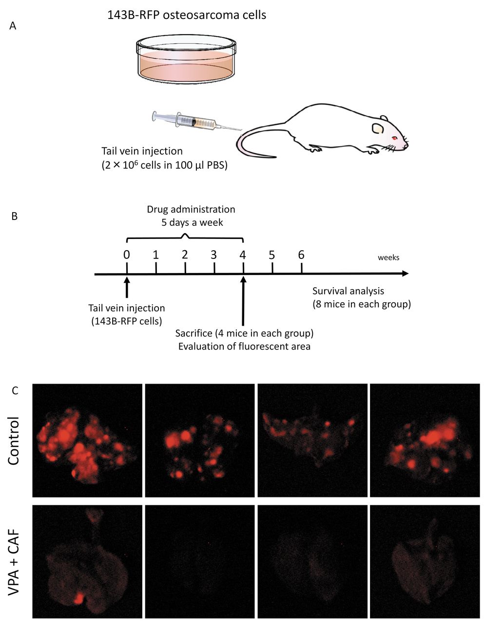
Experimental lung metastasis model of osteosarcoma. Four- to six-week-old nude mice were used. 143B-RFP cells (2×10⁶ cells in 100 μl PBS) were injected into the tail vein of 24 nude mice (day 0).
Treatment design. For the experimental metastasis model, on day 0 of tail-vein cell injection, mice were randomized into 2 groups; G1: control without treatment (n=12); G2: treated with the combination of caffeine (100 mg/kg) and valproic acid (500 mg/kg) by i.p. injection five times a week for 4 weeks (n=12). On day 28, eight mice (4 mice each group) were sacrificed and the lungs were imaged to observe RFP-expressing lung metastases and to determine the efficacy of therapy. The fluorescent areas of the lung metastasis were recorded using the OV100 variable magnification small animal imaging system (Olympus, Tokyo, Japan) (28). An additional 16 mice comprising 8 control mice, 8 caffeine-combined-with-valproic-acid-treated mice were observed for survival analysis.
For the spontaneous-metastasis model, one week after orthotopic implantation, mice were randomized into 4 groups; G1: control without treatment (n=8); G2: treated with caffeine (100 mg/kg), i.p., five times a week for 4 weeks (n=8); G3: treated with valproic acid (500 mg/kg), i.p., five times a week for 4 weeks (n=8); G4: treated with the combination of caffeine (100 mg/kg); and valproic acid (500 mg/kg), i.p., five times a week for 4 weeks (n=8). Tumor length, width, and mouse body weight were measured once in a week. Six weeks after implantation, all mice were sacrificed. The lungs were excised and the metastases on the surface were imaged and counted with the OV100.
Primary tumor-growth determination. Tumor volumes were calculated using the following equation: volume=4π (A/2)(B/2)(C/2)/3, where A is the width (average distance in the medial–lateral plane), B is the length (average distance in the proximal–distal plane), and C is the width (average distance in the anterior–posterior plane). Data are presented as mean±SD.
Results and Discussion
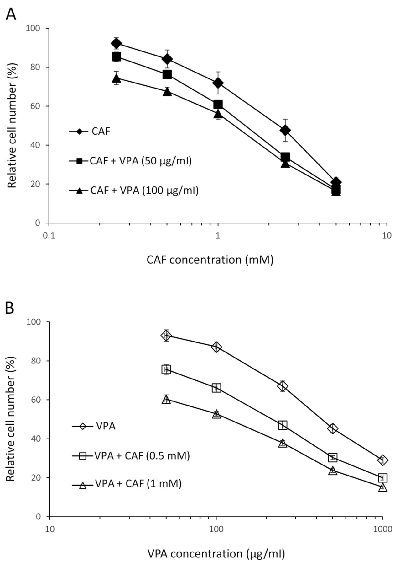
Cell killing by caffeine and valproic acid. The cell-kill activity of caffeine and valproic acid was determined on the 143B-RFP osteosarcoma cell line. Each compound significantly inhibited 143B-RFP cell growth in a dose-dependent manner (Figure 1). Addition of 50 and 100 μg/ml valproic acid to caffeine enhanced the cell kill of 143B-RFP cells (Figure 1, Table I). In addition, the CI values were significantly <1 and thereby demonstrated synergy at almost all tested concentrations on 143B-RFP cells (Table II).

Growth inhibition assay of caffeine (CAF) and valproic acid (VPA) against 143B-RFP cells. Osteosarcoma cells were incubated with each drug for 72 hrs and then counted with the WST-8 assay. (A) Efficacy of VPA as a function of CAF concentration. (B) Efficacy of CAF as a function of VPA concentration.
IC50 value of caffeine (CAF) on 143B-RFP cells with and without valproic acid (VPA).
Efficacy of caffeine and valproic acid on primary osteosarcoma and spontaneous lung metastases in the orthotopic model. The volume of the primary tumor was 5085.1±1183 mm3 in the untreated group (G1); 3781.8±1303 mm3 in the caffeine group (G2) (p=0.09); 4088.6±1687 mm3 in the valproic acid group (G3) (p=0.584); and 1335.3±657 mm3 in the caffeine and valproic-acid combination group (G4) (p=0.001); (Figure 2b). The number of lung metastasis was 9.0±2.6 in the untreated group (G1); 10.8±2.9 in the caffeine group (G2); 10.0±3.1 in the valproic acid group (G3); and 3.0±1.1 in the combination group (G4); (p=6.78E-5 control vs. combination; p=0.006 valproic acid vs. combination; p=0.003 caffeine vs. combination); (Figure 3b).

Efficacy determination of caffeine (CAF) and valproic acid (VPA) on an orthotopic mouse model of 143B-RFP osteosarcoma. (A) Treatment scheme of CAF and VPA on the orthotopic model of osteosarcoma. (B) 143B-RFP cells were orthotopically transplanted into the proximal tibia of nude mice and allowed to form tumors. Mice were treated with CAF, VPA, or the combination of CAF and VPA, i.p. CAF (100 mg/kg), VPA (500 mg/kg) five times a week for four weeks. N=8 mice/group.
CI values of the combination of CAF and VPA.

Efficacy of valproic acid (VPA) and caffeine (CAF) on spontaneous lung metastases of 143B-RFP osteosarcoma (31, 32). (A) Fluorescence imaging of spontaneous lung metastases in the control and mice treated with the combination of CAF and VPA. (B) Number of lung metastases in control and mice treated with CAF, VPA or VPA combined with CAF.
Efficacy of caffeine and valproic acid on osteosarcoma experimental lung metastasis. Fluorescence imaging demonstrated that the combination of caffeine and valproic acid strongly inhibited experimental lung metastases (Figure 4c). The mean fluorescence intensity of lung metastases of the control mice was 1.82×10⁶ and for the combination of caffeine and valproic acid treated mice was 3.17×10⁴, (p=0.023); (Figure 4d). The fluorescent area of the lung metastases of control mice was 78.6±27.2 mm2 and for the caffeine-valproic acid treated mice was 2.1±1.6 mm2, (p=0.001); (Figure 4e).


Efficacy of valproic acid (VPA) combined with caffeine (CAF) on experimental lung metastases. (A) Mouse model of osteosarcoma experimental lung metastases. (B) Treatment scheme. (C) Fluorescence imaging of lung metastases with and without VPA+CAF treatment (31, 32). (D) Fluorescence area of osteosarcoma lung metastases with and without VPA+CAF treatment. (E) Fluorescent intensity of 143B-RFP osteosarcoma lung metastases with and without VPA+CAF treatment. (F) Kaplan-Meier survival curve of mice with 143B-RFP osteosarcoma treated with VPA+CAF compared to control untreated mice.
Kaplan–Meier analysis with the log rank test demonstrated that the combination of caffeine and valproic acid significantly improved the survival of the treated mice (p=0.0005); (Figure 4f).
The present study demonstrates the potential of caffeine and valproic acid for effective treatment of osteosarcoma using an established cell line. Future studies will focus on patient-derived orthotopic xenograft (PDOX) models of osteosarcoma (29, 30).
- Received January 24, 2017.
- Revision received February 23, 2017.
- Accepted February 24, 2017.
- Copyright© 2017, International Institute of Anticancer Research (Dr. George J. Delinasios), All rights reserved


