


Abstract
The embryonic stem cell factors Oct3/4 and Sox2 are essential for pluripotency and self-renewal of embryonic stem cells. Cancer cells, especially in poorly differentiated or undifferentiated tumours, have been characterized by many phenotypic traits similar to undifferentiated embryonic cells, indicating that Oct3/4 and Sox2 may be expressed in solid tumours. With the methods of real-time PCR, Western blotting and immunocytochemistry/immunohistochemistry, the expression of these two genes in the esophageal squamous cancer cell lines Kyse70, Kyse140 and Kyse450 were characterized, in addition to a virus-transformed “normal” esophageal epithelial cell line, Ket-1A. Both Oct3/4 and Sox2 were variably expressed in the cancer cell lines, but were either negative or very weakly expressed in the normal cell line. Further examinations in a series of 162 consecutive esophageal squamous cancer patients showed that 17.90% and 22.84% of the tumours highly expressed Oct3/4 and Sox2 proteins, respectively, and the expressions of these two factors were significantly associated with higher histological grade and poorer clinical survival. Since the function of pluripotency and self-renewal of these factors has been characterized in human embryonic stem cells, these data may indicate that the expression of these factors enables the tumours to have higher degree of stemness tumour cells, which in turn results in poorer clinical outcome for patients with esophageal squamous cell carcinomas.
Oct3/4, also known as Oct3 or Oct4, belongs to the POU (Pit-Oct-Unc) transcription factor family (1). The POU family of transcription factors can activate the expression of their target genes through binding an octameric sequence motif of an AGTCAAAT consensus sequence (2, 3). The expression of this gene is necessary for the maintenance of pluripotentiality in embryonic stem cells (ESCs) and primordial germ cells and is down-regulated in all differentiated somatic cell types in vitro as well as in vivo (2).
Sox2 is an important member of the Sox gene family. Sox (SRY box) genes have been identified through their homology to the high mobility group (HMG) box (79 amino acids) of sex-determining factor SRY (4-7). The Sox genes encode transcription factors that interact with DNA through their highly conserved HMG domain (8, 9). The Sox genes are expressed in a wide variety of tissues, and play important roles in the regulation of organ development and cell type specification (5, 7). They also have a crucial function in ESC development (10).
Previous studies have demonstrated that many cancers express Oct3/4 and Sox2 and that their expression appears to be important for cancer cell survival. Rodriguez et al. (11) reported Sox2 expression in 16.7% of 226 node-negative sporadic breast carcinomas by immunostaining of tissue array sections. Hattab et al. (12) discovered Oct3/4 immunoreactivity in 100% of 25 primary intracranial germinomas. Sanada et al. (13) suggested Sox2 involvement in later events of carcinogenesis after histopathologically and immunohistochemically analyzing 14 invasive pancreatic carcinomas. Jin et al. (14) discovered that the human breast cancer cell line, MCF7, expressed at least four POU gene products including Oct3/4. Sung et al. (15) proposed that with its superior sensitivity and easy interpretation compared with other markers, OCT3/4 immunostaining is a powerful tool for confirming the diagnosis of retroperitoneal seminoma. The Oct3/4 and Sox2 genes have also been shown to be expressed in human tumours including pancreatic and gastric carcinomas (16, 17).
Recent studies argue that the transcription factors Oct3/4 and Sox2 exhibit the hallmarks of global regulators during mammalian embryogenesis. Oct3/4 and Sox2 work together during embryogenesis to co-ordinate their own transcriptions (18, 19), via the Oct3/4/Sox2 complex in ESCs (20). The two genes constitute part of an important gene regulatory network and are essential for embryogenesis and/ or the pluripotency and self-renewal of ESC (10).
Cancer cells, especially in poorly differentiated or undifferentiated tumours, have been characterized by many phenotypic traits similar to undifferentiated embryonic cells (21-23). These similarities suggest the expression of genes determining cell renewal/stemness.
Esophageal cancer is one of the leading causes of cancer-related death throughout the world, especially in some high-risk areas in China, such as Linxian (Western Anyang, Henan Province). In the present study, it was intended to firstly characterize the expression status of Oct3/4 and Sox2 genes in four esophageal cell lines and then explore their clinicopathological and survival correlations in a series of 162 esophageal carcinomas from patients from Anyang, Henan, China. It was found that both Oct3/4 and Sox2 were expressed in the esophageal squamous cancer cell lines Kyse70, Kyse140 and Kyse450, but negative or very weakly expressed in the virus-transformed “normal” esophageal epithelial cell line Het-1A. Furthermore, both Oct3/4 and Sox2 protein expressions were significantly associated with higher histological grade and poorer clinical survival, indicating that these embryonic self-renewal factors may exert a positive influence on tumour cell stemness, which in turn results in a negative clinical course for the tumours.
Materials and Methods
Patients and cell lines. In this retrospective study, 162 consecutive esophageal cancer patients who underwent potentially curative surgery without preoperative chemotherapy or radiotherapy during the period of 1989-1994 at the Anyang Tumour Hospital, Henan, China were randomly selected. Among them, 99 were men and 63 were women, ranging from 33-73 years of age with a mean age of 56.4 years. According to International Union against Cancer (UICC) 1997 standard, 107 were classified as stage II and 55 cases as stage III. The patients' information and tumour parameters are listed in Table I. All the patients were followed at the Anyang Tumour Hospital until May 2004 supported by an international collaboration project between Anyang Tumour Hospital and The Norwegian Radium Hospital (24). Among these patients, the follow-up data were missing for 21. A total of 97 (59.9%) patients died during the follow-up period. Surgically removed specimens were routinely fixed in buffered formalin and embedded in paraffin blocks for clinical diagnosis and reclassification for this study.
The human esophageal squamous cell carcinoma cell lines Kyse70, Kyse140, Kyse450 (DSMZ, Germany) and a virus transformed human normal esophageal epithelial cell line Het-1A (ATCC, USA) were cultured in RPMI-1640 medium supplemented with 10% fetal bovine serum at 37°C under 5% CO2 and saturated moisture. The sample cells were collected and counted by trypan blue exclusion using a hemocytometer.
Immunocytochemistry. Immunocytochemistry was performed on all the four cell lines. The cultured cells were detached and the cell suspension was centrifuged at 2000 rpm for 10 minutes. The supernatant was removed before the sediment was mixed with 2-3 drops plasma and 2 drops of thrombin for 1 minute, followed by addition of 4 % buffered formalin to the coagulated cell mass, which was then fixed for 30 minutes and placed in linen paper for further conventional paraffin cytoblock preparation. Sections (4 μm-thick) were cut from the paraffin-embedded cytoblocks and immunostained according to the same procedure as tissue blocks described below.
Tissue arrays. Multitissue array blocks were made with the MTA-1 manual tissue arrayer (Beecher Instruments Inc., Sun Prairie, WI, USA). Briefly, 5 μm sections from the routinely made paraffin blocks were stained with H&E and reevaluated to confirm the diagnosis and to identify two representative tumour areas and one stromal area. The related paraffin blocks were oriented and marked. From these blocks, tissue cores with a diameter of 0.6 mm were punched and arrayed in triplicate on a recipient paraffin block. After the block construction was completed, the block was placed into a 40°C oven overnight to tighten the cylinders by slightly melting the paraffin. Subsequently, 5 μm sections of these tissue array blocks were cut and placed on charged Super-Frost Plus glass slides and dried at 60°C oven for 2-4 hours. These sections were used for immunohistochemical analysis. For those samples whose tissue array materials were not representative or not available, the paraffin-embedded conventional sections were used for additional immunohistochemistry analyses as well.
Immunohistochemistry. Immunohistochemical analysis for Oct3/4 and Sox2 was performed on 5 μm sections that were made from tissue microarray blocks. The Envision Plus detection system (Dako, Carpinteria, CA, USA) was used for the immunostaining. The sections were deparaffinized in xylene and microwaved in 10 mM citrate buffer (pH 6.0) to unmask the epitopes. Endogenous peroxidase activity was blocked by incubation with 0.03% hydrogen peroxide in methanol for 5 minutes. Slides were incubated with anti-human Oct3/4 antibody (microwave retrieval in citrate buffer at 1: 40 concentration; catalog no. AF1759, R&D Systems) and anti-human/mouse Sox2 antibody (microwave retrieval in low PH buffer at 1:200 concentration; catalog no. MAB2018, R&D Systems), respectively. For the detection of Oct3/4, the sections were incubated with the polyclonal goat anti-Oct3/4 antibody for 30 min at room temperature. Mouse anti-goat IgG (sc-2489; Santa Cruz Biotechnology Inc, Santa Cruz, CA, USA) diluted at 1:100 was added for incubation for another 30 min followed by a gentle rinse with washing buffer three times. Thereafter, the sections were incubated with a peroxidase labeled polymer conjugated to goat anti-mouse IgG (Dako, Carpinteria, CA, USA) for 30 min before stained for 5 min with 3′3 diaminobenzidine tetrahydrochloride (DAB), counterstained by hematoxylin, dehydrated and mounted in Diatex. For the detection of Sox2, the sections were incubated with monoclonal mouse antibody at 4°C overnight. The following procedures were same as Oct3/4 except addition of mouse anti-goat IgG. Known Oct3/4- and Sox2-positive seminoma was used as positive control while the same concentration of non-immune goat IgG or mouse IgG was applied as negative control for Oct3/4 or Sox2, respectively.
Correlation of Oct3/4 and Sox2 expressions to clinicopathological features of esophageal squamous cancer.
Evaluation of staining for Oct3/4 and Sox2. For both Oct3/4 and Sox2, only nuclear staining was considered as positive. The intensity and percentage of immunostained carcinoma cells were all taken into consideration according to the previous published method with modification (25). Briefly, the extent of positivity was scored as 0 when no positive cells were observed; 1 when the percentage of positive cells was <10%; 2 when it was 10-50%; and 3 when it was >50% . The intensity was scored as 0 when no positive cells were identified; 1, weak; 2, moderate; and 3, strong staining. Multiplying the extent by intensity gave the following immunohistochemical staining grades as 0, 1, 2, 3, 4, 6 and 9. For statistical analyses, the grades 0, 1 and 2 were considered as no or weakly stained and scored as 0, the grades 3 and 4 were considered as moderately stained and scored as 1, and the grades 6 and 9 were considered as strongly stained and scored as 2.
Total RNA and protein isolation. Total RNA and protein isolation was performed by using Total RNA and Protein Isolation kit (Macherey-Nagel, Düren, Germany) according to the user manual. About 5×10⁶ cultured cells were collected and lysed. Through the NucleoSpin RNA/Protein column, RNA and DNA were bound to the column and protein was contained in the flow-through. After digestion of DNA, total RNA was isolated by washing the column. Protein was isolated from the flow-through and incubated for 3 min at 98°C for dissolving and denaturation and stored at -20°C until used. All of the preparations and handling steps of RNA took place in a laminar flow hood, under RNase-free conditions.
cDNA synthesis. RNA quality and quantity were determined by absorbance readings at 260 and 280 nm with the Nano Drop (ND-1000) spectrophotometer (Wilmington, DE, USA). RNA integrity was tested by PCR amplification of the GAPDH gene. Reverse transcription of RNA was performed by using Transcriptor First Strand cDNA Synthesis Kit (Roche, Mannheim, Germany). cDNA was synthesized from 5 μg of total RNA isolated from the cell lines according to the manufacturer's handbook.
Primer/probe design. The primer pairs and hydrolysis probes for quantitative real-time PCR of Oct3/4, Sox2 and GAPDH were designed by Universal ProbeLibrary Assay Design Center (Roche, Mannheim, Germany). For Oct3/4, the sense primer sequence was 5′-AGCAAAACCCGGAGGAGT-3′ and the antisense primer sequence was 5′-CCACATCGGCCTGTGTATATC-3′, giving a product of 114 pb. For Sox2, the Sense primer sequence was 5′-TTGCTGCCTCTTTAAGACTAGGA-3′ and the antisense primer sequence was 5′-CTGGGGCTCAAACTTCTCTC-3′, giving a product of 75 bp. For GAPDH, the sense primer sequence was 5′-AGCCACATCGCTCAGACA-3′ and the antisense primer sequence was 5′-GCCCAATACGACCAAATCC-3′, giving a product of 66 bp. The hydrolysis probes (#35, cat no 04687680001 for both Oct3/4 and Sox2, and #60, cat no 04688589001, for GAPDH) were bought from Roche Diagnostics (Mannheim, Germany). In addition to the primer pairs for real-time RT-PCR, the specific primer pair for Oct3/4 gene which had been verified avoiding pseudogene amplification by Suo et al. (26) was also applied in this study for conventional RT-PCR analyses.
Quantitative RT-PCR and conventional RT-PCR. Relative quantification RT-PCR was performed with the LightCycler 2.0 Real-Time PCR System (Roche) in a total volume of 20 μL in glass-capillaries containing 2 μl of cDNA, 0.5 μM of each primer, 0.1 μM of hydrolysis probe and 4 μl of LightCycler TaqMan Master Mix (Roche, Mannheim, Germany). Quantification was performed according to a published method with some modification (27). After the expression ratio value for each target gene (Oct3/4 or Sox2) versus housekeeping gene (GAPDH) was obtained, the ratio of Het-1A was set as 1, and all other ratio values were compared with this value and normalized as relative quantities. The PCR reaction was initiated with a 12-min denaturation at 95°C and terminated with a 30-s cooling step at 40°C. The cycling protocol consisted of denaturation at 95°C for 10 s, annealing at 54°C for 10 s and extension at 72°C for 10 s and was cycled 45 times. Fluorescence detection was performed at the end of each extension step. The housekeeping gene GAPDH and DEPC-H2O were set as internal control and negative control, respectively.
The specific conventional RT-PCR reaction for Oct3/4 gene was initiated with a 5-min denaturation at 95°C. Amplification was carried out for 30 cycles consisting of 30 s at 95°C, 50 s at 55°C and 50 s at 72°C. An additional extension step of 5 min at 72°C was added at the end of 30 cycles. GAPDH was used as an internal control to confirm the success of reverse-transcription action and the identical quantity of different cDNA templates. PCR products were analyzed by electrophoresis on 1.5% agarose gel.
Western Blotting analysis. Twenty μl of protein samples were separated on a 10% SDS-acrylamide gel (Bio-Rad) for 1 h at 150 V and the proteins were transferred to nitrocellulose membrane (Whatman, Kent, UK). After blocking in 5% fat-free milk, the membrane was treated with the dilution of the primary antibody overnight at 4°C and the dilution of the secondary IgG-horseradish peroxidase (HRP) conjugated antibody for 1 h at room temperature. The antibodies used for immunohistochemistry were applied for Western blotting. All the antibodies were diluted in phosphate-buffered saline containing 5% Blotto and 0.1% Tween-20. The stained membranes were visualized by enhanced chemiluminescence reaction using the ECL Plus (GE Healthcare, Fairfild, CT, USA). Western blot experiments were repeated at least three times on every sample with similar results.
Statistical analysis. Statistical evaluation was performed using chi-square tests to compare positive staining rates and the Spearman correlation test to analyze rank data. Kaplan-Meier survival plots were generated and comparisons between survivals were made with log-rank statistics. SPSS 13.0 software was employed to analyze all data and p<0.05 was considered as statistically significant.
Results
Characterization of Oct3/4 and Sox2 in esophageal cell lines. Quantitative real-time PCR was employed to analyze Oct3/4 and Sox2 mRNA levels in three human esophageal squamous cell carcinoma cell lines (Kyse70, Kyse140 and Kyse450), and one human normal esophageal epithelial cell line (Het-1A). Comparatively, the ratios of Oct3/4 versus GAPDH in Kyse70, Kyse140 and Kyse450 were 7.1, 4.5 and 2.6 times higher than that in Het-1A (Figure 1a). The ratios of Sox2 versus GAPDH in these cell lines were 917, 612 and 147 times higher than that in Het-1A, respectively (Figure 1a). To further examine the protein expression of Oct3/4 and Sox2, Western blot analysis was performed on these four cell lines. Figure 1b shows 38 kDa Oct3/4 bands, which are in agreement with the NP_002692 Oct3/4 characterization on NCBI. The band of Oct3/4 in Het-1A was very weak, in Kyse450, strong and in Kyse140 and Kyse70, even stronger. Figure 1b also shows 34 kDa Sox2 bands which are in agreement with the NP_003097 Sox2 description on NCBI. The expression of Sox2 in Het-1A was too low to be detected. But in Kyse70, derived from a poorly differentiated tumour, a high intensity Sox2 band was revealed and Kyse145 and Kyse450 showed relatively weaker Sox2 bands. Immunocytochemistry of the cytoblock paraffin sections of the cell lines gave similar results as shown by RT-PCR and Western blotting (Figure 2).

Levels of mRNA for Oct3/4 and Sox2 in the esophageal cancer cell lines Kyse70, Kyse140, Kyse450 and Het-1A were measured by quantitative real-time RT-PCR. (a), (b) Western blot analyses of Oct3/4 and Sox2 in these cell lines, and (c) conventional RT-PCR results using the Oct3/4-specific primer pair (408bp upper part) and GAPDH primer pair (66bp, lower part).
Six Oct3/4 pseudogenes had been proposed to exist by using a bioinformatics approach to analyze the genomic nucleotide sequences (28). To confirm the specific transcription of Oct3/4, specific primer pair for Oct3/4 gene RT-PCR examination was designed and the specificity of it was verified by Suo et al. (26). This primer pair was also applied in this study for additional verification of Oct3/4 expression. As shown in Figure 1c, the predicated PCR product (408 bp) with the specific primer pair could be detected in these cell lines: highly in Kyse70 and Kyse140 and weakly in Kyse450 and Het-1A cell lines. This verifies the results of immunocytochemistry and Western blot that Oct3/4 gene was variably expressed in the human esophageal squamous cell carcinoma cell lines and the virus-transformed human normal esophageal epithelial cell line.
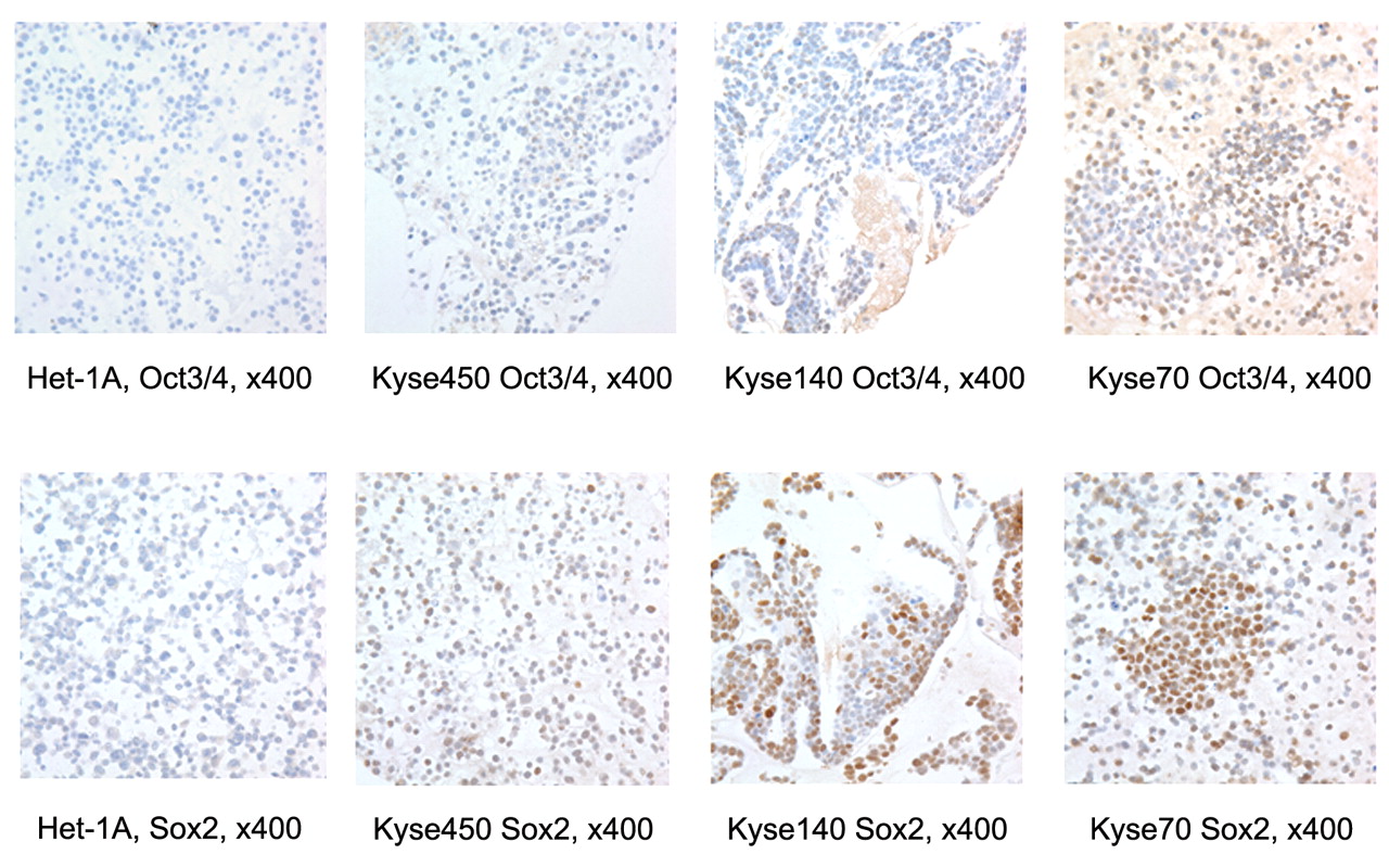
Oct3/4 and Sox2 expressions in tumour samples. Immunohistochemically, Oct3/4 positive immunostaining was observed in the cancer cell nuclei (Figure 3). Among the 162 tumours, 107 were negative for Oct3/4 staining, and 5 were scored as 1 or 2. These tumours were again classified as either negative or weakly positive for Oct3/4 immunostaining (112 in total, 69.14%). Twenty-one (12.96%) were scored as 3 or 4 and classified as intermediately positive for Oct3/4 immunostaining. Twenty-nine (17.90%) were scored as 6 or 9 and classified as strongly positive for Oct3/4 immunostaining. For Sox2, positive immunostaining was also observed in the cancer cell nuclei (Figure 3). In total, 89 were negative for Sox2 staining and 15 were scored as 1 or 2, giving a total of 104 (64.20%) tumours classified as either negative or weakly positive; twenty-one (12.96%) were scored as 3 or 4 and classified as intermediately positive for Sox2 immunostaining, while 37 (22.84%) were scored as 6 or 9 and classified as strongly positive for Sox2 immunostaining.
Immunocytochemistry of Oct3/4 and Sox2 in Kyse70, Kyse140, Kyse450 and Het-1A cell lines.

Immunohistochemistry of Oct3/4 and Sox2 in tissue array materials of esophageal squamous cell carcinomas (×400).
Correlation between Oct3/4 and Sox2 protein expressions. It was possible to observe that Oct3/4 and Sox2 expressions co-localized in the same tumour areas, with similar staining patterns in some tumours, although their expressions were also independently heterogeneous. In order to determine whether the expressions of Oct3/4 and Sox2 in these tumours were associated, a crosstable analysis was performed, which showed that their expressions were significantly correlated (Pearson Chi-square analysis, two sided, p<0.001, Table II).
Clinicopathological correlations. The correlations between the clinicopathological features and Oct3/4 and Sox2 expressions in the primary tumours are summarized in Table I. Neither Oct3/4 nor Sox2 expression was correlated to age, sex, tumour location, tumour size and clinical stage. However, higher levels of Oct3/4 or Sox2 expressions were significantly associated with higher histological grade (p<0.001 for both factors), indicating their correlation to dedifferentiation in these tumours.
Relation to survival. Follow-up information was available for 141 patients for a period of minimum 10 years. Survival curves were analyzed using the Kaplan-Meier method. In univariate analysis, patients with tumours moderately or strongly immunostained for Oct3/4 had an overall poorer survival than patients with negative or weakly stained tumours (p=0.001). A significant correlation between increasing levels of Sox2 immunostaining and decreasing survival for the patients (p<0.001) was observed. After being stratified by histological grade, Oct3/4 and Sox2 expressions were still significantly associated with unfavorable overall survival (p=0.008 and p=0.003, respectively).
Discussion
The expressions of Oct3/4 and Sox2 were firstly discovered in human esophageal squamous cancer cell lines with the antibody AF1759 and MAB2018 from R&D System for immunocytochemistry. Since controversial results concerning the expression of Oct3/4 in tumours exist (29), the expression status of these two genes in these cell lines, in addition to a virus-transformed “normal” esophageal epithelial cell line Het-1A was investigated. The expressions were repeatedly demonstrated immunocytochemially, with different positive and negative controls. To verify these findings, real-time RT-PCR with the hydrolysis probes was carried out, and repeatedly demonstrated similar results in these cell lines as well. Considering the fact that Oct4 pseudogenes are transcribed in cancers (26), the Oct3/4 gene- specific PCR primer pair (poct-4a, 5′-TCCCTTCGCA AGCCCTCAT-3′ and poct-4b, 5′-TGACGGTGCAGGGCTCCGGGGAGGCCC CATC-3′) was applied for conventional RT-PCT for these four cell lines, obtaining similar results. All these experiments were repeated at least twice. The additional Western blot analyses also disclosed similar results to those shown by immunocytochemistry and RT-PCT. Therefore, it was concluded that both Oct3/4 and Sox2 were variably expressed in these cell lines.
Oct3/4 and Sox2 expression crosstabulation.
Based on the findings on cell lines, this work was extended to clinical esophageal cancer materials (24) for which official follow-up data were obtained during the international collaboration. To date, this study is the first report investigating the expression patterns of the embryonic stem cell factors Oct3/4 and Sox2 in esophageal squamous carcinomas.
Esophageal cancer is one of the most aggressive neoplasms and the overall prognosis for esophageal cancer patients is poor (30). One of the reasons for the low survival rate is the tumour's intrinsic resistance to many clinical therapies, especially chemotherapy. Chemotherapy often removes the bulk of a tumour mass without preventing tumour recurrence, suggesting the survival of a subset of cancer stem cells. Recent studies have provided experimental evidence for the concept that human tumour growth may depend on a small portion cancer stem cells (31).
The present investigation on esophageal squamous carcinomas revealed several novel observations. Firstly, it was discovered that 17.90% and 22.84% of these esophageal carcinomas had a strong positive expression for Oct3/4 and Sox2, respectively. Secondly, the expression of Sox2 and Oct3/4 was coordinated. Pearson or Spearman correlated analyses showed a positive correlation between the two proteins. Thirdly, although the protein expression of either Oct3/4 or Sox2 did not correlate to age, sex, tumour size, location, histological type or UICC stage, both protein expressions were significantly associated with higher histological grade of the tumours and poorer clinical outcome of the patients. However, it should be pointed out that although these two proteins were significantly associated in expression and co-localization, their expressions in these samples were heterogeneous.
It has been proposed that Sox2 and Oct3/4 are key regulators of stem cell pluripotency and differentiation, which means that they are the primary factors determining the fate of ESCs by controlling cell self-renewal and differentiation (32). It is also known that the two factors act in cooperation in the ESCs (33). Phenotypically, human preimplantation embryonic cells resemble cancer cells in many ways, especially in their ability to grow indefinitely. Both types of cells undergo deprogramming to a proliferating state and become immortal, self-renewable and invasive. It has been reported that Oct3/4 and Sox2 are expressed in human tumours but not in normal somatic tissues (34), in agreement with the hypothesis that embryonic genes are re-activated in tumour cells.
In this series, Oct3/4 and Sox2 nuclear expressions were strongly associated with the histological grade of the tumours and survival of the patients. These data support the previous observations (13, 35, 36). Sox2 was frequently and strongly expressed in poorly differentiated and neurally invasive components of invasive pancreatic carcinoma (13). Oct3/4 was expressed in poorly differentiated (insular) carcinoma of the thyroid gland (35). The detection of anti-Sox2 T-cells may predict the clinical outcome in patients with asymptomatic plasma proliferation disorders (36). Given the previously reported functions of Oct3/4 and Sox2, the results suggest that their expressions may play a role in conferring a less differentiated phenotype or inactivating the ability to differentiate in esophageal carcinomas.
It has been demonstrated that human somatic cells can be reprogrammed into pluripotent stem cells by either a combination of Oct3/4, Sox2, Nanog and Lin28 factors (37), or a combination of Oct3/4, Sox2, Klf4 and c-Myc factors (38). A common feature of these studies is the contribution of Oct3/4 and Sox2 to pluripotency, indicating that tumour cells with Oct3/4 and Sox2 expression may behave as or similarly to tumour stem cells. It is also known that SP-positive tumour cells, which most probably harvest tumour stem cells, are resistant to chemotherapy and radiotherapy (39, 40). Thus the present investigation on esophageal epithelial cell lines and squamous esophageal cancer materials may indicate that Oct3/4 and Sox 2 convey stemness features on the tumour cells, so that these tumours could easily develop and relapse, resulting in poorer clinical outcome.
The current results demonstrate that ESC markers Oct3/4 and Sox2 could be detected in human esophageal squamous (cancer and virus-transformed “normal”) cell lines and related cancer tissues. The poorly differentiated esophageal squamous cancer cell lines expressed higher levels of Oct3/4 and Sox2. The higher levels of Oct3/4 and Sox2 protein expression in the esophageal carcinomas were correlated with higher histological grade and poorer patient overall survival. Since the function of pluripotency and self-renewal of these factors have previously been characterized in adult human cells (37, 38), the role of Oct3/4 and Sox2 in esophageal carcinogenesis, especially in consideration of tumour cell stemness, merits further studies.
Footnotes
-
↵* Both authors contributed equally to this work. This work was supported by The Norwegian Cancer Society and The National Nature Science Foundation of China (30470464, 30670532).
- Received August 18, 2008.
- Revision received December 19, 2008.
- Accepted January 19, 2009.
- Copyright© 2009 International Institute of Anticancer Research (Dr. John G. Delinassios), All rights reserved
References
In this issue


Jump to section
Related Articles
Cited By...
- Induced Pluripotent Stem Cell-related Genes Correlate With Poor Prognoses of Oral Squamous Cell Carcinoma
- Induced Pluripotent-stem-cell Related Genes Contribute to De-differentiation in Oral Squamous Cell Carcinoma
- Expression Analysis of iPS Cell - Inductive Genes in Esophageal Squamous Cell Carcinoma by Tissue Microarray
- Hyaluronan-CD44v3 Interaction with Oct4-Sox2-Nanog Promotes miR-302 Expression Leading to Self-renewal, Clonal Formation, and Cisplatin Resistance in Cancer Stem Cells from Head and Neck Squamous Cell Carcinoma