


Abstract
Background: Malignancies affecting the central nervous system are intractable to conventional therapies thereby requiring an alternative strategy, such as ultrasound irradiation. Materials and Methods: We originally designed a transducer for intracranial insonation and investigated the effect of 210.4 kHz ultrasound on malignant glioma cells. Results: The insonation of 2.61 W/cm2 effectively disrupted the malignant cells. This effect was reinforced by the echo-contrast agent, Levovist. The condition was applied to tumor-bearing animals and external insonation inhibited subcutaneous tumor growth. It also repressed the growth of intracranially implanted tumors and prolonged survival of the animals. When Levovist was stereotactically injected into the tumors, the effect of insonation was significantly enhanced. Conclusion: A neuronavigation system or stereotactic device has been used commonly for patients with brain tumor. Administration of combination therapy consisting of insonation and a local echo-contrast agent will have a role in improving the treatment for malignant gliomas.
Malignant glioma is the most common primary tumor in the central nervous system. Since the central nervous system is a critical organ, total resection of the tumor is often difficult. In such cases, a residual tumor recurs and the prognosis for recovery is poor because of the infiltrating nature of the tumor growth. Despite recent advances in adjuvant therapies, tumors still resist various therapies and, therefore, the development of an alternative therapy is required. In this study aimed at utilizing ultrasound, we created an ultrasound transducer for the central nervous system and applied it for the treatment of malignant glioma.
Ultrasound has been commonly used in the clinical field as a diagnostic tool. Compared to other diagnostic equipment, ultrasonic devices are handy and less costly. In addition, they do not emit radiation, making them suitable for non-invasive screening for lesions of superficial organs. They are not only used to observe organs by brightness or the B-mode, but also for dynamic studies, such as the measurement of blood flow or cardiac movement. Other applications of elastography, color Doppler, and 3D sonography are possible. To date, ultrasound has been used widely in the clinical field and many physicians and medical staff are familiar with the technology. The existence of many people who are aware of ultrasound properties is a great advantage for further development and widespread use of treatment using ultrasound.
While ultrasound has long been used as a diagnostic tool, therapeutic applications also have been spotlighted. Several such applications are already commercially available. High-intensity focused ultrasound is a typical example.
High-intensity focused ultrasound (HIFU) is used to treat malignancies such as prostate, uterine, or breast carcinoma. By focusing the high-intensity ultrasound beam onto the lesion, the acoustic force of the ultrasound delivers lethal energy to the tumor tissues. Another application is sonodynamic therapy, which is used for neovascularization and malignancies. By exposure to ultrasound after the administration of a sonosensitizing agent, only neosynthesized blood vessels or malignant cells that accumulate the agent are eliminated by the energy.
Previously, we investigated sonoporation as a therapeutic application of ultrasound and found a favorable effect on gene transfer for colon cancer (1), and on the central nervous system (2). The effect was enhanced by echo-contrast microbubbles. In the context of these experiments, we noticed that the combination of ultrasound and the echo-contrast agent, Levovist, could generate lethal energy to kill intracranial malignant glioma cells by perforating their cell membrane.
Several effects have been demonstrated in the combination of echo-contrast microbubbles and ultrasound (3-9). During insonation, microbubbles absorb physical energy and confer cavitation damage, sonochemistry and sonoluminescence to the neighboring structures. Other than gene transfer, this effect has been applied to drug delivery and thrombolysis (10-15). However, its actual application in clinical fields is relatively limited. The innate cavitation perforates the cellular membrane adjoining ruptured microbubbles and can cause cell death of the surrounding tissue. Ultrasound-induced apoptosis and cell lysis by echo-contrast agents for malignant cells is promising for future cancer therapy (16). Several issues including target tissues, ultrasound devices and conditions need to be addressed before development of clinical therapy by insonation for malignant diseases.
In the current paper, we propose therapeutic ultrasound irradiation for malignant glioma in combination with intratumoral or stereotactic injection of echo-contrast microbubbles. The ultrasonic condition was originally determined during in vitro three-dimensional cell culture experiments and applied to tumor-bearing animal models.
Materials and Methods
Insonation. Ultrasound was generated by the wave synthesizer, Wave Factory WF 1943 (NF electronic instruments, Yokohama, Japan). The amplifier and ultrasound probe were produced by Honda Electric Inc., Toyohashi, Japan. The transducer was made of piezo ceramics and the diameter of the probe was 5 mm. This apparatus can emit a continuous wave of 210.4 kHz ultrasound up to 5.08 W/cm2 measured by the force balance method in water using an ultrasound power meter (UPM-DT-1; Ohmic Instruments Co., Easton, MD, USA). The power intensity of 2.61 W/cm2 was selected for the study. The reason for this was that, unlike 3.80 W/cm2, this value was not directly toxic to brain culture slices even for 20 seconds without microbubbles (2). In this setting, the ultrasound intensity of 2.61 W/cm2 corresponded to 0.143 MPa.
In vitro three-dimensional cell culture of human brain tumor cells. Cells of the human malignant glioma cell line, T98G (ATCC, Rockville, MD, USA), were cultivated in Dulbecco's minimal essential medium supplemented with 10% fetal bovine serum. Cells were grown in a normal culture flask until the start of the experiments. Material for the scaffold of the three-dimensional cell culture was gelatin. The three-dimensional culture method has been described elsewhere (17). In brief, a gelatin sponge was cut into 5-mm cubes and dispersed cells (1×10⁴ cells/100 μl) were injected into the sponge. After adhesion of the cells to the scaffold, cubes containing brain tumor cells were transferred into a 10-cm dish and further cultivated at 37°C in a 5% CO2 incubator for 10 days. Levovist was purchased from Japan Schering, Osaka, Japan. Disruption of the cell membrane by insonation was measured by a chromate-releasing assay.
Chromate-releasing assay. Cells in the three-dimensional cube were labeled with 300 μCi of 51Cr-sodium chromate in 3 ml of culture medium for 4 hours at 37°C in a 5% CO2 incubator. After three-washes with phosphate-buffered saline (PBS) or by changing the buffers, the cube was insonated with 2.61 W/cm2 of power intensity (Figure 1). Proportions of the dead cells showing membrane disruption were determined from counts of chromate released into the culture supernatant. Before and after insonation, released radioisotopes were measured and after the experiments, total labeled chromates were measured after cell lysis by adding Triton®X to the inner chamber. Percentage cell death was calculated by dividing the net amount of released chromate by the total amount of labeled chromate ×100.
Animal studies. Four-week-old (50-70 g) of female Fischer 344 rats were purchased from Sankyo Labo-service (Tokyo, Japan). All the animal experiments were performed with permission from the Animal Care Committee at Jikei University School of Medicine. The RT2 glioblastoma cells (18) of 2×10⁶ cells/100 μl were implanted subcutaneously into the right flank of syngeneic Fischer 344 rats and then three days later, the rats were treated by insonation and microbubbles for 3 consecutive days. The efficacy of the treatment was assessed from the tumor growth rate.
In another experiment, RT2 cells were implanted stereotactically into the right caudate nucleus using a modification of the method of Kobayashi et al. (19). Four-week-old Fischer 344 rats were anesthetized and placed in a small animal stereotactic frame (David Kopf Instruments, Tujunga, CA, USA). A sagittal incision was made through the scalp to expose the skull and a small burr hole was made 1.3 mm posterior and 4 mm lateral to the right of the bregma. Ten thousand tumor cells suspended in 10 μl of PBS were injected with a 701 Hamilton syringe over 30 seconds to a depth of 4.5 mm. The needle was left in place for one minute and withdrawn slowly. The hole in the skull was plugged with bone wax and the incision was closed by surgical clips (Autoclip, 9 mm; Nippon Beckton Dickinson, Tokyo, Japan) (20, 21). On the following day, the rats were treated by insonation. When microbubbles were used, 5 mg in 10 μl of microbubbles were gently injected into the tumor through the needle track using the same stereotactic frame. The tip of the ultrasound beam was adjusted to the tumor-injected site and insonation was conducted through the skull. Rats were treated for 20 seconds for 3 consecutive days and their survival was monitored.
Statistical analysis. Statistical analysis was performed with two-sample t-tests. Significance was determined at p-values less than the 0.05 level. Statistics were computed using the Statview statistic package (ver. 5.0: SAS Institute Inc, Berkeley, CA, USA).

In vitro experiment of insonation with three-dimensional culture cells. Three-dimensionally cultured cells were radio-labeled with sodium chromate for 4 hours. After washing, cells in a sponge were directly insonated with or without microbubbles through the bottom of the cellophane membrane of the inner chamber. The chromate released by insonation was determined and the proportion of dead cells with membrane disintegration was calculated.
Results
Insonation disrupts the cell membrane by cavitation. Previously, we used 210.4 kHz of ultrasound and observed membrane disintegration of cells of the central nervous system by scanning electron microscopy (2). To confirm if insonation could induce cell membrane porosity in glioma cells with microbubbles of the echo-contrast agent, Levovist, the effect on cell viability was evaluated as the first step of the experiments. Three-dimensionally cultured glioma cells were insonated at 2.61 W/cm2 with 30 mg/ml of Levovist. The membrane integrity was assayed by chromate-releasing assay. Unless insonation was performed, there was barely any chromate released from the cells. Insonation disrupted the cell membrane and chromate was released into the culture supernatant. When the insonation time was extended, cell death as shown by chromate release also increased (Figure 2A). Since the ultrasound power intensity was constant, time elongation meant an increase in the total dose of insonation. As the insonation time was doubled, the amount of chromate released was approximately doubled. There was a direct correlation between cell death defined by the release of chromate and the duration of insonation in the range of 0 to 40 seconds.
In order to determine whether microbubbles could modify membrane disintegration, the dose effect of the microbubbles was examined. Cells were exposed to several dilutions of microbubbles and insonated at 2.61 W/cm2 for 20 seconds. The amount of chromate released under each condition was measured and the effect of the microbubbles determined. When the concentration of microbubbles increased, cell death also increased (Figure 2B). However, the correlation was not proportional. Cell death at 60 mg/ml was less than twice that at 30 mg/ml of microbubbles. Additionally, the effect almost plateaued at concentrations above 60 mg/ml.

Effect of ultrasound on glioma cell death. A, Relationship of insonation time to cell death. Glioma cells in three-dimensional culture were irradiated with various durations of insonation. Within the measured ranges, the percentage of dead cells increased when the insonation time was elongated. Note that since the power intensity of the ultrasound was constant 2.61W/ cm2, the total dose of acoustic energy increased when the insonation time was extended. Results are expressed as the mean of six experiments and the bars indicate sample standard deviation. B, Relationship of Levovist concentration to cell death. Glioma cells were irradiated with various concentrations of Levovist. Ultrasound of 2.61 W/cm2 for 20 seconds was used in the experiment. A correlation was seen between a low concentration of microbubbles and the amount of cell death. However at higher concentrations, the effect plateaued. Results are expressed as the mean of six experiments and the bars indicate sample standard deviation. C, Effect of the combination of insonation and Levovist on cell death. Glioma cells were treated or not treated with ultrasound and Levovist. Conditions of the ultrasound were 2.61 W/cm2 for 0 (US-) and 20 seconds (US+ Levovist+). The concentration of Levovist was 0 (Levovist-) and 30 mg/ml. Levovist by itself did not confer toxicity on the cells. On the contrary, insonation alone caused chromate release. The effect was significantly enhanced by Levovist. Results are expressed as the mean of six experiments and the bars indicate the sample standard deviation.
In these experiments, insonation induced chromate release and a low concentration, in the range of 0 to 60 mg/ml, of microbubbles facilitated the effect. To validate the effect of insonation and microbubbles, cells were treated with 0 or 30 mg/ml of microbubbles and insonation at 0 or 2.61 W/cm2 for 20 seconds and the differences in each group were examined. Unless insonated, the microbubbles themselves led to very little cell death in glioma and there was no difference between the treated and non-treated groups (Figure 2C). When cells were exposed to the ultrasound, insonation caused chromate release under such conditions. Differences were observed between the insonated and non-insonated groups (p<0.0001) as previously suggested. When cells were treated with microbubbles, presence of the echo-contrast agent enhanced cell death by ultrasound. Under ultrasound exposure, there was a difference in the death of cells treated with microbubbles and those without them (p<0.002).

Tumor growth retardation by insonation. After the inoculation of glioma cells, the subcutaneous tumor nodule was sequentially treated with insonation and Levovist for three days. Rats were treated with 0 seconds of 2.61 W/cm2 of insonation with 0 mg/ml of Levovist (●), 0 seconds of insonation with 30 mg/ml of Levovist (■), 20 seconds of insonation with 0 mg/ml of Levovist (○), and 20 seconds of insonation with 30 mg/ml of Levovist (◻). Results are expressed as the mean of each rat and the bars indicate sample standard deviation.
The results of the in vitro studies suggested a potential application of insonation in oncotherapy for brain tumor. Efficacy of the treatment was successively examined by in vivo experiments. RT2 glioma cells were implanted into the right flank of Fischer 344 rats. After confirmation that a small tumor nodule was established at the implanted site, the rats were treated with local microbubble injection followed by insonation for three days. When the rats were treated only by microbubbles and not insonated, the growth of the tumor was almost equal to that of the untreated control rats (Figure 3.) In contrast, ultrasound exposure inhibited tumor growth. The effect was significantly enhanced by co-treatment with microbubbles (p<0.01 day 9, 12, 16, and p<0.05 day 19). Unlike the other groups, the combination of insonation and microbubbles even led to a cure in some animals. Three out of six rats treated by insonation with microbubbles demonstrated complete tumor regression. These rats survived for more than 365 days without any recurrence.
Since combination treatment was effective for in vivo malignant glioma, tumor cells were stereotactically inoculated into the caudate nucleus of brain parenchyma and then the efficacy of treatment was evaluated. After implantation of RT2 glioma cells, rats were treated by insonation with or without echo-contrast microbubbles. As a control, other rats were mock insonated. Due to limited intracranial space and rapid growth of the tumor, the animals had a low survival. All the rats died by day 15 unless insonated, whether with or without microbubbles (Figure 4). Mean survival was 12.62±1.84 days for untreated animals, and 12.13±1.13 days for animals treated with microbubbles. When the rats were insonated, the treatment prolonged their survival (mean survival: 15.50±4.04 days). The effect was further reinforced by microbubbles (mean survival: 20.71±3.04 days). There were significant differences between the untreated group or that treated only by microbubbles and the group treated with both insonation and microbubbles (p-values <0.0001).
Discussion
Malignant glioma is one of the most intractable diseases in the human body. In spite of recent advancements of therapies, including surgery, radiotherapy, chemotherapy, immunotherapy and other adjuvant therapies, this disease is refractory and the prognosis is unsatisfactory, with median survival times of 5.6 years for low-grade astrocytoma, 1.6 years for anaplastic astrocytoma and 0.4 years for glioblastoma, respectively (22). Since most patients die within 2 to 5 years after their diagnosis and the efficacy of current therapy is limited, a potent alternative strategy is required. In spite of poor patient prognosis, the tumor seldom metastasizes to organs other than the central nervous system and since the main cause of death is local recurrence of the tumor, long-term survival or even complete remission can be expected only by modulation of local recurrence (23). In addition, a direct route for drug or material delivery into the tumor is available (24). The advantage of the central nervous system for developing a new therapy is that agents can be administered into brain parenchyma by stereotactic surgery, neuronavigation or utilization of the Ommaya reservoir. These methods have been commonly used as clinical procedures and lesions are accessible by these methods.
As a therapeutic tool, ultrasound has been highlighted since it induces cell membrane porosity (25). The impact of ultrasonic forces also induces mechanical damage to living tissues, stones, clots, or other organic and inorganic compounds. It has the capability for transmitting mechanical power or force to a distant area of the human body, therefore insonation can be applied not only to the superficial tissues, but also to deeper areas. Compared to therapies with energies of shorter wavelength, such as photons, this may be a benefit for the treatment of deep-seated lesions. Brain tumor is one of the most suitable targets for the application of insonation.

Therapeutic efficacy of insonation to intracerebral tumor. Therapeutic efficacy of insonation was evaluated by survival of the tumor-bearing animals. After stereotactic inoculation of glioma cells into the caudate nucleus of the right cerebrum, the animals were treated with local administration of Levovist and external insonation. Rats were treated with 0 seconds of 2.61 W/cm2 of insonation with 0 mg of Levovist (●), 0 seconds of insonation with 3 mg of Levovist (■), 20 seconds of insonation with 0 mg of Levovist (○), and 20 seconds of insonation with 3 mg of Levovist (◻). Results are expressed as the mean of 8 individuals and the bars indicate sample standard deviation.
Ultrasound with a continuous 210.4 kHz wave was used for the study. The reason for this was based on a previous finding that ultrasound with this frequency could perforate the cell membrane of central nervous glial cells effectively (2). Perforation of the cell membrane was observed by scanning electron microscopy. The effect was similar to that demonstrated by Tachibana et al. (25). The mechanical index of the current study was 0.32, which is less than 0.612 of a previous gene delivery study for neonatal glial cells that was also conducted at 210.4 kHz. Furthermore, for gene delivery, unlike the power intensity of 2.61 W/cm2 used in the current study, 5.0 W/cm2 was required to transduce DNA by ultrasound for gene expression, since DNA had to be delivered into the cell nucleus across cellular and nuclear membranes. In addition to the macromolecular nature of DNA, both the cell membrane and DNA were positively charged and electrically repelled. Therefore, a higher external force was required for gene transduction. Correspondingly, the thermal index category soft tissue, or TIS, was smaller (=0.489) than that of the previous study (=0.935). In the experiments, employment of microbubbles enabled further energy to reach the local area of the insonation field.
Kudo et al. reported that microbubbles exposed to ultrasound caused mechanical stress that acted on the cells and bubble collapse was responsible for cell membrane damage (6). Even gentle linear bubble oscillation is sufficient to achieve rupture of lipid membranes (9). Currently, air-filled albumin microspheres (Albunex), non-shell type granules composed of 99.9% galactose and 0.1% palmitic acid (Levovist), and an albumin-shelled agent composed of fluorocarbon gas octafluoropropane (Optison) have been used as echo-contrast agents in the clinical field. In this study, we tested Levovist, which is the only agent available in Japan. As the next step, a comparison of microbubbles will be required in order to optimize the combination.
The effect of ultrasound was first evaluated on three-dimensionally cultured glioma cells and then applied to in vivo studies. This approach was useful for the study since the effects evaluated by two-dimensional culture did not correlate with those obtained with animals. Aside from cell detachments from the flat and hard plastic or plastic substrate caused by insonation, ordinary cell culture generally did not present tissue-specific architecture, mechanical and biochemical cues or cell-to-cell communication and did not mimic the function of living tissues (26). While the quantitative analysis method as well as scaffold materials and pore sizes needed to be more sophisticated, the results obtained in the three-dimensional cell culture were comparable to the results of animal experiments.
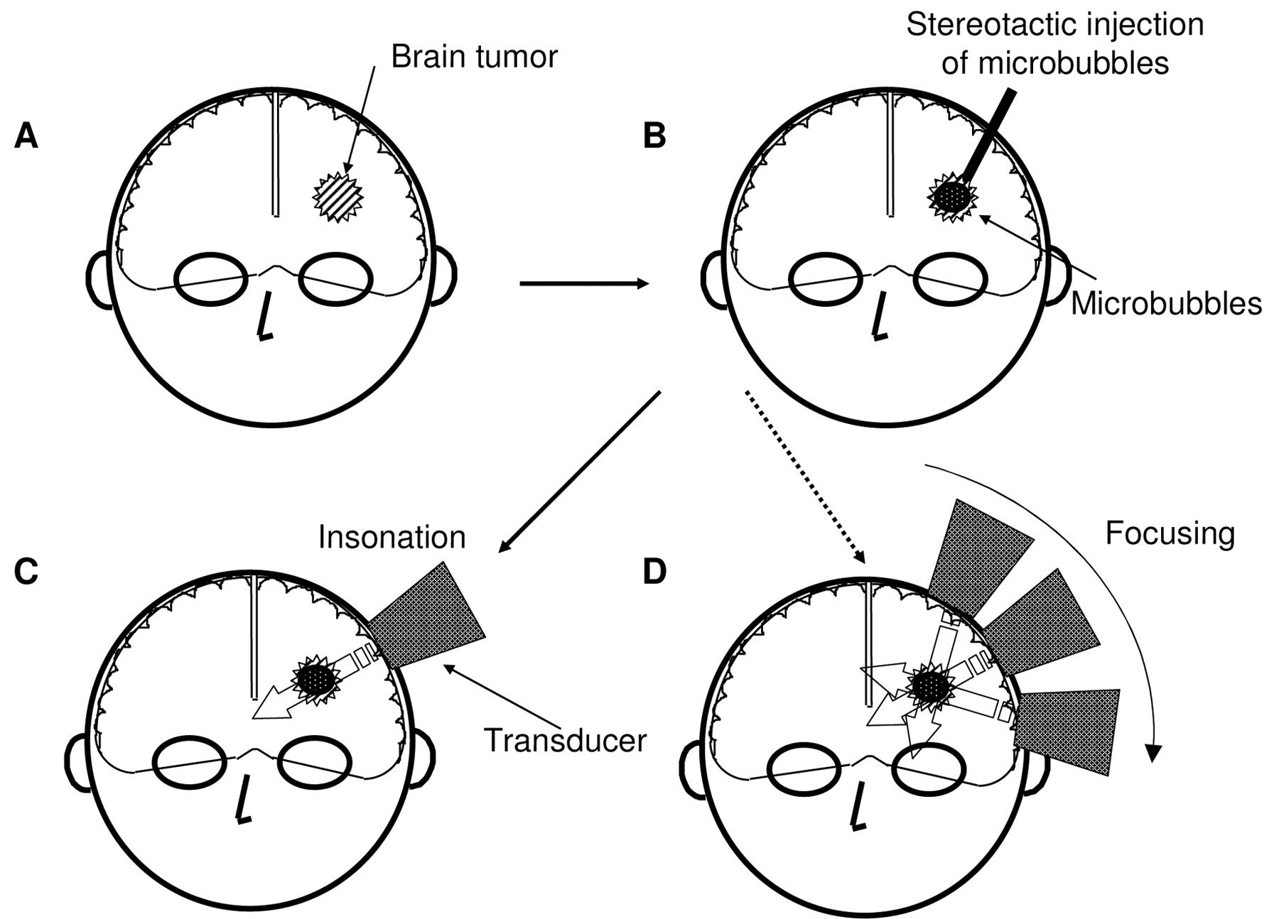
Schema of a possible approach to malignant glioma using intratumoral injection of microbubbles and insonation. A, When a patient is examined, medical imaging instruments show the location and size of the tumor. The patient undergoes treatment. B, Stereotactic injection of microbubbles into an intracranial tumor results in increased sensitivity of ultrasound and glioma cells become vulnerable to the ultrasound. C, External insonation of the injected site induces cell death in the tumor tissue. Although the power level of ultrasound is attenuated during passage through the cranium, insonation with 210.4 kHz transmits sufficient intensity to the deep area of the cerebrum. D, One of the characteristics of insonation is accumulation of acoustic energy in the insonated region. Ultrasound can be focused by moving the tip of the transducer of ultrasound and, by so doing, the conduction effect can be intensified without damaging unfocused areas of the brain.
The authors previously implanted a malignant brain tumor in the caudate nucleus of the cerebrum of rats, injected drugs or adenoviral vectors encoding β-galactosidase with a stereotactic device and found that agents can be delivered to the center of implanted brain tumor (18, 21, 27). One possible application of the strategy is shown in Figure 5. By focusing the ultrasound, intensified power energy can be concentrated on the target area. This possibility is valuable for such a therapeutic approach.
HIFU has been recently highlighted for the treatment of malignant tumors. The difference from our method is in the power intensity of the emission ultrasound. Echo-contrast agent, such as Levovist, oscillate by insonation and rupture due to inert cavitation. In this step, intense heat and mechanical forces are produced at the ruptured site. In combination to an echo-contrast agent, ultrasound can generate enough power to eradicate malignant glioma cells. For treatment of the central nervous system, avoiding the adverse effect of therapy is essential since the central nervous system is critical, as once neuronal cells are damaged, they seldom regenerate. The power intensity used in the present study was extraordinarily less than that of HIFU. Even though a lower intensity is set for the unfocused area of HIFU, utilizing as low an intensity as possible is desirable for treatment of intracranial lesions. Our previous toxicity study demonstrated minimal invasiveness to the central nervous system cells (2).
We have demonstrated therapeutic ultrasound irradiation for malignant glioma in combination with stereotactic intratumoral injection of echo-contrast microbubbles. The ultrasonic condition used for the study might have clinical relevancy. Further study is warranted.
Footnotes
- Received July 1, 2008.
- Revision received November 18, 2008.
- Accepted December 1, 2008.
- Copyright© 2009 International Institute of Anticancer Research (Dr. John G. Delinassios), All rights reserved


