


Abstract
Recent progress is described in an ongoing collaborative multidisciplinary research project directed towards the purification, structural characterization, chemical modification, and biological evaluation of new potential natural product anticancer agents obtained from a diverse group of organisms, comprising tropical plants, aquatic and terrestrial cyanobacteria, and filamentous fungi. Information is provided on how these organisms are collected and processed. The types of bioassays are indicated in which initial extracts, chromatographic fractions, and purified isolated compounds of these acquisitions are tested. Several promising biologically active lead compounds from each major organism class investigated are described, and these may be seen to be representative of a very wide chemical diversity.
Despite recent improvements in survival rates due to advances in early detection and the quality of treatment available, cancer remains a major public health hazard in countries all over the world (1, 2). For example, in the case of the United States in 2016, nearly 1.7 million new cases of cancer are predicted to occur, in addition to over 595,000 cancer deaths (1). The three most common types of cancer in females in the U.S. are those of the breast, uterine corpus, and colon and rectum, while those of males are prostate, colon and rectum, and melanoma (2). Unfortunately, death rates are increasing for cancer of the liver, pancreas, and uterine corpus (1). In the People's Republic of China, with a much larger population than the U.S., nearly three million cancer deaths were projected to occur in 2015, with lung cancer being the major cause, and stomach, esophageal and liver cancer also commonly occurring (3).
Natural products have played a major beneficial role in cancer chemotherapy for over 50 years, both in terms of providing established drugs and new lead compounds for synthetic optimization, and in furnishing substances for probing cellular and molecular mechanisms of action relevant to cancer inhibition (4-6). Thus, out of a total of 175 small-molecule anticancer drugs introduced into therapy in Western countries over an approximately 70-year period, about 49% were either obtained from organisms directly or were derived from natural products (5). Currently, a considerable number of natural products of terrestrial microbial origin are used in cancer chemotherapy, including actinomycin D, several anthracycline derivatives (inclusive of daunorubicin, doxorubicin, epirubicin, idarubicin, and valrubicin), bleomycin, actinomycin D, mitomycin C, and mitoxantrone HCl (4). In turn, four main classes of higher plant-derived anticancer agents are used clinically in the U.S. and Europe, namely, the vinca (Catharanthus) bisindole alkaloids (vinblastine, vincristine, vinorelbine, vinflunine), the semi-synthetic epipodophyllotoxins (etoposide, teniposide, and etoposide phosphate), the taxanes (paclitaxel and paclitaxel albumin-stabilized nanoparticule formulation, docetaxel, cabizitaxel), and the camptothecin derivatives (irinotecan and topotecan) (4). In the last few years, the first small-molecule anticancer agents of marine origin have been introduced into the market, including trabectedin (ET-743), a partially synthesized version of a marine tunicate constituent (a DNA-binding agent via an effect on transcription; for soft tissue sarcoma) (6, 7). Also included in this category are eribulin mesylate, a synthetic derivative of the marine polyether macrolide, halichondrin B (an inhibitor of microtubule dynamics; for metastatic breast cancer) (5, 6, 8), and brentuximab vedotin, a conjugate of an antibody–marine compound derivative (a binding agent to CD30 cells that interacts with tubulin; for Hodgkin's lymphoma and anaplastic large-cell lymphoma) (5, 6, 9). Additional recently approved natural product anticancer agents are romidepsin, from a soil bacterium (a histone deacetylase inhibitor; for T-cell lymphoma) (6, 10), and the terrestrial microbial semi-synthetic derivatives ixabepilone (a microtubulin inhibitor; for locally advanced and metastatic breast cancer) and temsirolimus [an inhibitor of the kinase mechanistic inhibitor of rapamycin (mTOR); for advanced renal cell carcinoma)] (6, 10). From plants, the cephalotaxine alkaloid, omacetaxine mepesuccinate (homoharringtonine), a protein translation inhibitor, was approved by the U.S. Food and Drug Administration (FDA) as a new antileukemic agent (5, 6, 11). Another new plant substance approved in 2012 was ingenol mebutate, for the topical treatment of actinic keratosis, a condition that can lead to squamous cell carcinoma (5, 6, 12). Following the approval of brentuximab vedotin mentioned above, a second antibody–drug conjugate (ADC) was approved recently, namely, ado-tratuzamab emtansine, which is based in part on the natural product maytansine, and used for patients with human epidermal growth factor receptor 2 (HER2)-positive metastatic breast cancer (5, 6, 13, 14). While initially reported as deriving from a plant, it appears that maytansine is actually of bacterial endophyte origin (15). There are a relatively large number of natural products and their derivatives (inclusive of ADCs) currently in clinical trials as potential new oncolytic agents (5, 6, 14), so further new drugs of this type from a taxonomically varied range of organisms should reach the market. Importantly, Cragg and colleagues have pointed out that natural products are enormously useful as laboratory probes for a large number of diverse targets involved with cancer cell cycle biology (4, 16).
In this review, recent progress in an ongoing multi-institutional collaborative project funded by the U.S. National Cancer Institute (NCI), National Institutes of Health (NIH), Bethesda, MD, USA will be described. This research program is funded through the ‘Program Project’ (P01) mechanism and has been reviewed previously (17, 18). Currently, there are three primary academic groups involved: The Ohio State University (OSU), the University of Illinois at Chicago (UIC), and the University of North Carolina at Greensboro UNCG), with the participation of a fungus biotechnology company, Mycosynthetix, Inc. (Hillsborough, NC, USA) and a pharmaceutical company (Eisai Inc., Andover, MA, USA). Several other senior investigators in the project team are based at other academic institutions and a private non-profit research institute. The overall administration of the program project is as previously published, with focus being on the isolation, structural characterization, and biological evaluation of lead anticancer compounds from tropical plants, freshwater and terrestrial cyanobacteria, and filamentous fungi (17). In the following paragraphs, the potential of each of these three major types of organisms is mentioned.
As indicated above, plants (of both temperate and tropical origin) have already afforded several clinically used oncological agents, and are a proven resource for further research in anticancer drug discovery. In addition to various camptothecin, podophyllotoxin, taxane, and vinblastine derivatives, compounds based on other structural types of plant-derived secondary metabolites are currently in clinical trials, including the stilbenoids combretastatin A-1 diphosphate and combretastatin A4 phosphate (4, 5). Other plant-derived compounds in phase I-III oncological clinical trials include curcumin, gossypol, genistein, resveratrol, and triptolide or their derivatives (5).
Cyanobacteria (also known as blue-green algae) have been cited as a promising and productive source for new natural products, with both marine and non-marine species having proven to be rich sources of diverse new metabolites (4, 19-21). Cyanobacteria are relatively unexplored as potential anticancer agents, particularly those from aquatic and terrestrial sources. An investigation of pharmaceutical and agrochemical agents from cyanobacteria revealed a discovery rate for bioactive compounds of approximately 7%, which is typical for microorganisms (22). The rate of rediscovery of known bioactive compounds, however, was significantly lower among the cyanobacteria than other sources with similar hit rates such as actinomycetes. A screening project of cultured cyanobacteria published by Patterson and Moore also demonstrated a similar hit rate (7%) against several cancer cell lines (23).
Fungi have led to the development of numerous drugs, including beta-lactam antibiotics, cholesterol-lowering agents, and immunosuppressants (5, 6). In cancer chemotherapy, the fungal-derived compound irofulven has been studied extensively, and is derived from illudin S, which was isolated from the fungus Clitocybe illudens (24). The results of a phase II trial of irofulven were reported in 2010 in women with recurrent epithelial ovarian cancer, where its potential utility as an adjuvant with angiogenesis inhibitors was suggested, but it was not recommended as a single agent (25). A review of 14 phase I/II trials on irofulven has been published (26). Other fungal-derived compounds have been developed as anticancer leads (18), including sonolisib, a derivative of wortmannin (27), which was reported recently to be in phase I/II clinical trials (5). The current rather limited laboratory investigation of fungi is surprising when it is considered how many medical breakthroughs their small-molecule organic secondary metabolites have afforded to date. Of the potentially over five million fungal species in existence on Earth (28), a much smaller percentage has been studied for bioactive secondary metabolites, and only a small proportion of these has been evaluated for anticancer activity. Moreover, while nearly 250,000 compounds have been described from nature, only about 14,000 (or 5%) of these are from fungi (29, 30). When coupled with genomic data that reinforce the biosynthetic potential of these organisms (31), fungi may be seen to continue to be promising sources for new medicines, including those for treating cancer.

New organizational scheme of the program project.
Natural products tend to cover a more extensive ‘chemical space’ than compounds produced by chemical synthesis and by combinatorial chemistry (32). This is a term used in chemoinformatics referring to the property space spanned by all possible molecules and chemical compounds under a given set of conditions. It is our contention that research into tropical plants, cultured cyanobacteria, and filamentous fungi will lead to the discovery of new and chemically diverse compounds that will serve as anticancer drug leads. To support this, a chemical diversity study was performed in which data on 343 bioactive compounds isolated from our Program Project from these three different types of source organisms were compared, both to each other and to a set of 96 FDA-approved anticancer agents, in terms of the chemical space covered, using principal component analysis. The conclusion of this study was that while our plant, cyanobacterial, and fungal bioactive compounds represent different areas of chemical space, as a group they have substantial overlap with the standard anticancer agents administered in the clinic (33), indicating an excellent overall prospect of discovering new medicinally valuable leads.
Program Project Organization
An organizational scheme of our program project research collaboration is shown in Figure 1. This figure shows the locations of each project and core, and the major technical activities carried out. The scheme has been refined in the present period of funding (2014-2019) in one major way when compared with the previous format (17), through the inclusion of a new Core B (Medicinal Chemistry and Pharmacokinetics), housed at OSU. Each core component is highly integrated with all three of the main projects, with Projects 1-3 focused on the investigation of tropical plants, aquatic cyanobacteria, and filamentous fungi, respectively. The fungi are provided for this collaboration by Mycosynthetix, Inc. (Hillsborough, NC, USA). The technical expertise represented by the senior investigators enables all of the following activities to be achieved: collection and taxonomic authentication of organisms; development of intellectual property agreements in order to access organisms; initial solvent extraction of organisms; testing of extracts against a wide panel of cell-based and mechanism-based primary in vitro assays; isolation and structure elucidation of in vitro-active principles of initial lead bioactive extracts; de-replication of bioactive extracts and chromatographic fractions for previously known bioactive compounds; testing of promising compounds in cancer-related inhibition assays; medicinal chemistry for enhancing the supply of certain natural product bioactive substances and generating new lead compounds; preliminary pharmacokinetics, including solubility testing and drug metabolism work; application of biostatistics for bioassays, and overall project administration. The senior investigators of the project have face-to-face meetings in different locations or group electronic meetings up to three times a year, including an annual meeting in the presence of the program project External Advisory Committee, comprised of Drs. William H. Gerwick (University of California-San Diego), Susan B. Horwitz (Albert Einstein College of Medicine), G. Robert Pettit (Arizona State University), and William C. Rose (formerly of Bristol-Myers Squibb). The program project team receives very helpful input from the NCI Program Coordinator (Dr. Yali Fu).
Collection and Processing of Organisms
The strategies being used to collect and process tropical plants, aquatic and terrestrial cyanobacteria, and filamentous fungi, the three basic types of organisms in our program project, are discussed briefly in turn below.
In the first organism collection approach taken in our program project, endemic species of tropical rainforest plants that have not been well studied are targeted. The greater biodiversity evident in tropical rainforests when compared with temperate species results in enhanced complexity of secondary metabolites (natural products), thus leading to a higher chance of discovering new pharmacologically active agents (34). There are approximately 298,000 terrestrial plant species, with about 215,000 of these presently catalogued (35). The tropical rainforests include many of the 25 biodiversity ‘hotspots’ that cover only 1.4% of the world's surface while containing nearly half of all vascular plant species (36). Our project team has collected plants previously from different tropical rainforests, with the intention of finding new anticancer agents, including in the Dominican Republic (37), Ecuador (37), Indonesia (37), Peru (37), Vietnam (38), and most recently Laos. Access to plant species in foreign countries must be gained through the development of detailed intellectual property agreements as a result of the provisions of the 1992 United Nations Convention on Biological Diversity (39) and the 2010 Nagoya Protocol (40). Members of our project team are well versed in formulating Memorandum of Agreement (MOA) documents with source countries of tropical plants of interest, conforming to contemporary requirements [e.g. see (41)].
Once an appropriate plant-collection agreement with a source country has been mutually signed, the NAPRALERT® database housed at UIC is used to aid in the selection of plant genera found in a given country for which there are relatively few phytochemical reports (38). It is worthwhile collecting more than one plant part from a given plant acquisition (e.g. the leaves, stems, bark, roots, fruits, and/or flowers), since their phytochemical profiles may be quite different. In a retrospective analysis of over 2500 plant acquisitions collected by our group in six tropical and semi-tropical locations, it was determined that roots and other below-ground organs tend to produce more potently cytotoxic extracts than those prepared from the aerial plant parts (37). During the period 2008-2011, over 800 primary plant collections for our program project were obtained from Vietnam, with the plant diversity available optimized by collecting species in different climatic zones, occurring in the north, central, and southern regions of the country (38). Dried plant material or locally produced plant extracts are shipped to the U.S. for subsequent phytochemical and biological studies. Plant taxonomic identifications are made by botanist members of our project team, and voucher specimens are deposited at the John G. Searle Herbarium of the Field Museum of Natural History, Chicago, IL, USA. Using a small amount (20-40 g) of each powdered plant for initial in vitro biological screening, a simple solvent extraction scheme is applied (42). Typically, only the organic-soluble extract (a chloroform extract treated with sodium chloride) for each plant acquisition is submitted for initial biological screening, with the remaining fractions (hexane wash and aqueous residue) retained in case of need. De-replication (the process of circumventing the re-isolation of previously known compounds from the same organism) using liquid chromatography-mass spectrometry (LC-MS) may be applied to plant extracts to avoid known cytotoxic compounds, such as cucurbitacins (43). Approximately 8,000 plant extracts have been prepared for our program project and earlier work on potential anticancer agents from plants.
In the work on cyanobacteria, a primary focus is on bioactive metabolites from both field-collected freshwater and terrestrial specimens obtained from the continental U.S., with a concentration on the Upper Midwest and Great Lakes regions, focusing on organisms in the orders Nostocales and Stigonometales. This region provides a large variety of microenvironments that are home to a diverse set of cyanobacteria (44-46). The culture collection is supplemented by blue-green algal strains from a number of commercial collections, of which many are also unstudied for their potential to provide anticancer agents. About 800 different cyanobacterial strains have been obtained for this project to date. Strain identification is based on a combination of microscopic evaluation and phylogenetic analysis using the 16S rDNA gene. The slow growth rate and low biomass yield has traditionally been a challenge for studying bioactive metabolites from cultured cyanobacteria, hence relatively few drug discovery efforts currently target these organisms. However, the sensitivity of nuclear magnetic resonance (NMR) spectroscopy as used in the identification of bioactive compounds from blue-green algae may be enhanced through use of a cryoprobe, which allows for the structure determination of natural products at a nanomolar scale (47).
Once there is sufficient algal growth, the biomass is used to inoculate larger-scale cultures, which are aerated with sterile filtered air and allowed to grow for 6-8 weeks in the appropriate inorganic medium under illumination, with an 18/6-hour light/dark cycle at a constant temperature of 22°C. Cells are separated by centrifugation and lyophilization, and the freeze-dried cell material is extracted by maceration with dichloromethane-methanol (1:1) to produce initial extracts, which are then dried (48). The extracts are pre-fractionated to create a fraction library prior to biological testing, which allows for the improved detection of minor active compounds in bioactivity screening (49). Cyanobacterial cells may be cryopreserved by freezing in their maintenance medium with 5% dimethyl sulfoxide. A well-established protocol for the de-replication of known bioactive compounds from cyanobacteria has been developed (18, 50).
As a final group of organisms, the Program Project has access to a library of over 55,000 filamentous fungi, available at Mycosynthetix, Inc. Until the initiation of our present Program Project, acquisitions in this collection had never been examined systematically as anticancer drug leads, although they have been investigated for several different pharmaceutical and agrochemical applications in the past. The Mycosynthetix fungal collection was established over the past two decades and was collected in the form of mycelia, rather than simply as spores. These samples were isolated and preserved from targeted ecosystems from around the world, after obtaining the necessary permissions. Mycosynthetix has developed approaches to culturing fungi that supports the production of unusual metabolites, including the use of both liquid and solid media. In many instances, it has been observed that a particular metabolite is produced specifically under only one set of conditions. Diverse growth conditions are utilized to optimize the chances of discovering promising biological activity. This ability to culture and optimize the growth conditions of an organism presents a means of supplying a promising lead compound in a scaled-up quantity. Prior to initial biological screening, fungal specimens are subjected to a two-stage culturing procedure, in both liquid seed and grain-based solid media. For initial biological testing, cultures are extracted with 1:1 chloroform-methanol, with organic and aqueous partitions being produced, and the organic extract partitioned into 1:1 acetonitrile-hexane (17, 18). Nearly 2,000 fungal strains have been extracted since the start of the project.
A major concentration in the work on fungi has been the development of procedures for secondary metabolite de-replication, to improve efficiency and avoid focusing resources on the re-identification of known compounds (51, 52). This includes the application of a new technique to analyze the chemistry of fungal cultures in situ via a droplet liquid micro-junction surface sampling probe, which has been utilized for our Program Project (53). This technique was optimized for the natural products environment via a close collaboration with a team of researchers at Oak Ridge National Laboratory (Oak Ridge, TN, USA).
Biological Evaluation of Samples
Once initial extracts are prepared from tropical plants, aquatic cyanobacteria, and filamentous fungi, they are subjected to evaluation in a battery of cell-based and mechanism-based assays housed at the various locations where the laboratory work in our program project is carried out. A key aspect of our group interactions is the prioritization of in vitro-active crude extracts for subsequent activity-guided purification (17, 18). Cytotoxicity assays using selected cancer cell lines are conducted currently mainly at our biological testing core component at UlC [for example, see (54-56)], with some solid tumor and leukemia cancer cell line testing also having been carried out at OSU [for example, see (54, 57, 58)]. Selected target-based in vitro bioassays that have been conducted for the program project include activation of nuclear factor κB (NFκB) (56, 59), mitochondrial transmembrane potential (MTP) (60), and semaphorin 3B assays (61). In addition, screening has begun for compounds that regulate ferroptosis, an emerging form of regulated, non-apoptotic cell death (62). Several of the bioactive compounds obtained in our program project have been followed up recently in more specialized oncology-related in vitro and in vivo bioassays performed by colleagues at the OSU Comprehensive Cancer Center, Nationwide Children's Hospital, Columbus, (OH, USA), and Georgia Regents University (Augusta, GA, USA) (63-65).
A prominent aspect of our approach to biological testing has been in the use of an in vivo hollow-fiber assay to follow up on selected in vitro-active compounds, and to help determine if such compounds are likely to be active in a subsequent murine xenograft assay. We have found that only a small percentage of natural products that are active in initial in vitro assays have in vivo biological activity. Members of our group have reviewed the use of the hollow-fiber assay for in vivo testing (66, 67). This procedure was developed initially at the U.S. NCI (68, 69) and then transferred to the UIC College of Pharmacy (70). In the hollow-fiber assay, human cancer cells are propagated within fibers that are implanted either intraperitoneally or subcutaneously in immunodeficient mice, with the method being quite rapid and relatively sparing of the quantity needed of each compound evaluated (67, 70). Recent examples of compounds active in the in vivo hollow-fiber assay from our program project are the sesquiterpene lactone, goyazensolide (71) and the diphyllin glycoside, phyllanthusmin D (72).
It is important to obtain information on the mechanism of action of promising in vivo- and in vitro-active natural products with potential anticancer activity. Recent mechanistic studies on compounds of interest to the program project team have been conducted at OSU (73, 74) and UIC (75). The COMPARE algorithm of the Developmental Therapeutics Program of the NCI is a means of identifying the pattern of growth inhibition for a lead compound among a standard panel of 60 human cancer cell lines and checking for similarities against clinically used cancer chemotherapeutic agents (76). We have accessed this very useful resource in the past, since it provides information on the possible cellular mechanism of action of a given compound under investigation. In addition, our project team includes expertise in chemical biology and determining the mechanism of action of natural products (77).
Our program project team is fortunate to have participation by colleagues from the OSU Center for Biostatistics, who have made many contributions to the statistical processing and interpretation of our data [for example, see (17, 33, 60, 61)]. Examples of such activities include determining sample size and power in experimental design, bioassay method development, statistical support for pharmacokinetics, and high-dimensional data analysis.
Examples of Bioactive Compounds Isolated from Tropical Plants, Aquatic and Terrestrial Cyanobacteria, and Filamentous Fungi
Since our Program Project was first funded by the U.S. NCI in 2007, we have purified and structurally characterized hundreds of compounds of interest from each of tropical plants, aquatic and terrestrial cyanobacteria, and filamentous fungi. Many of these were new compounds when first isolated, and are of wide chemical diversity. Several of the compounds have been subjected to more advanced biological testing, inclusive of in vivo evaluation. In view of space constraints, only a few examples in each case are given here. In selected cases, optimization of the lead natural products and the investigation of their pharmacokinetic parameters is underway through our Program Project joint medicinal chemistry and pharmacokinetics core component. As pointed out in an earlier review (17), compound development work has progressed the furthest on plant-derived secondary metabolites, when compared with bioactive substances from cyanobacteria and fungi.
Plant-derived compounds. The structures of six representative lead compounds (1-6) obtained in our research investigation on tropical plants are shown in Figure 2. Thus, two new [phyllanthusmins D (1) and E (2)] and several known glycosylated arylnaphthalene-type (diphyllin) lignan lactones [including phyllanthusmin C (3)] were isolated from different plant parts of Phyllanthus poilanei, obtained from Vietnam. The structure of phyllanthusmin D was confirmed by single-crystal X-ray crystallography, and this compound was found to be selectively cytotoxic towards HT-29 human colon cancer cells when compared with CCD-112CoN normal human colon cells [half-maximal concentration (IC50)] 170 nM vs. >100 μM, respectively. Compound 1 was shown to be active in the hollow fiber assay at UIC, using HT-29 cells (dose 20 mg/kg) implanted into immunodeficient NCr nu/nu mice. Phyllanthusimins C and D were evaluated by Dr. Jack Yalowich, Division of Pharmacology, OSU College of Pharmacy, and found not to be topoisomerase II poisons, despite their general similarity in structure to etoposide. However, phyllanthusmin D was shown to mediate its cytotoxic effects through the activation of caspase-3 (72). Selected compounds from P. poilanei were evaluated for their effects on human natural killer (NK) cells in collaborative work with Drs. Jianhua Yu and Michael Caligiuri (OSU College of Medicine and Comprehensive Cancer Center). It was found that phyllanthusmin C (IC50 for HT-29 cells, 3.2 μM) enhanced interferon-gamma (IFN-γ) production by human NK cells through up-regulation of toll-like receptor (TLR)-mediated NFκB signaling (63).
From the root bark of Sphenostylis marginata ssp. erecta, collected in Zimbabwe, 11 new isoflavonoid derivatives have been isolated (sphenostylisins A-K). Three of these compounds, sphenostylisins A-C, are based on two different novel carbon skeletons. The most interesting of the new compounds biologically, sphenostylisin A (4), exhibited IC50 values, in turn, of 6 nM in a NFκB (p65) inhibition enzyme-linked immunosorbent assay (ELISA) and 1.6 μM against HT-29 human colon cancer cells. Sphenostylin A is complex dimeric compound for which the structure elucidation process required a detailed spectroscopic data analysis (78).
For some time, our project team has been interested in the ‘rocaglate’ constituents of the Asian tropical plant genus Aglaia as potential anticancer agents (79). To this end, 21 compounds in total were isolated and characterized structurally from various plant parts of a specimen of Aglaia perviridis, collected in Vietnam. These included two new cyclopenta[b]benzofuran and two new cyclopenta[b] benzofuran rocaglate derivatives, three new triterpenoids, and one new apocarotenoid. Several known rocaglates exhibited potent inhibitory activity against HT-29 cells. Among the known compounds were a series of cyclopenta[b]benzofuran derivatives, including rocaglaol, which was the most potent compound when tested against HT-29 cells (IC50 7 nM). One of the new cyclopenta[b]benzofuran analogs (perivisidin B, 5), exhibited inhibitory activity against both HT-29 cells and in the NFκB (p65) ELISA (IC50 values of 0.46 and 2.2 μM, respectively), but was not cytotoxic towards CCD-112CoN normal human colon cells (IC50 >50 μM). From the various members of the cyclopenta[b] benzofuran group of rocaglate derivatives present, it was shown that when the typical C-1 tertiary hydroxy group is replaced with a methoxy group, the resultant cytotoxicity potency is reduced drastically, although not completely lost (80).
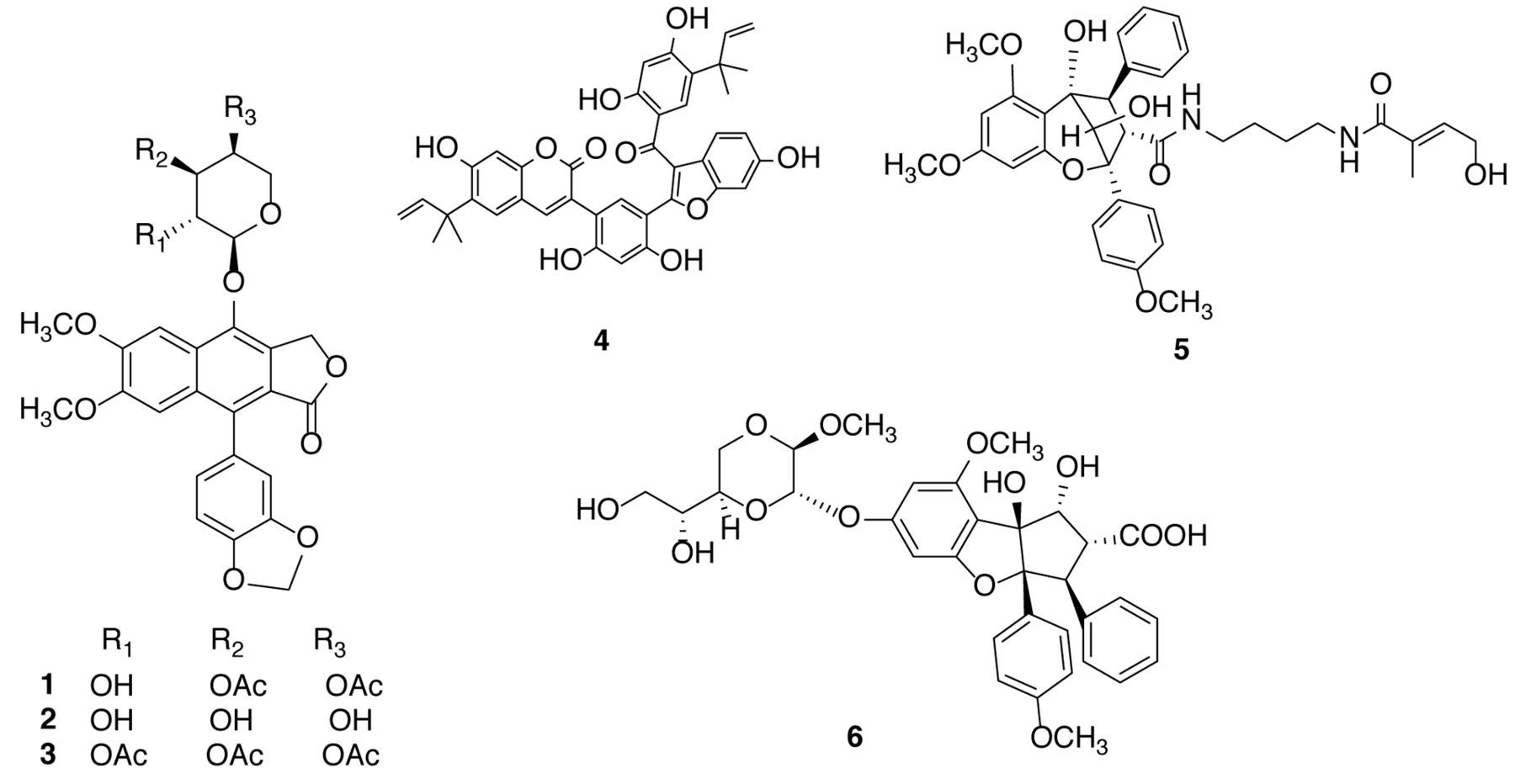
Structures of compounds isolated from tropical plants. 1: Phyllanthusmin D, 2: phyllanthusmin E, 3: phyllanthusmin C, 4: sphenostylisin A, 5: perviridisin B, 6: silvestrol.
The rocaglate cyclopenta[b]benzofuran derivative with which our Program Project team has been the most deeply involved is (–)-silvestrol (6). Furthermore, important work on the synthesis, biological evaluation, and mechanism of action of silvestrol has been conducted by other investigators. Our collaborative group first reported the structure and stereochemistry of this substance in 2004 as a dioxanyl ring-containing constituent of both the fruits and twigs of Aglaia foveolata collected in Kalimantan, Indonesia. Thus, the structure and absolute configuration of silvestrol were determined by detailed spectroscopic data interpretation and by X-ray crystallography (81). At about the same time as our work was performed, Meurer-Grimes et al. disclosed in a patent an antineoplastic compound with the same planar structure as silvestrol as an antineoplastic constituent of A. leptantha (82), a species shown subsequently to be A. stellatopilosa (83). This compound was found to be highly cytotoxic towards a panel of solid tumor human cell lines, with a potency range (IC50 1.2 to 1.5 nM) similar to that observed for a positive control substance, paclitaxel (IC50 0.7 to 4.7 nM). Next, silvestrol was tested in the in vivo hollow-fiber assay at UIC at doses up to 5 mg/kg, and inhibited proliferation of all three cell lines used when administered intraperitoneally, while showing no detectable gross toxicity to the mice (81). Silvestrol was also tested in the P388 murine lymphocytic leukemia model at Bristol-Myers Squibb (Princeton, NJ, USA), a former industrial partner in our Program Project group, and was determined as being active at a maximal tolerated dose of 2.5 mg/kg when administered as five daily intraperitoneal injections, corresponding to maximum increase in lifespan with a treatment/control (T/C) ratio of 150%. In addition, when silvestrol was injected intravenously twice daily for five days in the P388 model, a treated/control (T/C) ratio of 129% was obtained at a cumulative dose of 2 mg/kg/day (81).
At UIC, preliminary studies were conducted to elucidate the cellular mechanism of action of silvestrol (6) in LNCaP cells. This rocaglate derivative was found to down-regulate p53 at the RNA and protein level within 15 minutes of exposure, and, after 6 hours, no p53 could be detected. Down-regulation of mouse double minute 2 homolog (MDM2), the E3 ligase specific for p53, was also obtained, which was not prevented by lactacystin, suggesting that silvestrol-induced degradation of p53 is not mediated by the proteasome. In addition, on cell-cycle analysis by flow cytometry, silvestrol induced a block in the cycle at the G2/M checkpoint (84). The cell-cycle arrest induced by silvestrol led to cell death by apoptosis, and it was found that in LNCaP cells this substance disrupted the mitochondrial transmembrane potential and caused cytochrome c release into the cytoplasm. In these cells, silvestrol induces the apoptosome/mitochondrial pathway to stimulate apoptosis (85). In a very recent mechanistic study also performed at UIC, using MDA-MB-435 melanoma cells, silvestrol was found to enhance autophagosome formation and to induce cell death through induction of early autophagy and caspase-mediated apoptosis (75).
In vivo testing and mechanism of action studies carried out at McGill University, Montreal, Canada, by Pelletier and colleagues have shown that silvestrol is a protein translation inhibitor. More specifically, this group demonstrated that silvestrol promotes aberrant interaction of mRNA with the RNA helicase eukaryotic initiation factor (eIF) 4A to prevent its productive association with eIF4F, thus inhibiting the initiation step of translation (86, 87). The structure of silvestrol was confirmed though total synthesis by two groups independently, namely, Porco, Jr. and co-workers at Boston University (88), and Rizzacasa and colleagues at the University of Melbourne in Australia (89, 90). The Rizzacasa group also prepared a biotinylated derivative of episilvestrol [the C-5‴ epimer of silvestrol (81)], and showed this to be highly selective in binding eIF4A, further supporting this factor as the principal cellular target of silvestrol (79, 90).
One of the first activities performed by our Program Project team when it was first funded in 2007 was the organization of a large-scale recollection of A. foveolata from Kalimantan, Indonesia, so that a larger quantity of pure silvestrol could be generated for additional biological testing. Accordingly, approximately 45 kg of the stem bark of the plant was collected, and this led to about 2 g of the compound being produced in highly pure form. This large-scale isolation work was conducted with the cooperation of Dr. David Newman, then at the NCI (Frederick, MD, USA) (92). As a result of an effort spearheaded by Drs. Michael Grever and David Lucas, of the OSU College of Medicine, a series of studies was performed to show the potential of silvestrol for treating human B-cell malignancies. This work, supported also through a NCI Specialized Programs of Research Excellence (SPORE) project directed by Dr. John Byrd (OSU College of Medicine), suggested the differential activity of silvestrol in immune cell subsets as well as P-glycoprotein expression as a mechanism of resistance (93-97). Silvestrol has also shown efficacy against hepatocellular carcinoma (98) (OSU College of Medicine) and neurofibromatosis (99) (Nationwide Children's Hospital) targets, and has an overall an favorable pharmacokinetic profile in the mouse (100).
In October, 2007, the Drug Development Group of the U.S. NCI selected silvestrol (6) for preclinical evaluation at the IIA level (including additional sourcing of the plant of origin and preliminary formulation, pharmacokinetics, and toxicology), potentially leading to clinical development. This was later transferred to the NExT pipeline of NCI's Division of Experimental Therapeutics (http://next.cancer.gov/), where it was evaluated for preclinical toxicology. Unfortunately, unexpected toxicity was observed in one of the test animal models using silvestrol, and further developmental work at NCI was suspended. However, depending on supply, it is anticipated that silvestrol will be valuable both as a standard cytotoxic agent in the laboratory, as well as a specific probe for the cellular eIF4A protein translation target.
Cyanobacterial-derived compounds. In Figure 3, the structures of six purified cyanobacterial compounds (7-12) are shown and these were obtained from several in vitro-active strains of cyanobacteria from the collection developed at UIC. Two new [carbamidocyclophane F (7) and carbamidocyclophane G (8)] and several known carbamidocyclophanes were isolated from a cultured freshwater cyanobacterium, Nostoc sp. (UIC 10274), obtained from a sample collected at Des Plaines, IL, USA. Both carbamidocyclophanes F and G were cytotoxic against MDA-MB-435 (melanoma) and HT-29 (colon cancer) cells, with IC50 values ranging from 0.5-0.7 μM (101).
Two new cyclic lipopeptides, trichormamide A (9) and trichormamide B (10), were isolated from the cultured freshwater cyanobacterium, Trichormus sp. (UIC 10339). This strain was obtained from a sample collected in Raven Lake in Northern Wisconsin. Trichormamide B, a cyclic dodecapeptide, displayed cytotoxic activity against the MDA-MB-435 and HT-29 cell lines with IC50 values of 0.8 and 1.5 μM, respectively. Trichormamide A, a cyclic undecapeptide, was less potent than trichormamide B and exhibited IC50 values of 9.9 and 16.9 μM against MDA-MB-435 and HT-29 cells, respectively (102).
Several pure compounds obtained from cyanobacteria were evaluated in mechanistic assays (NFκB; MTP; semaphorin-3B) at OSU. For example, ambiguine I isonitrile (11), originally obtained from the cultured cyanobacterium Fischerella ambigua (103), was identified as a potent NFκB inhibitor (IC50 30 nM), and was also found to be cytotoxic towards the MCF-7 breast cancer cell line (IC50 1.7 μM). Further studies indicated that the apoptotic effect on MCF-7 cells was associated with blocking the G1 phase of the cell cycle and that cell death was induced independently of caspase-7 (74). In addition, hapalindole H (12), also obtained from the cultured Fischerella ambigua (103), was found to be cytotoxic towards hormone-independent prostate cancer PC-3 cells (IC50 0.02 μM). Hapalindole H (12) also inhibited transcription factor NFκB activity (IC50 0.76 μM). A further study has indicated hapalindole H (12) induces apoptosis through the intrinsic mitochondrial pathway in hormone-independent prostate PC-3 cells (unpublished data).

Structures of compounds isolated from freshwater (7-10) and cultured (11, 12) cyanobacteria. 7: Carbamidocyclophane F, 8: carbamidocyclophane G, 9: trichormamide A, 10: trichormamide B 11: ambiguine I isonitrile; 12: hapalindole H.
Fungal-derived compounds. In Figure 4, compounds 13-20 exemplify the considerable structural variation among substances that have been isolated and characterized thus far in our Program Project work from filamentous fungi, using strains from the extensive culture collection available at Mycosynthetix, Inc. For the first of these, a 1:1 chloroform-methanol crude extract of an unidentified bioactive strain (code-named MSX 70741) of the order Hypocreales, Ascomycota, was found to show cytotoxic activity against H460 human non-small cell carcinoma cancer cells. The peptaibol derivative, atroviridin D (13) was purified as one of 12 structural analogs in this isolation chemistry investigation, and its structure was determined with the use of higher-energy collisional dissociation high-resolution MS/MS and high-field NMR spectroscopy at 950 MHz. Using an ultra-performance liquid chromatographic (UPLC) method, the absolute configuration of atroviridin D (13) was determined by Marfey's analysis of the individual amino acid components present. This compound was found to contain an unusual Ala1 residue. While atroviridin D was not cytotoxic (IC50 >10 μM) in a small panel of cancer cell lines employed, a very closely related congener also isolated, alamethicin F50 (containing an Aib1 residue in place of the Ala1 reside, but otherwise structurally identical), showed broad cytotoxic potency in the low micromolar range (104).
A new o-pyranonaphthoquinone decaketide, obionin B (14), was isolated and structurally characterized from a cytotoxic crude extract of an unidentified fungus (strain: MSX 63619) from the order Pleosporales. This compound showed cytotoxic potencies of 3.1, 7.3, and 7.6 μM against the HT-29 (colon cancer), MDA-MB-435 (breast cancer), and H460 (non small-cell carcinoma) human cell lines, respectively (105).
Thielavin B methyl ester (15), a new benzoate trimer, was purified as a cytotoxic constituent of the unidentified fungal strain MSX 55526 of the order Sordiariales. This was obtained along with two ergostane derivatives of known structure, with its characterization aided by high-resolution MS coupled to an atmospheric pressure photoionization source. Thielavin B methyl ester exhibited cytototoxic activity against the H460 (non-small cell carcinoma), MCF-7 (breast cancer) and SF268 (astrocytoma) human cancer cell lines, with IC50 values of 6.6, 7.3, and 8.1 μM, respectively (106).
7-Epi-zeaenol (16) was obtained as one of two new resorcylic acid lactones that were chromatographically fractionated to purity from a cytotoxic extract of the cultured strain MSX 63935. DNA analysis was performed on the producing organism, and the D2 variable region of the large subunit (LSU) rRNA was sequenced and compared to a database, with the closest match being to the fungal genus Phoma (107). The absolute configuration of 7-epi-zeaenol was established by reduction with sodium borohydride of the related compound, (5E)-7-oxozeaenol, which was also isolated from the same fungal source. 7-Epi-zeaenol was not active in any of several in vitro bioassays applied. However, several previously known substances with only minor structural variations from this compounds were broadly active against the cancer cell line panel used, having IC50 values in the low micromolar range (107). As this strain produced a large supply of some of the resorcylic acid lactones (ca. 400 mg per solid phase on 150 g of rice), their structures and biological activities have been probed further, via a semi-synthetic chemistry approach, including the generation of analogs that incorporate a difluoro moiety.

Structures of compounds isolated from cultured filamentous fungi. 13: Atroviridin D, 14: obionin B, 15: thielavin B methyl ester, 16: 7-epizeaenol, 17: trichodepsipeptide B, 18: acremoxanthone D, 19: verticillin H, 20, verticillin A.
From the filamentous fungus strain MSX 51320, representative of the genus Trichothecium, compounds of both the cyclodepsipeptide and sesquiterpenoid types were isolated and identified, attesting to the biosynthetic complexity of the producing species. Of several new compounds obtained, the new cyclodepsipeptide trichodepsipeptide B (17) was determined structurally using spectroscopic data interpretation, with its absolute configuration established using chiral HPLC. None of the eight compounds purified was responsible for the cytotoxic effects of the initial crude extract observed against cancer cell lines, but it was hypothesized that such activity may be due to uncharacterized roseotoxin and trichothecene derivatives (108).
Acremoxanthone D (18) was isolated as one of two new xanthone-anthraquinone heterodimers from an unidentified fungus in the order Hypocreales (strain MSX 17022). The structure of this compound was determined using NMR spectroscopic and MS methods, with the relative configuration established using a rotating-frame Overhauser spectroscopy (ROESY) NMR experiment. Acremoxanthone D exhibited an IC50 value of 14.0 μM against the MCF-7 human breast cancer cell line and 58% inhibition at 20 μg/ml in a 20S protease assay (109).
As a final example of a new compound from filamentous fungi, the epipolythiodioxopiperazine (ETP) alkaloid verticillin H (19) was isolated and elucidated structurally from strain MSX 64546 of the family Bionectriaceae. The structure of this compound was determined by direct comparison with its known analog, verticillin A (20), which was purified in the same investigation. When compared with the latter compound, verticillin H was shown to contain an additional methylene group in each of its monomeric units. The absolute configuration of verticillin H was determined by comparison of its specific rotation and circular dichroism spectrum with those of reference compounds. Like verticillin A, verticillin H was found to be potently cytotoxic against the various cancer cell lines for which it was tested, with the most susceptible example being the HT-29 human colon cancer cell line (IC50 40 nM) (110).
Owing to their cytotoxic potency against cancer cell lines, the ETP alkaloidal class of fungal secondary metabolites seems worthy of more extensive biological evaluation, including more detailed testing in in vivo models and mechanism of action work. Since the new compound, verticillin H (19) was obtained only in small amounts, it has been decided additional work should be performed on its more abundant analog, verticillin A. In a published collaborative study, verticillin A was found to be a selective inhibitor of histone methyltransferases SUV39H1, SUV39H2, and G9a/GLP in metastatic human colon carcinoma cells. Genome-wide gene expression analysis identified FAS as a target gene for verticillin A, and this compound showed greater efficacy than decitabine and vorinostat in overcoming colon carcinoma resistance to Fas-ligand (FASL)-induced apoptosis. Moreover, verticillin A overcame metastatic colon carcinoma resistance to 5-fluorouracil in vitro and in vivo, using SW62-FFU-R cells injected into nude mice (65). Further in vivo testing of verticillin A (20) is ongoing.
Conclusion
From the structural examples given in this review, it can be seen that when combined into a single integrated research program, tropical plants, aquatic and terrestrial cyanobacteria, and filamentous fungi together offer the possibility of harnessing small-molecular weight molecules of considerable chemical diversity as potential anticancer agents. Collectively, it has been shown statistically by our group that the types of compounds obtained by activity-guided fractionation from these classes of organisms occupy the same chemical space as already approved oncolytic agents, but are complementary to one another. A wide variety of analytical techniques has been applied in determining the structures of the new compounds selected as representative examples in this review. While stringent efforts are made to identify taxonomically all organisms on which work has been conducted, this is a more straightforward proposition for higher plants than for cyanobacteria and fungi, which require the use of molecular biology techniques. By pooling project resources for in vitro and preliminary biological testing, medicinal chemistry, pharmacokinetics, and biostatistics, this has enabled greater progress of the group than would otherwise have been the case. Our project has benefited greatly through the collaboration of colleagues both within our own institutions and at those at outside affilations, such as the U.S. NCI, who can offer specialized in vivo evaluation and other testing on our best lead compounds. Although a preliminary notion of the cellular target(s) of a given bioactive compound of interest may be obtained from standard in vitro tests, for substances based on a new carbon skeleton, this type of investigation is probably conducted optimally after in vivo activity in a murine xenograft tumor inhibition model has been demonstrated.
Acknowledgements
The Authors wish to acknowledge financial support through program project grant P01-CA125066 and CCC support grant P30-CA016058 from the National Cancer Institute, National Institutes of Health, Bethesda, MD, USA. We are grateful to our taxonomic collaborators in several countries for their kind cooperation with plant collections. We wish to thank many present and former faculty and staff colleagues, postdoctoral associates, and graduate and undergraduate students who have participated in this collaborative project, and whose names are included in the bibliography below.
Footnotes
This article is freely accessible online.
- Received August 24, 2016.
- Revision received September 17, 2016.
- Accepted September 20, 2016.
- Copyright© 2016 International Institute of Anticancer Research (Dr. John G. Delinassios), All rights reserved
References
In this issue


Jump to section
Related Articles
Cited By...
- Targeting Protein Translation by Rocaglamide and Didesmethylrocaglamide to Treat MPNST and Other Sarcomas
- Papuamine Inhibits Viability of Non-small Cell Lung Cancer Cells by Inducing Mitochondrial Dysfunction
- Verticillin A Causes Apoptosis and Reduces Tumor Burden in High-Grade Serous Ovarian Cancer by Inducing DNA Damage
- Oxidized products of {alpha}-linolenic acid negatively regulate cellular survival and motility of breast cancer cells
- The Combination of Flavokawain B and Daunorubicin Induces Apoptosis in Human Myeloid Leukemic Cells by Modifying NF-{kappa}B