


Abstract
Background: Fifteen 3-styrylchromones were subjected to quantitative structure–activity relationship (QSAR) analysis based on their cytotoxicity, tumor selectivity and anti-HIV activity, in order to explore their biological activities. Materials and Methods: Cytotoxicity against four human oral squamous cell carcinoma (OSCC) cell lines and three human oral normal cells was determined by the 3-(4,5-dimethylthiazol-2-yl)-2,5-diphenyltetrazolium bromide (MTT) method. Tumor-selectivity was evaluated by the ratio of the mean CC50 (50% cytotoxic concentration) against normal human oral cells to that against OSCC cell lines. Anti-HIV activity was evaluated by the ratio of CC50 to EC50 (50% cytoprotective concentration from HIV infection). Physicochemical, structural and quantum-chemical parameters were calculated based on the conformations optimized by the LowModeMD method followed by the density functional theory (DFT) method. Results: All 3-styrylchromone derivatives showed moderate-to-high tumor selectivity. Especially, compounds that have a methoxy group at 6-position of the chromone ring and hydroxyl group at 4’-position of phenyl group in styryl moiety [11] showed the highest tumor-selectivity. On the other hand, their cytotoxicity against normal cells showed good correlation to the descriptors that reflect hydrophobic interaction and molecular shapes. Conclusion: Multivariate statistics with chemical descriptors for the location of substituted group, molecular shape and electrostatic interaction may be useful for designing the most favorable compound with higher tumor selectivity.
4H-1-benzopyran-4-ones (chromones) are an important class of oxygenated heterocyclic compounds since the chromone core structure is found in flavones, isoflavones and 2-styrylchromones. As compared with flavones and isoflavones, 2-styrylchromones constitute a much smaller group of naturally occurring chromones. Synthetic 2-styrylchromones showed antioxidant (1), anti-allergic (2), anti-inflammatory (3), anti-tumor (4-6), anti-viral (7, 8) activities. In contrast, as far as we know there were only two papers of 3-styrylchromones that investigated the antiviral (9) and antibacterial activity (10).
In order to further explore novel biological activities of 3-styrylchromones, we recently synthesized a series of 3-styrylchromone derivatives and reported antioxidant and α-glucosidase inhibition activities (11). In the present study, we investigated the cytotoxicity and anti-HIV activity of fifteen 3-styrylchromones and then performed the quantitative structure–activity relationship (QSAR) analysis.
For the cytotoxicity assay, both human normal oral cells (gingival fibroblast, HGF; pulp cells, periodontal ligament fibroblast, HPLF; pulp cell, HPC) and human oral squamous cell carcinoma (OSCC) cell lines (Ca9-22, HSC-2, HSC-3, HSC-4) were used as target cells. The anti-tumor potential was evaluated by the tumor-selectivity index (TS), calculated by dividing the mean 50% cytotoxic concentration (CC50) against normal oral cells by that against OSCC cell lines. We have already confirmed that the TS value determined by this method reflects the anti-tumor potential of test samples, although these normal oral cells and OSCC cell lines are classified as different types of cells (mesenchymal or epithelial) (12).
For the anti-HIV assay, mock- and HIV-infected-human T-cell lymphotropic virus-I (HTLV-I) carrying human T-cell line MT4 was used. The selectivity index (SI) was calculated by dividing the CC50 by the 50% cytoprotective concentration from HIV infection (EC50).
Materials and Methods
Materials. The following chemicals and reagents were obtained from the indicated companies: Dulbecco's modified Eagle's medium (DMEM), from GIBCO BRL, Grand Island, NY, USA; fetal bovine serum (FBS), 3-(4,5-dimethylthiazol-2-yl)-2,5-diphenyltetrazolium bromide (MTT), azidothymidine and 2’,3’-dideoxycytidine from Sigma-Aldrich Inc., St. Louis, MO, USA; dimethyl sulfoxide (DMSO), dextran sulfate (molecular mass, 5 kDa) from Wako Pure Chem. Ind., Osaka, Japan; 5-fluorouracil (5-FU) from Kyowa, Tokyo, Japan; curdlan sulfate (molecular mass, 79 kDa) from Ajinomoto Co. Ltd., Tokyo, Japan. Culture plastic dishes and plates (96-well) were purchased from Becton Dickinson (Franklin Lakes, NJ, USA).
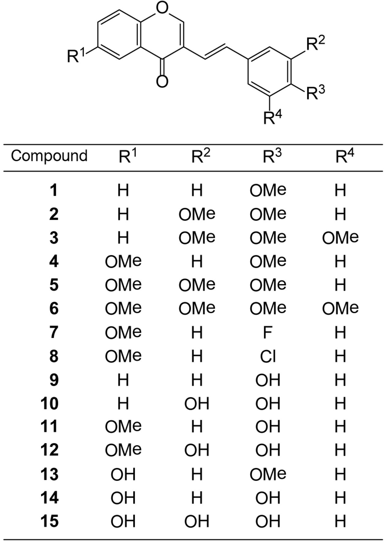
Synthesis of test compounds. (E)-3-(4-Methoxystyryl)-4H-chromen-4-one [1], (E)-3-(3,4-Dimethoxystyryl)-4H-chromen-4-one [2], (E)-3-(3,4,5-Trimethoxystyryl)-4H-chromen-4-one [3], (E)-6-Methoxy-3-(4-methoxystyryl)-4H-chromen-4-one [4], (E)-6-Methoxy-3-(3,4-dimethoxystyryl)-4H-chromen-4-one [5], (E)-6-Methoxy-3-(3,4,5-trimethoxystyryl)-4H-chromen-4-one [6], (E)-3-(4-Fluorostyryl)-6-methoxy-4H-chromen-4-one [7], (E)-3-(4-Chlorostyryl)- 6-methoxy-4H-chromen-4-one [8], (E)-3-(4-Hydroxystyryl)-4H-chromen-4-one [9], (E)-3-(3,4-Dihydroxystyryl)-4H-chromen-4-one [10], (E)-3-(4-Hydroxystyryl)- 6-methoxy-4H-chromen-4-one [11], (E)-3-(3,4-Dihydroxystyryl)- 6-methoxy-4H-chromen-4-one [12], (E)-6-Hydroxy-3-(4-methoxystyryl)-4H-chromen-4-one [13], (E)-6-Hydroxy-3-(4-hydroxystyryl)-4H-chromen-4-one [14] and (E)-6-Hydroxy-3-(3,4-dihydroxystyryl)-4H-chromen-4-one [15] (Figure 1) were synthesized by Knoevenagel condensation of the appropriate 3-formylchromone with selected phenylacetic acid derivatives, according to previous methods (11). All compounds were dissolved in DMSO at 80 mM and stored at −20°C before use.
Cell culture. HGF, HPLF and HPC cells, established from the first premolar tooth extracted from the lower jaw of a 12-year-old girl (13), and OSCC cell lines (Ca9-22, HSC-2, HSC-3, HSC-4), purchased from Riken Cell Bank, Tsukuba, Japan were cultured at 37°C in DMEM supplemented with 10% heat-inactivated FBS, 100 units/ml, penicillin G and 100 μg/ml streptomycin sulfate under a humidified 5% CO2 atmosphere. Cells were then harvested by treatment with 0.25% trypsin-0.025% EDTA-2Na in PBS(−) and either subcultured or used for experiments.
Assay for cytotoxic activity. Cells were inoculated at 2.5×10³ cells/0.1 ml in a 96-microwell plate (Becton Dickinson Labware, Franklin Lakes, NJ, USA). After 48 hours, the medium was removed by suction with an aspirator and replaced with 0.1 ml of fresh medium containing different concentrations of single test compounds. Control cells were treated with the same amounts of DMSO present in each diluent solution. Cells were incubated for 48 hours and the relative viable cell number was then determined by the MTT method. In brief, the treated cells were incubated for another three hours in fresh culture medium containing 0.2 mg/ml MTT. Cells were then lysed with 0.1 ml of DMSO and the absorbance at 540 nm of the cell lysate was determined using a microplate reader (Biochromatic Labsystem, Helsinki, Finland). The CC50 was determined from the dose–response curve and the mean value of CC50 for each cell type was calculated from three independent experiments.

Structure of 3-styrylchromones.
Calculation of TS. The tumor-selectivity index (TS) was calculated by the following equation: TS=mean CC50 against normal cells/mean CC50 against tumor cells [(D/B) in Table I]. Since Ca9-22 cells were derived from gingival tissue (14), the relative sensitivity of Ca9-22 and HGF was also compared [(C/A) in Table I].
Assay for HIV activity. HTLV-I-carrying human T-cell line MT4 cells, highly sensitive to human immunodeficiency virus-1 (HIV-1), were infected with HIV-1IIIB at a multiplicity of infection (m.o.i.) of 0.01. HIV- and mock-infected (control) MT-4 cells were incubated for five days with different concentrations of samples and the relative viable cell number was determined by the MTT assay. The CC50 and EC50 were determined from the dose–response curve for mock-infected and HIV-infected cells, respectively (15). All data represent the mean values of triplicate measurements. The anti-HIV activity was evaluated by SI (=CC50/EC50).
Estimation of CC50 values. Original data contain the sign of inequality such as ”>”. For the convenience of analysis, these values were changed into forms suitable for arithmetic calculation. Since “>400” is equal to “from 400 to ∞”, we calculated the harmonic mean as follows: 1/[average(1/400,1/∞)]=800. Since the CC50 values had a distribution pattern close to a logarithmic normal distribution, we used the pCC50 (i.e., the −log CC50) for the comparison of the cytotoxicity between the compounds. The mean pCC50 values for normal cells and tumor cell lines were defined as N and T, respectively (16).
Cytotoxic activity of fifteen 3-styrylchromones. Each value represents the mean of triplicate determinations.
Calculation of the representative value for tumor selectivity. Tumor selectivity is defined by the balance between pCC50 values for normal (N) and tumor (T) cells. The difference (T–N) was used as a tumor-selectivity index in the following analyses.
Calculation of chemical descriptors. Each chemical structure was optimized by the LowModeMD method (17), a suitable search method for minimum energy conformers of flexible molecules, with Merck Molecular Force Field (MMFF94x) in Molecular Operating Environment (MOE) 2013.08 (Chemical Computing Group Inc., Quebec, Canada). Each structure was refined with density functional theory (DFT-B3LYP/6-31G**) by using Spartan10 for Windows (Wavefunction, Inc., Irvine, CA, USA) (18). The descriptors used were: R1 OMe (methoxy substitution at the 6-position on the chromone ring group), R3 OH (4’-hydroxy substitution in the phenyl group of styryl moiety), vsurf_DD23 (the interaction with hydrophobic probe assumed surrounding the molecule), G1u (the first component symmetry directional WHIM index/unweighted encoding molecular symmetry that extracts the global symmetry information), G2u (the second component symmetry directional WHIM index/unweighted encoding molecular symmetry that extracts the global symmetry information) (19), BCUT_SMR_3 (BCUT descriptor using atomic contribution to molar refractivity), rgvr (radius of gyration), diameter (largest value in the distance matrix defined by the elements Dij, where Dij is the length of the shortest path from atoms i to j). The BCUT descriptors are calculated from the eigenvalues of a modified adjacency matrix (20).
Statistical treatment. The relation among cytotoxicity, tumor specificity index, anti-UV activity and chemical descriptors was investigated using simple regression analyses by JMP Pro version 10.0.2 (SAS Institute Inc., Cary, NC, USA). The significance level was set at p<0.05.
Results
Cytotoxicity. Among fifteen 3-styrylchromone derivatives, compound [11] showed the highest cytotoxicity against four human OSCC cell lines (mean CC50=2.0±1.2 μM) followed by [4] (6.4±2.4 μM) and [6] (13±6.1 μM) (Table I). Since these compounds showed much lower cytotoxicity against three human oral normal cells (mean CC50=138±116, 258±126 and 339±183 μM, respectively), they showed the highest tumor-selectivity [TS (D/B)=69.0, 40.3 and 26.1, respectively], comparable with that of doxorubicin (TS=>26) and 5-FU (TS=>55.6) (Table I). When tumor-selectivity was calculated by different equation using Ca9-22 and HGF cells, both derived from gingival tissues ([TS(C/A)], these three compounds again showed very high tumor-selectivity (31.9, 52.0 and 54.4, respectively) (Table I).

Effects of functional groups on cytotoxicity of 3-styrylchromones against tumor cells (defined as T). The mean (pCC50 i.e., the −log CC50) values for tumor cell lines were defined as T.
Anti-HIV activity. In contrast to popular anti-HIV agents (dextran sulfate, curdlan sulfate, azidothymidine, 2’,3’-dideoxycytidine) (SI=2445-20421), none of 3-styrylchromones 1-15 protected the cells from the cytopathic effect of HIV infection (SI<1) (Table II). Based on these data, the following QASR analysis was focused on the cytotoxicity of 3-styrylchromones.
Computational analysis. Cytotoxicity of 3-styrylchromones against tumor cells (defined by T) correlated with descriptors based on the substituent group. R1 OMe (methoxy substitution at the 6-position on the chromone ring) (p=0.100) and R3 OH (4’-hydroxy substitution in the phenyl group of styryl moiety) (p=0.0182) are crucial for the enhancement of cytotoxicity against OSCC cell lines (Figure 2).
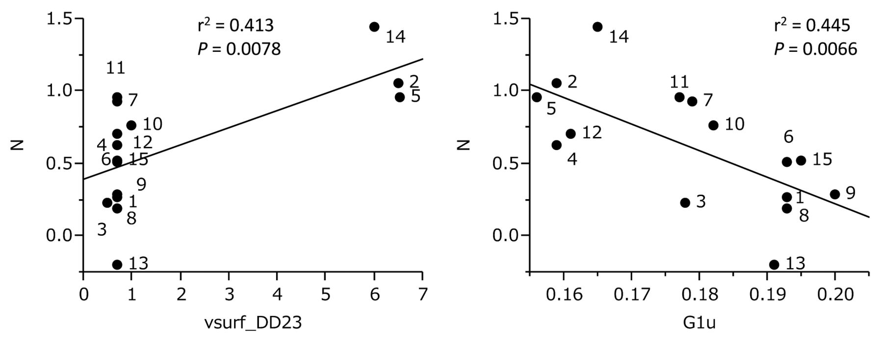
On the other hand, cytotoxicity of 3-styrylchromones against normal cells (defined by N) was correlated with vsurf_DD23 (the interaction with hydrophobic probe assumed surrounding the molecule) (r2=0.413, p=0.0078) and G1u (the first component symmetry directional WHIM index/unweighted encoding molecular symmetry that extracts the global symmetry information) (r2=0.445, p=0.0066) (Figure 3).
Tumor selectivity of 3-styrylchromones (defined by T-N) correlated with R3 OH (p=0.0616), vsurf_DD23 (r2=0.280, p=0.0426) and G2u (the second component symmetry directional WHIM index/unweighted encoding molecular symmetry that extracts the global symmetry information) (r2=0.406, p=0.0106) (Figure 4).
Anti-HIV activity of 3-styrylchromones and chemotherapeutic agents. Each value represents the mean of triplicate determinations.
A model for the estimation of T, N and T-N values was constructed by multiple regression analysis using the descriptors calculated in this study. T can be estimated by R1 OMe and R3 OH (r2=0.702, Q2=0.532, s=0.409) (left). N can be estimated by BCUT_SMR_3, rgyr (radius of gyration) and vsurf_DD23 (R2=0.834, Q2=0.695, s=0.194) (left). T-N can be estimated by diameter (largest value in the distance matrix defined by the elements Dij), vsurf_DD23 and R3 OH (R2=0.764, Q2=0.570, s=0.308) (right) (Figure 5).

Determination coefficient of chemical descriptors and cytotoxicity of 3-styrylchromones against normal cells (defined as N). The mean (pCC50 i.e., the −log CC50) values for normal cells were defined as N.

Determination coefficient of chemical descriptors and tumor specificity of 3-styrylchromones (defined as T-N).

Multiple regression models for the estimation of T, N and T-N.
Discussion
The present study demonstrated for the first time that fifteen 3-styrylchromones showed moderate-to-potent tumor-specificity and no detectable anti-HIV activity (Table I). Three compounds [4, 6, 11] showed the highest tumor-selectivity, comparable to doxorubicin and 5-FU (Table I). The lack of anti-HIV activity is not in agreement with a previous study where 4H-chromen-4-one and 2H-chromene derivatives inhibited the replication of picornavirus in vitro (9). The discrepancy between our and their results may be due to the fact that they did not produce the chemotherapeutic index (safety margin).
The present study is the first report of QSAR analysis of 3-styrylchromones. We used the following soft Spartan (34 quantum chemistry-related descriptors), MOE (330 quantum chemistry, structure and physical chemistry-related descriptors), dragon (114 three-dimensional molecular shape-related descriptors) and 9 descriptors (that relate the location of substituent groups and types). The valuables used are T, N and T-N. Descriptors that showed significant difference or significant trends (p≤0.1) were R1 OMe (methoxy substitution at the 6-position on the chromone ring), R3 OH (4’-hydroxy substitution in the phenyl group of styryl moiety), vsurf_DD23, G1u and G2u.
Van der Waals interaction through the methoxy substitution at the 6-position and the hydrogen bonding through the 4’-hydroxy substitution may be important for the interaction with key proteins engaged in the cytotoxicity against OSCC cells (Figure 2). Hydrophobic interaction and molecular shape may be important for the cytotoxicity against normal cells (Figure 3). Molecular shape and 4’-hydroxy substitution in the phenyl group of styryl moiety affected the tumor-selectivity (Figure 4).
For the multiple regression analysis model, the following descriptors were used: BCUT_SMR_3 (that relates to the topological shape of molecule and the interaction between the low molecular weight molecule and protein interaction), rgvr (that relates to the molecular size and shape) and diameter (that reflects the topological size of molecule). Leave-one-out cross verification method confirmed that cytotoxicity against OSCC cells (T), oral normal cells (N) and tumor-specificity (T-N) can be estimated by the combination of descriptors for substituent groups, molecular shape and hydrophobic interaction (Figure 5).
We have previously reported that 2-styrylchromone derivatives having methoxy group in the phenyl group of styryl moiety showed relatively higher tumor-selectivity and induced apoptosis in the human promyelocytic leukemia HL-60 cell line and non-apoptosis in the human oral squamous cell carcinoma HSC-2 cell line (4). In contrast, tumor-selectivity of 3-styrylchromones was significantly enhanced by the introduction of methoxy group on the chromone ring and hyxroxyl group in the phenyl group of styryl moiety. It remains to be investigated which type of cell death is induced by 3-styrylchromones in OSCC cell lines.
In conclusion, the present study demonstrated that 3-styrylchromones substituted with methoxy group on the chromone ring and hydroxy group in the phenyl group of styryl moiety are promising for the exploration of new anti-tumor agents. Multivariate statistics with chemical descriptors for the location of substituted group, molecular shape and electrostatic interaction may be useful for designing the most favorable compound with higher tumor selectivity.
Footnotes
-
This article is freely accessible online.
- Received June 9, 2014.
- Revision received July 16, 2014.
- Accepted July 17, 2014.
- Copyright© 2014 International Institute of Anticancer Research (Dr. John G. Delinassios), All rights reserved
References
In this issue


Jump to section
Related Articles
Cited By...
- Further Quantitative Structure-Cytotoxicity Relationship Analysis of 3-Styrylchromones
- Quantitative Structure-Cytotoxicity Relationship of 2-Arylazolylchromones and 2-Triazolylchromones
- Quantitative Structure-Cytotoxicity Relationship of 3-(N-Cyclicamino)chromone Derivatives
- Quantitative Structure-Cytotoxicity Relationship of Pyrano[4,3-b]chromones
- Quantitative Structure-Cytotoxicity Relationship of 2-(N-cyclicamino)chromone Derivatives
- Quantitative Structure-Cytotoxicity Relationship of Furo[2,3-b]chromones
- Quantitative Structure-Cytotoxicity Relationship of Cinnamic Acid Phenetyl Esters
- Search for New Type of Anticancer Drugs with High Tumor Specificity and Less Keratinocyte Toxicity