
Inflammation-Related Biomarkers for the Prediction of Prognosis in Colorectal Cancer Patients


1
Department of Surgery, Graduate School of Medicine, Kyoto University, Kyoto 606-8501, Japan
2
Department of Gastroenterological Surgery, Kitano Hospital, The Tazuke Kofukai Medical Research Institute, Osaka 530-8480, Japan
*
Author to whom correspondence should be addressed.
Int. J. Mol. Sci. 2021, 22(15), 8002; https://doi.org/10.3390/ijms22158002
Submission received: 7 July 2021
/
Revised: 21 July 2021
/
Accepted: 23 July 2021
/
Published: 27 July 2021
(This article belongs to the Special Issue Biomarkers of Colorectal Cancer)
Abstract
:Colorectal cancer (CRC) is the leading cause of cancer deaths around the world. It is necessary to identify patients with poor prognosis or with high risk for recurrence so that we can selectively perform intensive treatments such as preoperative and/or postoperative chemotherapy and extended surgery. The clinical usefulness of inflammation-related prognostic biomarkers available from routine blood examination has been reported in many types of cancer, e.g., neutrophil–lymphocyte ratio (NLR), lymphocyte–C-reactive protein ratio (LCR), platelet–lymphocyte ratio (PLR), lymphocyte–monocyte ratio (LMR), and so on. Moreover, some scoring systems based on circulating blood cell counts and albumin concentration have been also reported to predict cancer patients’ prognosis, such as the Glasgow prognostic score (GPS), systemic inflammation score (SIS), and prognostic nutritional index (PNI). The optimal biomarker and optimal cutoff value of the markers can be different depending on the cancer type. In this review, we summarize the prognostic impact of each inflammation-related marker in CRC.
1. Introduction
Colorectal cancer (CRC) is one of the main causes of cancer death in the world. Despite recent improvements in multidisciplinary approaches, including surgery, chemotherapy, and radiotherapy, the mortality rate of CRC is still high, especially in patients with distant metastasis or postoperative recurrence even after curative surgery. Development of the optimal biomarker useful for the prediction of recurrence or poor prognosis is clinically important in order to identify patients who can benefit from intensive treatment including chemotherapy, chemoradiotherapy, and extended surgery.
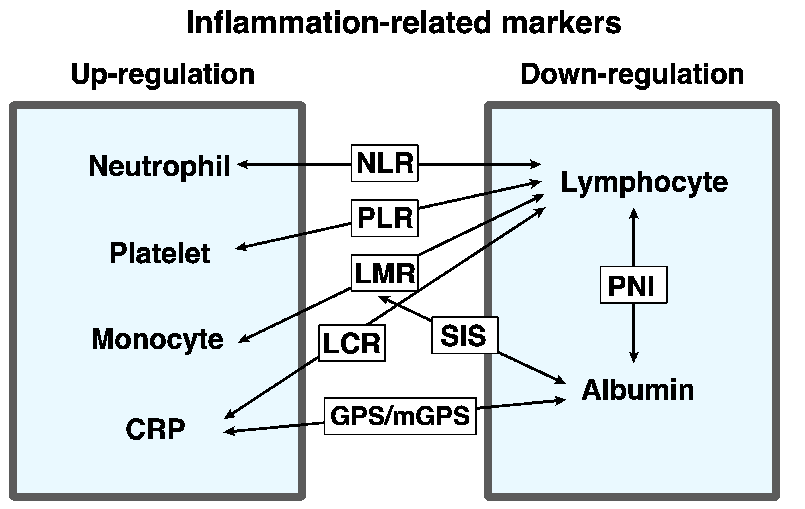
The cancer-associated systemic inflammatory response is one of the critical indicators of tumor progression. Numerous previous studies have reported serum systemic inflammatory markers that can be useful for the prediction of prognosis, e.g., neutrophil–lymphocyte ratio (NLR), lymphocyte–monocyte ratio (LMR), platelet–lymphocyte ratio (PLR), Glasgow prognostic score (GPS), lymphocyte–C-reactive protein ratio (LCR), systemic inflammation score (SIS), and prognostic nutritional index (PNI) (Figure 1). These markers are easily available by use of routine blood examinations for cancer patients.
However, the best combination and optimal cutoff value can be different depending on the cancer type. Moreover, various anti-cancer treatments can differently affect the systemic inflammation status. Therefore, it is mandatory to focus on a certain cancer type so that we can evaluate the prognostic impact of inflammation-related markers.
Previously, we reported that the combination of GPS and NLR could effectively predict the prognosis of CRC patients who underwent curative resection [1]. In addition, we focused on rectal cancer patients (Stage II and III) and showed that the combination of circulating lymphocyte count and serum albumin concentration could be a useful predictor for prognosis [2]. However, there are many other inflammation-related biomarkers investigated regarding CRC. Thus, in the present report, we review previous papers about inflammation-related biomarkers focusing on CRC.
2. Neutrophil-Related Markers
The cancer-associated systemic inflammatory response is often correlated with an increase in circulating neutrophil counts. Neutrophils secrete cytokines and chemokines, which play important roles in cancer progression. Yang et al. reported that a high serum neutrophil count itself was independently associated with poor overall survival (OS) and progression-free survival (PFS) in metastatic CRC patients with wild-type RAS [3]. On the other hand, lymphocytes can promote a cytotoxic immune response to cancer. Some previous papers reported that a decrease in serum lymphocyte counts negatively affected the prognosis of CRC patients [3,4,5,6]. Noh et al. analyzed 231 colon cancer patients who underwent curative resection and adjuvant chemotherapy, and found that high post-chemotherapy lymphocyte counts (≥1.78 × 10⁹/L) were associated with better disease-free survival (DFS) [6].
The neutrophil–lymphocyte ratio (NLR) is one of the most robust biomarkers to predict prognosis in various types of cancer. Proctor et al. analyzed 12,118 patients including 1413 CRC patients, and indicated that NLR was a significant surrogate marker for OS and cancer-specific survival (CSS) [7]. There were numerous reports about NLR that focused on CRC (Table 1). The cutoff values were different depending on papers; the smallest one was 2, while the largest was 5, and they were determined in a different manner, i.e., by use of receiver-operating characteristic (ROC) curve, referring to previous reports, or using the median/mean value among the study population. Li et al. enrolled the most CRC patients (n = 5336) following curative resection (Stage I-III) and reported that NLR > 2.72 was an independent predictor for OS and DFS [8].
The patient population enrolled in each study was also different, i.e., CRC patients who underwent curative resection, metastatic CRC (Stage IV), colorectal liver metastasis (CRLM) following liver resection, CRC with peritoneal carcinomatosis, and so on. For example, Dell’Aquila et al. analyzed 413 metastatic CRC who received FOLFOXIRI or FOLFIRI plus bevacizumab enrolled in a TRIBE trial [55] and reported that NLR > 3.0 independently affected OS but not PFS [40]. Feliciano et al. analyzed 2470 Stage I-III CRC patients who underwent curative resection and found that the combination of high NLR (≥3.0) and sarcopenia determined by computed tomography scan was an independent predictor for OS [30].
Most of the papers evaluated “preoperative NLR”, while some assessed NLR in a different way. Yasui et al. stratified 568 CRC patients into three groups depending on inflammation status: preoperatively low (normal group), preoperatively high but postoperatively low (normalized group), and persistently high (elevated group) [52]. They indicated that the normal group showed a better prognosis than the elevated group regarding several inflammation-related markers, including NLR. Similarly, Li et al. also focused on the change between pre- and post-operative NLR [38]. They defined “post-NLR minus pre-NLR” as the delta-NLR and reported that the delta-NLR ≥ 0 group had better prognosis than the delta-NLR < 0 group, and that delta-NLR independently affected OS in CRC patients who underwent curative resection (Stage I-III). Chan et al. analyzed 2280 CRC patients who underwent surgery and found that the combination of pre- and post-NLR was a significant predictor for OS, i.e., the patients with high preoperative and high postoperative NLR (>3.75) exhibited the worst OS, whereas those with low preoperative and low postoperative NLR (≤3.75) exhibited the best OS [39].
On the other hand, Thiagarajan et al. analyzed 436 CRC patients with peritoneal carcinomatosis, and evaluated NLR on several postoperative timings (0, 1 to 3, 4 to 7, 8 to 21, 22 to 56, and 57 to 90 days) [53]. They reported that high NLR (postoperative days 8–21) >13.26 was most strongly associated with poor OS, while high NLR (postoperative 57 to 90) >1.57 was most strongly associated with poor RFS.
Some previous reports indicated that NLR could be predictive for recurrence patterns after liver resection against CRLM. Giakoustidis et al. reported that the patients with preoperatively elevated NLR (>2.5) had a higher risk for extrahepatic/multifocal recurrence [23]. Similarly, Verter et al. indicated that a high NLR (>3) was significantly associated with extrahepatic recurrence after surgery against CRLM [54].
Thus, numerous papers have proven that NLR can be used as a significant predictor for the survival of CRC patients, although the characteristics of the patients enrolled and the optimal cutoff values of NLR varied from report to report.
3. Albumin-Related Markers
Albumin is one of the acute-phase proteins and decreases in response to inflammation. Moreover, a low albumin concentration reflects cancer-induced malnutrition and can have negative impact on prognosis.
The GPS, introduced by Forrest et al., is a well-known inflammation-related marker that includes serum C-reactive protein (CRP) levels and serum albumin levels [56]. GPS is determined as follows: GPS of 2, CRP > 1.0 mg/dL and albumin < 3.5 g/dL; GPS of 1, either CRP > 1.0 mg/dL or albumin < 3.5 g/dL; and GPS of 0, neither CRP > 1.0 mg/dL nor albumin < 3.5 g/dL. Namely, high GPS reflects both systemic inflammation (elevated CRP) and low nutritional state (hypoalbuminemia).
A number of previous studies have revealed the postoperative prognostic potential of GPS (Table 2). As mentioned above, we previously reported that a high score of GPS was significantly associated with CSS and DFS from the analysis of 448 CRC patients who underwent curative resection [1]. Lee et al. also analyzed 1590 CRC patients who underwent curative resection, and found that a GPS of 1 or 2 independently affected survival [57].
GPS is also useful for metastatic CRC. Kobayashi et al. collected data from 63 CRLM patients who underwent curative resection and reported that GPS 1 or 2 was an independent prognostic factor for CSS [61]. Similarly, Kobayashi et al. investigated 99 CRC patients with lung metastasis following resection and indicated that GPS was an independent predictor for OS [62].
GPS has been modified by numerous recent reports so that it could be more sensitive for the prediction of prognosis; this is often called “modified GPS” (mGPS) (Table 3). For example, Park et al. constructed mGPS as follows: CRP ≤ 1.0 mg/dL is classified as a score of 0, CRP > 1.0 mg/dL and albumin ≥ 3.5 g/dL is a score of 1, and a CRP > 1.0 mg/dL and albumin < 3.5 g/dL is a score of 2. They analyzed 1000 CRC patients who underwent curative surgery and proved that the combination of mGPS and TNM stage was useful for the prediction of postoperative prognosis [63]. Guthrie et al. adopted the same definition of mGPS as Park et al. [63] and found that both preoperative and postoperative mGPS independently could affect the CSS of 206 CRC patients who underwent curative surgery [15].
On the other hand, Inoue et al. defined mGPS as follows: CRP ≤ 0.5 mg/dL and albumin ≥ 3.5 mg/dL was a score of 0, CRP > 0.5 mg/dL or albumin < 3.5 mg/dL was a score of 1, and CRP > 0.5 mg/dL and albumin < 3.5 mg/dL was a score of 2. They indicated the prognostic potential of this mGPS by analyzing 245 Stage IV and recurrent CRC patients treated with chemotherapy [67].
Thus, there were a number of different mGPSs, and the optimal definition is still controversial.
Other than GPS, there are also several albumin-related prognostic markers. For example, the prognostic nutritional index (PNI), first reported by Onodera et al. [71], is a well-known prognostic marker in Japan. PNI is calculated as “albumin level (g/L) + 0.005 × lymphocyte count”, and decreased PNI can affect cancer-related prognosis. Tokunaga et al. evaluated 468 CRC patients who underwent curative resection and indicated that PNI was independently associated with OS and RFS, and that PNI was more sensitive than mGPS if combined with TNM stages [69]. They adopted the cutoff value of 45 of PNI. Similarly, Mohri et al. analyzed 365 CRC patients who underwent curative resection, and reported that PNI < 45 independently affected OS [72]. On the other hand, Tominaga et al. established the optimal cutoff value of PNI as 42.4 by using ROC analysis [73]. They evaluated 84 elderly CRC patients (≥85 years) who underwent curative resection and reported that low PNI was independently associated with OS and RFS.
We have recently reported the novel prognostic marker “LA”, which means “lymphocyte count × albumin level (g/dL) [2]. We retrospectively collected data from a total of 448 rectal cancer patients (Stage II and III) from two large centers, and found that low LA (≤5950) was independently associated with both OS and RFS. Theoretically, LA is similar to PNI but LA is more easily calculated and simply used for the stratification of patients. However, our cohort was focused on rectal cancer patients and whether it can be applied to colorectal cancer patients as a whole remains to be elucidated.
Wang et al. analyzed 877 CRC patients who underwent curative resection (Stage I-III) and built a scoring system, “NLR–albumin”. They reported that the combination of high NLR (≥2.39) and hypoalbuminemia (<39.75 g/L) was a negative prognostic factor for OS in the multivariate analysis [74]. Hong et al. named “NLR–albumin” as IPI (inflammation related prognostic index) and evaluated the prognostic impact of IPI [75]. In their definition, patients with both NLR ≤ 3.0 and albumin ≥ 35 g/L were allocated an IPI score of 0, patients with either NLR ≤ 3.0 or albumin < 35 g/L were given a score of 1, and patients with both NLR > 3.0 and albumin < 35 g/L were given a score of 2. They evaluated 571 CRC patients following curative resection and indicated that IPI was a statistically better predictor for CSS than mGPS.
Furthermore, Shibutani et al. showed the usefulness of ALI (advanced lung cancer inflammation index) for CRC patients [76]. ALI was calculated as BMI × albumin concentration/NLR. They set 28.9 as the cutoff value for ALI by using ROC analysis and reported that low ALI independently affected OS, based on the retrospective analysis for 159 unresectable metastatic CRC patients. As described above, sarcopenia has a negative impact on cancer-related prognosis. Low BMI is related to the sarcopenic status. Therefore, ALI can be a more sensitive marker than IPI, although the calculation is somewhat complicated.
The albumin–globulin ratio (AGR) is also reported as contributive for the prediction of cancer-related prognosis. Low AGR reflects the combination of hypoalbuminemia and hyperglobulinemia. Hyperglobulinemia is also correlated to systemic inflammation, which can promote cancer development. Li et al. investigated 5336 CRC patients who underwent curative resection (Stage I-III) and indicated that AGR was an independent factor of OS and DFS [8]. Similarly, Fujikawa et al. conducted a retrospective analysis for 248 colon cancer patients who underwent curative resection (Stage I-III) and found that AGR was independently associated with OS and DFS [77].
4. Monocyte-Related Markers
In the tumor microenvironment, monocytes play an important role in tumor progression. Monocytes differentiate into tumor-associated macrophages (TAMs) and can contribute to tumor infiltration and metastasis. An increase in serum monocyte count can reflect the activity of TAMs.
Zhang et al. reported that an elevated monocyte count (>595/mm3) was significantly associated with poor OS and DFS in a cohort of 270 pathological T3N0M0 rectal cancer patients who underwent curative resection [78]. Sasaki et al. reported that an elevated monocyte count (>300/mm3) was independently associated with poor CSS in a cohort of 97 CRLM patients who underwent liver resection [79]. Haruki et al. also investigated 64 CRLM patients who underwent liver resection and indicated that an increase in serum monocyte count of less than two times before and after surgery was independently associated with DFS [80]. Thus, the monocyte count itself can be an important predictor for prognosis.
The prognostic potential of the lymphocyte–monocyte ratio (LMR) has been evaluated in several previous reports (Table 4); that is, lower LMR, which means lower lymphocyte counts and higher monocyte counts, can reflect an active inflammation status.
Chan et al. evaluated 1623 CRC patients who underwent curative surgery (all stages) and found that elevated LMR (>2.38) was independently associated with better OS, although NLR and PLR were not [83]. They also indicated that the rate of histologically high-grade tumors was higher in the patients with low LMR than in those with high LMR, and that the tumors with low LMR were more likely to be found in the left-sided colon. Li et al. investigated 5336 CRC patients with curative resection (Stage I-III) and reported that lower LMR (≤2.83) was independently associated with worse OS and DFS [8].
Dolan et al. compared the prognostic value between composite ratios and cumulative scores of inflammation markers [85]. They devised a “lymphocyte–monocyte score (LMS)” as follows: patients with lymphocyte count ≥1.5 × 10⁹/L and monocyte count ≤0.80 × 10⁹/L were allocated a score of 0, patients with lymphocyte count <1.5 × 10⁹/L and monocyte count >0.80 × 10⁹/L were assigned a score of 2, and the others were assigned a score of 1. They reported that both LMR and LMS were significantly associated with OS and CSS in multivariate analysis. On the other hand. Chan et al. evaluated the perioperative change in inflammation markers in CRC patients and found that the patients with consistently high LMR (>2.86) both before and after surgery had significantly better prognosis [39].
SIS is a comparatively new prognostic marker that was first introduced by Chang et al. in patients with renal cell carcinoma [86]. SIS consists of serum albumin level and LMR. SIS stratifies patients as follows: patients with albumin > 4.0 g/dL and LMR > 4.44 were classified as a score of 0, patients with albumin < 4.0 g/dL and LMR < 4.44 were given a score of 2, and the others were given a score of 1. Suzuki et al. evaluated 727 CRC patients who underwent curative resection (all stages), and reported that both increased SIS and increased mGPS scores were independently associated with poorer prognosis [70]. Furthermore, they compared the predictive performance for prognosis between SIS and mGPS by the use of the time-dependent ROC curve, and found that SIS was superior to mGPS for the prediction of OS.
5. CRP-Related Markers
CRP is one of the most useful parameters to evaluate the inflammation status of cancer patients preoperatively or postoperatively. In many countries, however, CRP is not routinely tested in general clinical settings, although not in Japan; CRP is evaluated in almost all cases in a routine blood examination in Japan. Table 5 shows 12 previous papers that investigated the prognostic potential of CRP-related markers for CRC patients, 11 of which were reported by Japanese researchers.
CRP itself has been reported as a prognostic marker for CRC patients. For example, Koike et al. evaluated 300 CRC patients and reported that preoperative high CRP (>0.5 mg/dL) was an independent predictor of prognosis [87].
The combination of higher CRP and hypoalbuminemia can be also a sensitive biomarker for cancer-related prognosis. Matsuoka et al. evaluated 133 Stage III CRC patients who underwent curative resection and found that postoperative C-reactive protein–albumin ratio (CAR) (≥0.035) was significantly associated with worse OS and RFS [90]. Ide et al. analyzed 115 rectal cancer patients following neoadjuvant chemoradiothrapy (nCRT) and surgery (Stage I-III), and indicated that CAR ≥ 0.049 before CRT was an independent prognostic factor for OS and DFS [88]. Dolan et al. analyzed 801 CRC patients following curative resection (Stage I-III) and reported that CAR > 0.22 was one of the independent predictors for OS and CSS [36].
Lymphocyte–C-reactive protein ratio (LCR) is the combination of lymphocyte count and CRP, and is also a robust prognostic marker. Suzuki et al. investigated 16 inflammation-related markers, namely NLR, LMR, PLR, CAR, PNI, LCR, neutrophil–albumin ratio (NAR), monocyte–albumin (MAR), platelet–albumin ratio (PAR), neutrophil × monocyte, neutrophil × platelet, neutrophil × CRP, monocyte × platelet, monocyte × CRP, platelet × CRP, and LA, for 1303 CRC patients who underwent curative resection (Stage II–III) [91]. They indicated that LCR (≤12,980) was most significantly and independently correlated with worse OS and DFS. Okugawa et al. also found the prognostic potential of LCR in the discovery cohort (n = 373) and validated the result in the validation cohort (n = 104) [89]. They proved that low LCR (≤6000) was independently related to worse OS and DFS. Of note, they found that low LCR was also a significant predictor for postoperative infectious complication, which means that LCR can be a useful biomarker for both short- and long-term postoperative outcomes of CRC patients.
Most of the studies about CRP-related makers adopted preoperative parameters. However, as mentioned above, Yasui et al. analyzed the inflammation status between before and after surgery, and showed that postoperative, but not preoperative, inflammation-based markers (i.e., CAR, LCR, and NLR) were significantly associated with OS and RFS [52]. The best timing for the evaluation of CRP-related markers remains to be elucidated.
6. Platelet-Related Markers
As with neutrophils, platelets are also a typical blood cell component responsible for the inflammatory response, and thrombocytosis is often observed in solid tumor patients with chronic inflammation [93,94]. In the tumor microenvironment, platelets can promote tumorigenesis by facilitating angiogenesis by releasing proangiogenic proteins such as the vascular epidermal growth factor and transforming growth factor-beta. Platelet-derived growth factor, produced by platelets, also play an important role in promoting tumor growth and invasion. Furthermore, cytokines and chemokines produced by platelets can promote cancer-associated inflammation.
In several previous reports, serum platelet count was reported to be a prognostic factor in CRC. Ishizuka et al. analyzed 453 CRC patients (all stages) who underwent surgery and reported that an elevated platelet count (>300 × 109/L) was independently associated with poor OS [95]. Similarly, Pedrazzani et al. indicated that an elevated platelet count (>350 × 109/L) could be a significant predictor for poor OS and CSS in Stage IV CRC patients [34].
Platelet–lymphocyte ratio (PLR) has been reported as a well-known prognostic marker in CRC (Table 6). A high PLR reflects both an increase in platelet count and a decrease in lymphocyte count.
Kim et al. investigated 1868 CRC patients and reported that high PLR (≥160) and high NLR (≥3.0) were independent predictors for poor OS and DFS in Stage III and IV cases, although not in Stage I and II cases [32]. Erstad et al. evaluated 151 patients with CRLM who underwent liver resection and found that PLR ≥ 220 and NLR ≥ 5 were independent factors that negatively affected OS [47].
Mercier et al. recently introduced a new prognostic marker, the platelet–neutrophil–lymphocyte ratio (PNLR) [96]. High PNLR reflects an increase in both platelet and neutrophil counts. They analyzed 305 metastatic CRC patients and indicated that high PNLR (≥2000) was significantly associated with poor OS and RFS in multivariate analysis. Chen et al. named PNLR as the systemic immune–inflammation index (SII) and evaluated the prognostic impact of this marker in 1383 CRC patients who underwent surgery [97]. They indicated that SII was an independent predictor for OS and that the diagnostic performance of SII was superior to that of NLR and PLR.
7. Conclusions
Accumulating evidence has shown the usefulness of inflammation-related biomarkers in CRC patients. In this review, we divided the markers into five categories: neutrophil-related markers, albumin-related markers, monocyte-related markers, CRP-related markers, and platelet-related markers. Of note, the characteristics of the enrolled patients and the cutoff values of each inflammation-related marker varied widely from study to study. Multicenter prospective studies will be required to find the optimal cutoff values.
Author Contributions
Conceptualization, T.Y. and K.K.; methodology, T.Y. and K.K.; software, T.Y.; investigation, T.Y. and K.K.; data curation, T.Y.; writing—original draft preparation, T.Y.; writing—review and editing, K.K. and K.O.; supervision, K.K. All authors have read and agreed to the published version of the manuscript.
Funding
This work was supported by grants from the Ministry of Education, Culture, Sports, Science and Technology of Japan (to Kawada. K).
Institutional Review Board Statement
The study was conducted according to the guidelines of the Declaration of Helsinki and approved by the Institutional Review Board of Kyoto University Hospital (reference No. R1958).
Informed Consent Statement
Not applicable.
Conflicts of Interest
The authors declare no conflict of interest.
References
- Inamoto, S.; Kawada, K.; Okamura, R.; Hida, K.; Sakai, Y. Prognostic impact of the combination of neutrophil-to-lymphocyte ratio and Glasgow prognostic score in colorectal cancer: A retrospective cohort study. Int. J. Colorectal Dis. 2019, 34, 1303–1315. [Google Scholar] [CrossRef]
- Yamamoto, T.; Kawada, K.; Hida, K.; Matsusue, R.; Itatani, Y.; Mizuno, R.; Yamaguchi, T.; Ikai, I.; Sakai, Y. Combination of lymphocyte count and albumin concentration as a new prognostic biomarker for rectal cancer. Sci. Rep. 2021, 11, 5027. [Google Scholar] [CrossRef] [PubMed]
- Yang, J.; Guo, X.; Wang, M.; Ma, X.; Ye, X.; Lin, P. Pre-treatment inflammatory indexes as predictors of survival and cetuximab efficacy in metastatic colorectal cancer patients with wild-type RAS. Sci. Rep. 2017, 7, 17166. [Google Scholar] [CrossRef] [PubMed] [Green Version]
- Dou, X.; Wang, R.B.; Yan, H.J.; Jiang, S.M.; Meng, X.J.; Zhu, K.L.; Xu, X.Q.; Mu, D.B. Circulating lymphocytes as predictors of sensitivity to preoperative chemoradiotherapy in rectal cancer cases. Asian Pac. J. Cancer Prev. 2013, 14, 3881–3885. [Google Scholar] [CrossRef]
- Kitayama, J.; Yasuda, K.; Kawai, K.; Sunami, E.; Nagawa, H. Circulating lymphocyte number has a positive association with tumor response in neoadjuvant chemoradiotherapy for advanced rectal cancer. Radiat. Oncol. 2010, 5, 47. [Google Scholar] [CrossRef] [PubMed] [Green Version]
- Noh, O.K.; Oh, S.Y.; Kim, Y.B.; Suh, K.W. Prognostic Significance of Lymphocyte Counts in Colon Cancer Patients Treated with FOLFOX Chemotherapy. World J. Surg. 2017, 41, 2898–2905. [Google Scholar] [CrossRef]
- Proctor, M.J.; Morrison, D.S.; Talwar, D.; Balmer, S.M.; Fletcher, C.D.; O’Reilly, D.S.; Foulis, A.K.; Horgan, P.G.; McMillan, D.C. A comparison of inflammation-based prognostic scores in patients with cancer. A Glasgow Inflammation Outcome Study. Eur. J. Cancer. 2011, 47, 2633–2641. [Google Scholar] [CrossRef]
- Li, Y.; Jia, H.; Yu, W.; Xu, Y.; Li, X.; Li, Q.; Cai, S. Nomograms for predicting prognostic value of inflammatory biomarkers in colorectal cancer patients after radical resection. Int. J. Cancer. 2016, 139, 220–231. [Google Scholar] [CrossRef] [PubMed] [Green Version]
- Halazun, K.J.; Aldoori, A.; Malik, H.Z.; Al-Mukhtar, A.; Prasad, K.R.; Toogood, G.J.; Lodge, J.P. Elevated preoperative neutrophil to lymphocyte ratio predicts survival following hepatic resection for colorectal liver metastases. Eur. J. Surg. Oncol. 2008, 34, 55–60. [Google Scholar] [CrossRef] [PubMed]
- Kishi, Y.; Kopetz, S.; Chun, Y.S.; Palavecino, M.; Abdalla, E.K.; Vauthey, J.N. Blood neutrophil-to-lymphocyte ratio predicts survival in patients with colorectal liver metastases treated with systemic chemotherapy. Ann. Surg. Oncol. 2009, 16, 614–622. [Google Scholar] [CrossRef] [Green Version]
- Ding, P.R.; An, X.; Zhang, R.X.; Fang, Y.J.; Li, L.R.; Chen, G.; Wu, X.J.; Lu, Z.H.; Lin, J.Z.; Kong, L.H.; et al. Elevated preoperative neutrophil to lymphocyte ratio predicts risk of recurrence following curative resection for stage IIA colon cancer. Int. J. Colorectal Dis. 2010, 25, 1427–1433. [Google Scholar] [CrossRef]
- Hung, H.Y.; Chen, J.S.; Yeh, C.Y.; Changchien, C.R.; Tang, R.; Hsieh, P.S.; Tasi, W.S.; You, J.F.; You, Y.T.; Fan, C.W.; et al. Effect of preoperative neutrophil-lymphocyte ratio on the surgical outcomes of stage II colon cancer patients who do not receive adjuvant chemotherapy. Int. J. Colorectal Dis. 2011, 26, 1059–1065. [Google Scholar] [CrossRef] [PubMed]
- Chiang, S.F.; Hung, H.Y.; Tang, R.; Changchien, C.R.; Chen, J.S.; You, Y.T.; Chiang, J.M.; Lin, J.R. Can neutrophil-to-lymphocyte ratio predict the survival of colorectal cancer patients who have received curative surgery electively? Int. J. Colorectal Dis. 2012, 27, 1347–1357. [Google Scholar] [CrossRef] [PubMed]
- Carruthers, R.; Tho, L.M.; Brown, J.; Kakumanu, S.; McCartney, E.; McDonald, A.C. Systemic inflammatory response is a predictor of outcome in patients undergoing preoperative chemoradiation for locally advanced rectal cancer. Colorectal Dis. 2012, 14, e701–e707. [Google Scholar] [CrossRef] [PubMed]
- Guthrie, G.J.; Roxburgh, C.S.; Farhan-Alanie, O.M.; Horgan, P.G.; McMillan, D.C. Comparison of the prognostic value of longitudinal measurements of systemic inflammation in patients undergoing curative resection of colorectal cancer. Br. J. Cancer. 2013, 109, 24–28. [Google Scholar] [CrossRef] [PubMed] [Green Version]
- Mallappa, S.; Sinha, A.; Gupta, S.; Chadwick, S.J. Preoperative neutrophil to lymphocyte ratio >5 is a prognostic factor for recurrent colorectal cancer. Colorectal Dis. 2013, 15, 323–328. [Google Scholar] [CrossRef] [PubMed]
- Krauthamer, M.; Rouvinov, K.; Ariad, S.; Man, S.; Walfish, S.; Pinsk, I.; Sztarker, I.; Charkovsky, T.; Lavrenkov, K. A study of inflammation-based predictors of tumor response to neoadjuvant chemoradiotherapy for locally advanced rectal cancer. Oncology 2013, 85, 27–32. [Google Scholar] [CrossRef]
- Malietzis, G.; Giacometti, M.; Askari, A.; Nachiappan, S.; Kennedy, R.H.; Faiz, O.D.; Aziz, O.; Jenkins, J.T. A preoperative neutrophil to lymphocyte ratio of 3 predicts disease-free survival after curative elective colorectal cancer surgery. Ann. Surg. 2014, 260, 287–292. [Google Scholar] [CrossRef]
- Kim, I.Y.; You, S.H.; Kim, Y.W. Neutrophil-lymphocyte ratio predicts pathologic tumor response and survival after preoperative chemoradiation for rectal cancer. BMC Surg. 2014, 14, 94. [Google Scholar] [CrossRef] [Green Version]
- Shen, L.; Zhang, H.; Liang, L.; Li, G.; Fan, M.; Wu, Y.; Zhu, J.; Zhang, Z. Baseline neutrophil-lymphocyte ratio (>/=2.8) as a prognostic factor for patients with locally advanced rectal cancer undergoing neoadjuvant chemoradiation. Radiat. Oncol. 2014, 9, 295. [Google Scholar] [CrossRef] [PubMed] [Green Version]
- Choi, W.J.; Cleghorn, M.C.; Jiang, H.; Jackson, T.D.; Okrainec, A.; Quereshy, F.A. Preoperative Neutrophil-to-Lymphocyte Ratio is a Better Prognostic Serum Biomarker than Platelet-to-Lymphocyte Ratio in Patients Undergoing Resection for Nonmetastatic Colorectal Cancer. Ann. Surg. Oncol. 2015, 22 (Suppl. S3), S603–S613. [Google Scholar] [CrossRef]
- Shibutani, M.; Maeda, K.; Nagahara, H.; Ohtani, H.; Iseki, Y.; Ikeya, T.; Sugano, K.; Hirakawa, K. The prognostic significance of a postoperative systemic inflammatory response in patients with colorectal cancer. World J. Surg. Oncol. 2015, 13, 194. [Google Scholar] [CrossRef] [PubMed] [Green Version]
- Giakoustidis, A.; Neofytou, K.; Khan, A.Z.; Mudan, S. Neutrophil to lymphocyte ratio predicts pattern of recurrence in patients undergoing liver resection for colorectal liver metastasis and thus the overall survival. J. Surg. Oncol. 2015, 111, 445–450. [Google Scholar] [CrossRef] [PubMed]
- Neal, C.P.; Cairns, V.; Jones, M.J.; Masood, M.M.; Nana, G.R.; Mann, C.D.; Garcea, G.; Dennison, A.R. Prognostic performance of inflammation-based prognostic indices in patients with resectable colorectal liver metastases. Med. Oncol. 2015, 32, 144. [Google Scholar] [CrossRef] [PubMed]
- Tohme, S.; Sukato, D.; Chalhoub, D.; McDonald, K.A.; Zajko, A.; Amesur, N.; Orons, P.; Marsh, J.W.; Geller, D.A.; Tsung, A. Neutrophil-lymphocyte ratio is a simple and novel biomarker for prediction of survival after radioembolization for metastatic colorectal cancer. Ann. Surg. Oncol. 2015, 22, 1701–1707. [Google Scholar] [CrossRef]
- Nagasaki, T.; Akiyoshi, T.; Fujimoto, Y.; Konishi, T.; Nagayama, S.; Fukunaga, Y.; Ueno, M. Prognostic Impact of Neutrophil-to-Lymphocyte Ratio in Patients with Advanced Low Rectal Cancer Treated with Preoperative Chemoradiotherapy. Dig. Surg. 2015, 32, 496–503. [Google Scholar] [CrossRef] [PubMed]
- Ishizuka, M.; Nagata, H.; Takagi, K.; Iwasaki, Y.; Shibuya, N.; Kubota, K. Clinical Significance of the C-Reactive Protein to Albumin Ratio for Survival After Surgery for Colorectal Cancer. Ann. Surg. Oncol. 2016, 23, 900–907. [Google Scholar] [CrossRef] [PubMed]
- Zou, Z.Y.; Liu, H.L.; Ning, N.; Li, S.Y.; Du, X.H.; Li, R. Clinical significance of pre-operative neutrophil lymphocyte ratio and platelet lymphocyte ratio as prognostic factors for patients with colorectal cancer. Oncol. Lett. 2016, 11, 2241–2248. [Google Scholar] [CrossRef] [Green Version]
- Song, Y.; Yang, Y.; Gao, P.; Chen, X.; Yu, D.; Xu, Y.; Zhao, J.; Wang, Z. The preoperative neutrophil to lymphocyte ratio is a superior indicator of prognosis compared with other inflammatory biomarkers in resectable colorectal cancer. BMC Cancer 2017, 17, 744. [Google Scholar] [CrossRef] [Green Version]
- Feliciano, E.M.C.; Kroenke, C.H.; Meyerhardt, J.A.; Prado, C.M.; Bradshaw, P.T.; Kwan, M.L.; Xiao, J.; Alexeeff, S.; Corley, D.; Weltzien, E.; et al. Association of Systemic Inflammation and Sarcopenia With Survival in Nonmetastatic Colorectal Cancer: Results From the C SCANS Study. JAMA Oncol. 2017, 3, e172319. [Google Scholar] [CrossRef]
- Oh, S.Y.; Kim, Y.B.; Suh, K.W. Prognostic significance of systemic inflammatory response in stage II colorectal cancer. J. Surg. Res. 2017, 208, 158–165. [Google Scholar] [CrossRef]
- Kim, J.H.; Lee, J.Y.; Kim, H.K.; Lee, J.W.; Jung, S.G.; Jung, K.; Kim, S.E.; Moon, W.; Park, M.I.; Park, S.J. Prognostic significance of the neutrophil-to-lymphocyte ratio and platelet-to-lymphocyte ratio in patients with stage III and IV colorectal cancer. World J. Gastroenterol. 2017, 23, 505–515. [Google Scholar] [CrossRef]
- Balde, A.I.; Fang, S.; He, L.; Cai, Z.; Han, S.; Wang, W.; Li, Z.; Kang, L. Propensity score analysis of recurrence for neutrophil-to-lymphocyte ratio in colorectal cancer. J. Surg. Res. 2017, 219, 244–252. [Google Scholar] [CrossRef]
- Pedrazzani, C.; Mantovani, G.; Fernandes, E.; Bagante, F.; Luca Salvagno, G.; Surci, N.; Campagnaro, T.; Ruzzenente, A.; Danese, E.; Lippi, G.; et al. Assessment of neutrophil-to-lymphocyte ratio, platelet-to-lymphocyte ratio and platelet count as predictors of long-term outcome after R0 resection for colorectal cancer. Sci. Rep. 2017, 7, 1494. [Google Scholar] [CrossRef] [PubMed] [Green Version]
- Dimitriou, N.; Felekouras, E.; Karavokyros, I.; Alexandrou, A.; Pikoulis, E.; Griniatsos, J. Neutrophils to lymphocytes ratio as a useful prognosticator for stage II colorectal cancer patients. BMC Cancer 2018, 18, 1202. [Google Scholar] [CrossRef] [PubMed]
- Dolan, R.D.; McSorley, S.T.; Park, J.H.; Watt, D.G.; Roxburgh, C.S.; Horgan, P.G.; McMillan, D.C. The prognostic value of systemic inflammation in patients undergoing surgery for colon cancer: Comparison of composite ratios and cumulative scores. Br. J. Cancer 2018, 119, 40–51. [Google Scholar] [CrossRef] [Green Version]
- Patel, M.; McSorley, S.T.; Park, J.H.; Roxburgh, C.S.D.; Edwards, J.; Horgan, P.G.; McMillan, D.C. The relationship between right-sided tumour location, tumour microenvironment, systemic inflammation, adjuvant therapy and survival in patients undergoing surgery for colon and rectal cancer. Br. J. Cancer 2018, 118, 705–712. [Google Scholar] [CrossRef] [PubMed] [Green Version]
- Li, Z.; Zhao, R.; Cui, Y.; Zhou, Y.; Wu, X. The dynamic change of neutrophil to lymphocyte ratio can predict clinical outcome in stage I-III colon cancer. Sci. Rep. 2018, 8, 9453. [Google Scholar] [CrossRef] [Green Version]
- Chan, J.C.Y.; Diakos, C.I.; Chan, D.L.H.; Engel, A.; Pavlakis, N.; Gill, A.; Clarke, S.J. A Longitudinal Investigation of Inflammatory Markers in Colorectal Cancer Patients Perioperatively Demonstrates Benefit in Serial Remeasurement. Ann. Surg. 2018, 267, 1119–1125. [Google Scholar] [CrossRef]
- Dell’Aquila, E.; Cremolini, C.; Zeppola, T.; Lonardi, S.; Bergamo, F.; Masi, G.; Stellato, M.; Marmorino, F.; Schirripa, M.; Urbano, F.; et al. Prognostic and predictive role of neutrophil/lymphocytes ratio in metastatic colorectal cancer: A retrospective analysis of the TRIBE study by GONO. Ann. Oncol. 2018, 29, 924–930. [Google Scholar] [CrossRef]
- Ward, W.H.; Goel, N.; Ruth, K.J.; Esposito, A.C.; Lambreton, F.; Sigurdson, E.R.; Meyer, J.E.; Farma, J.M. Predictive Value of Leukocyte- and Platelet-Derived Ratios in Rectal Adenocarcinoma. J. Surg. Res. 2018, 232, 275–282. [Google Scholar] [CrossRef] [PubMed]
- Climent, M.; Ryan, É.J.; Stakelum, Á.; Khaw, Y.L.; Creavin, B.; Lloyd, A.; Alhassan, D.; Mohan, H.M.; Kennelly, R.; Sheahan, K.; et al. Systemic inflammatory response predicts oncological outcomes in patients undergoing elective surgery for mismatch repair-deficient colorectal cancer. Int. J. Colorectal Dis. 2019, 34, 1069–1078. [Google Scholar] [CrossRef]
- Dupré, A.; Jones, R.P.; Diaz-Nieto, R.; Fenwick, S.W.; Poston, G.J.; Malik, H.Z. Preoperative Leucocyte-Based Inflammatory Scores in Patients with Colorectal Liver Metastases: Can We Count on Them? World J. Surg. 2019, 43, 1351–1359. [Google Scholar] [CrossRef] [PubMed]
- Mao, R.; Zhao, J.J.; Bi, X.Y.; Zhang, Y.F.; Li, Z.Y.; Huang, Z.; Zhou, J.G.; Zhao, H.; Cai, J.Q. A Low Neutrophil to Lymphocyte Ratio Before Preoperative Chemotherapy Predicts Good Outcomes After the Resection of Colorectal Liver Metastases. J. Gastrointest. Surg. 2019, 23, 563–570. [Google Scholar] [CrossRef] [PubMed]
- Dudani, S.; Marginean, H.; Tang, P.A.; Monzon, J.G.; Raissouni, S.; Asmis, T.R.; Goodwin, R.A.; Gotfrit, J.; Cheung, W.Y.; Vickers, M.M. Neutrophil-to-lymphocyte and platelet-to-lymphocyte ratios as predictive and prognostic markers in patients with locally advanced rectal cancer treated with neoadjuvant chemoradiation. BMC Cancer 2019, 19, 664. [Google Scholar] [CrossRef] [PubMed] [Green Version]
- Mazaki, J.; Katsumata, K.; Kasahara, K.; Tago, T.; Wada, T.; Kuwabara, H.; Enomoto, M.; Ishizaki, T.; Nagakawa, Y.; Tsuchida, A. Neutrophil-to-lymphocyte ratio is a prognostic factor for colon cancer: A propensity score analysis. BMC Cancer 2020, 20, 922. [Google Scholar] [CrossRef]
- Erstad, D.J.; Taylor, M.S.; Qadan, M.; Axtell, A.L.; Fuchs, B.C.; Berger, D.L.; Clancy, T.E.; Tanabe, K.K.; Chang, D.C.; Ferrone, C.R. Platelet and neutrophil to lymphocyte ratios predict survival in patients with resectable colorectal liver metastases. Am. J. Surg. 2020, 220, 1579–1585. [Google Scholar] [CrossRef]
- Cimino, M.M.; Donadon, M.; Giudici, S.; Sacerdote, C.; Di Tommaso, L.; Roncalli, M.; Mavilio, D.; Hudspeth, K.; Torzilli, G. Peri-tumoural CD3+ Inflammation and Neutrophil-to-Lymphocyte Ratio Predict Overall Survival in Patients Affected by Colorectal Liver Metastases Treated with Surgery. J. Gastrointest. Surg. 2020, 24, 1061–1070. [Google Scholar] [CrossRef]
- Yoshida, D.; Minami, K.; Sugiyama, M.; Ota, M.; Ikebe, M.; Morita, M.; Matsukuma, A.; Toh, Y. Prognostic Impact of the Neutrophil-to-Lymphocyte Ratio in Stage I-II Rectal Cancer Patients. J. Surg. Res. 2020, 245, 281–287. [Google Scholar] [CrossRef] [PubMed] [Green Version]
- Zhang, Y.; Liu, X.; Xu, M.; Chen, K.; Li, S.; Guan, G. Prognostic value of pretreatment systemic inflammatory markers in patients with locally advanced rectal cancer following neoadjuvant chemoradiotherapy. Sci. Rep. 2020, 10, 8017. [Google Scholar] [CrossRef]
- Xia, L.J.; Li, W.; Zhai, J.C.; Yan, C.W.; Chen, J.B.; Yang, H. Significance of neutrophil-to-lymphocyte ratio, platelet-to-lymphocyte ratio, lymphocyte-to-monocyte ratio and prognostic nutritional index for predicting clinical outcomes in T1-2 rectal cancer. BMC Cancer 2020, 20, 208. [Google Scholar] [CrossRef]
- Yasui, K.; Shida, D.; Nakamura, Y.; Ahiko, Y.; Tsukamoto, S.; Kanemitsu, Y. Postoperative, but not preoperative, inflammation-based prognostic markers are prognostic factors in stage III colorectal cancer patients. Br. J. Cancer. 2021, 124, 933–941. [Google Scholar] [CrossRef]
- Thiagarajan, S.; Tan, J.W.; Zhou, S.; Tan, Q.X.; Hendrikson, J.; Ng, W.H.; Ng, G.; Liu, Y.; Tan, G.H.C.; Soo, K.C.; et al. Postoperative Inflammatory Marker Surveillance in Colorectal Peritoneal Carcinomatosis. Ann. Surg. Oncol. 2021. [Google Scholar] [CrossRef] [PubMed]
- Verter, E.; Berger, Y.; Perl, G.; Peretz, I.; Tovar, A.; Morgenstern, S.; Brenner, B.; Benchimol, D.; Kashtan, H.; Sadot, E. Neutrophil-to-Lymphocyte Ratio Predicts Recurrence Pattern in Patients with Resectable Colorectal Liver Metastases. Ann. Surg. Oncol. 2021. [Google Scholar] [CrossRef]
- Cremolini, C.; Loupakis, F.; Antoniotti, C.; Lupi, C.; Sensi, E.; Lonardi, S.; Mezi, S.; Tomasello, G.; Ronzoni, M.; Zaniboni, A.; et al. FOLFOXIRI plus bevacizumab versus FOLFIRI plus bevacizumab as first-line treatment of patients with metastatic colorectal cancer: Updated overall survival and molecular subgroup analyses of the open-label, phase 3 TRIBE study. Lancet Oncol. 2015, 16, 1306–1315. [Google Scholar] [CrossRef]
- Forrest, L.M.; McMillan, D.C.; McArdle, C.S.; Angerson, W.J.; Dunlop, D.J. Evaluation of cumulative prognostic scores based on the systemic inflammatory response in patients with inoperable non-small-cell lung cancer. Br. J. Cancer. 2003, 89, 1028–1030. [Google Scholar] [CrossRef] [Green Version]
- Lee, S.C.; Huh, J.W.; Lee, W.Y.; Yun, S.H.; Kim, H.C.; Cho, Y.B.; Park, Y.A.; Shin, J.K. Prognostic value of serum inflammatory markers in colorectal cancer. Int. J. Colorectal Dis. 2020, 35, 1211–1219. [Google Scholar] [CrossRef]
- Ishizuka, M.; Nagata, H.; Takagi, K.; Horie, T.; Kubota, K. Inflammation-based prognostic score is a novel predictor of postoperative outcome in patients with colorectal cancer. Ann. Surg. 2007, 246, 1047–1051. [Google Scholar] [CrossRef]
- Ishizuka, M.; Nagata, H.; Takagi, K.; Iwasaki, Y.; Kubota, K. Inflammation-based prognostic system predicts survival after surgery for stage IV colorectal cancer. Am. J. Surg. 2013, 205, 22–28. [Google Scholar] [CrossRef] [PubMed]
- Choi, K.W.; Hong, S.W.; Chang, Y.G.; Lee, W.Y.; Lee, B.; Paik, I.W.; Lee, H. Inflammation-based score (Glasgow prognostic score) as an independent prognostic factor in colorectal cancer patients. Ann. Surg. Treat. Res. 2014, 86, 309–313. [Google Scholar] [CrossRef] [Green Version]
- Kobayashi, T.; Teruya, M.; Kishiki, T.; Endo, D.; Takenaka, Y.; Miki, K.; Kobayashi, K.; Morita, K. Elevated C-reactive protein and hypoalbuminemia measured before resection of colorectal liver metastases predict postoperative survival. Dig. Surg. 2010, 27, 285–290. [Google Scholar] [CrossRef] [PubMed]
- Kobayashi, S.; Karube, Y.; Nishihira, M.; Inoue, T.; Araki, O.; Sado, T.; Chida, M. Usefulness of Inflammation-Based Prognostic Score in Patients Undergoing Lung Metastasectomy for Colorectal Carcinoma. World J. Surg. 2016, 40, 1632–1637. [Google Scholar] [CrossRef]
- Park, J.H.; Watt, D.G.; Roxburgh, C.S.; Horgan, P.G.; McMillan, D.C. Colorectal Cancer, Systemic Inflammation, and Outcome: Staging the Tumor and Staging the Host. Ann. Surg. 2016, 263, 326–336. [Google Scholar] [CrossRef] [PubMed]
- Leitch, E.F.; Chakrabarti, M.; Crozier, J.E.; McKee, R.F.; Anderson, J.H.; Horgan, P.G.; McMillan, D.C. Comparison of the prognostic value of selected markers of the systemic inflammatory response in patients with colorectal cancer. Br. J. Cancer 2007, 97, 1266–1270. [Google Scholar] [CrossRef]
- Roxburgh, C.S.; Salmond, J.M.; Horgan, P.G.; Oien, K.A.; McMillan, D.C. Comparison of the prognostic value of inflammation-based pathologic and biochemical criteria in patients undergoing potentially curative resection for colorectal cancer. Ann. Surg. 2009, 249, 788–793. [Google Scholar] [CrossRef] [PubMed] [Green Version]
- Toiyama, Y.; Miki, C.; Inoue, Y.; Tanaka, K.; Mohri, Y.; Kusunoki, M. Evaluation of an inflammation-based prognostic score for the identification of patients requiring postoperative adjuvant chemotherapy for stage II colorectal cancer. Exp. Ther. Med. 2011, 2, 95–101. [Google Scholar] [CrossRef] [Green Version]
- Inoue, Y.; Iwata, T.; Okugawa, Y.; Kawamoto, A.; Hiro, J.; Toiyama, Y.; Tanaka, K.; Uchida, K.; Mohri, Y.; Miki, C.; et al. Prognostic significance of a systemic inflammatory response in patients undergoing multimodality therapy for advanced colorectal cancer. Oncology 2013, 84, 100–107. [Google Scholar] [CrossRef]
- Park, J.H.; van Wyk, H.; Roxburgh, C.S.D.; Horgan, P.G.; Edwards, J.; McMillan, D.C. Tumour invasiveness, the local and systemic environment and the basis of staging systems in colorectal cancer. Br. J. Cancer 2017, 116, 1444–1450. [Google Scholar] [CrossRef] [PubMed] [Green Version]
- Tokunaga, R.; Sakamoto, Y.; Nakagawa, S.; Izumi, D.; Kosumi, K.; Taki, K.; Higashi, T.; Miyata, T.; Miyamoto, Y.; Yoshida, N.; et al. Comparison of systemic inflammatory and nutritional scores in colorectal cancer patients who underwent potentially curative resection. Int. J. Clin. Oncol. 2017, 22, 740–748. [Google Scholar] [CrossRef]
- Suzuki, Y.; Okabayashi, K.; Hasegawa, H.; Tsuruta, M.; Shigeta, K.; Kondo, T.; Kitagawa, Y. Comparison of Preoperative Inflammation-based Prognostic Scores in Patients With Colorectal Cancer. Ann. Surg. 2018, 267, 527–531. [Google Scholar] [CrossRef] [PubMed]
- Onodera, T.; Goseki, N.; Kosaki, G. Prognostic nutritional index in gastrointestinal surgery of malnourished cancer patients. Nihon Geka Gakkai Zasshi 1984, 85, 1001–1005. [Google Scholar]
- Mohri, Y.; Inoue, Y.; Tanaka, K.; Hiro, J.; Uchida, K.; Kusunoki, M. Prognostic nutritional index predicts postoperative outcome in colorectal cancer. World J. Surg. 2013, 37, 2688–2692. [Google Scholar] [CrossRef] [PubMed]
- Tominaga, T.; Nonaka, T.; Hisanaga, M.; Fukuda, A.; Tanoue, Y.; Yoshimoto, T.; Hidaka, S.; Sawai, T.; Nagayasu, T. Prognostic value of the preoperative prognostic nutritional index in oldest-old patients with colorectal cancer. Surg. Today 2020, 50, 449–459. [Google Scholar] [CrossRef]
- Wang, F.; He, W.; Jiang, C.; Guo, G.; Ke, B.; Dai, Q.; Long, J.; Xia, L. Prognostic value of inflammation-based scores in patients receiving radical resection for colorectal cancer. BMC Cancer 2018, 18, 1102. [Google Scholar] [CrossRef] [Green Version]
- Hong, T.; Shen, D.; Chen, X.; Cai, D.; Wu, X.; Hua, D. A novel systematic inflammation related index is prognostic in curatively resected non-metastatic colorectal cancer. Am. J. Surg. 2018, 216, 450–457. [Google Scholar] [CrossRef]
- Shibutani, M.; Maeda, K.; Nagahara, H.; Fukuoka, T.; Matsutani, S.; Kimura, K.; Amano, R.; Hirakawa, K.; Ohira, M. The prognostic significance of the advanced lung cancer inflammation index in patients with unresectable metastatic colorectal cancer: A retrospective study. BMC Cancer 2019, 19, 241. [Google Scholar] [CrossRef] [PubMed] [Green Version]
- Fujikawa, H.; Toiyama, Y.; Inoue, Y.; Imaoka, H.; Shimura, T.; Okigami, M.; Yasuda, H.; Hiro, J.; Yoshiyama, S.; Saigusa, S.; et al. Prognostic Impact of Preoperative Albumin-to-Globulin Ratio in Patients with Colon Cancer Undergoing Surgery with Curative Intent. Anticancer Res. 2017, 37, 1335–1342. [Google Scholar] [PubMed] [Green Version]
- Zhang, L.N.; Xiao, W.; OuYang, P.Y.; You, K.; Zeng, Z.F.; Ding, P.R.; Pan, Z.Z.; Xu, R.H.; Gao, Y.H. The prognostic impact of preoperative blood monocyte count in pathological T3N0M0 rectal cancer without neoadjuvant chemoradiotherapy. Tumour Biol. 2015, 36, 8213–8219. [Google Scholar] [CrossRef] [Green Version]
- Sasaki, A.; Kai, S.; Endo, Y.; Iwaki, K.; Uchida, H.; Tominaga, M.; Okunaga, R.; Shibata, K.; Ohta, M.; Kitano, S. Prognostic value of preoperative peripheral blood monocyte count in patients with colorectal liver metastasis after liver resection. J. Gastrointest. Surg. 2007, 11, 596–602. [Google Scholar] [CrossRef]
- Haruki, K.; Shiba, H.; Fujiwara, Y.; Furukawa, K.; Wakiyama, S.; Ogawa, M.; Ishida, Y.; Misawa, T.; Yanaga, K. Perioperative change in peripheral blood monocyte count may predict prognosis in patients with colorectal liver metastasis after hepatic resection. J. Surg. Oncol. 2012, 106, 31–35. [Google Scholar] [CrossRef]
- Stotz, M.; Pichler, M.; Absenger, G.; Szkandera, J.; Arminger, F.; Schaberl-Moser, R.; Samonigg, H.; Stojakovic, T.; Gerger, A. The preoperative lymphocyte to monocyte ratio predicts clinical outcome in patients with stage III colon cancer. Br. J. Cancer 2014, 110, 435–440. [Google Scholar] [CrossRef] [Green Version]
- Shibutani, M.; Maeda, K.; Nagahara, H.; Ohtani, H.; Sakurai, K.; Yamazoe, S.; Kimura, K.; Toyokawa, T.; Amano, R.; Tanaka, H.; et al. Prognostic significance of the lymphocyte-to-monocyte ratio in patients with metastatic colorectal cancer. World J. Gastroenterol. 2015, 21, 9966–9973. [Google Scholar] [CrossRef]
- Chan, J.C.; Chan, D.L.; Diakos, C.I.; Engel, A.; Pavlakis, N.; Gill, A.; Clarke, S.J. The Lymphocyte-to-Monocyte Ratio is a Superior Predictor of Overall Survival in Comparison to Established Biomarkers of Resectable Colorectal Cancer. Ann. Surg. 2017, 265, 539–546. [Google Scholar] [CrossRef]
- Chen, X.Q.; Xue, C.R.; Hou, P.; Lin, B.Q.; Zhang, J.R. Lymphocyte-to-monocyte ratio effectively predicts survival outcome of patients with obstructive colorectal cancer. World J. Gastroenterol. 2019, 25, 4970–4984. [Google Scholar] [CrossRef]
- Dolan, R.D.; Lim, J.; McSorley, S.T.; Horgan, P.G.; McMillan, D.C. The role of the systemic inflammatory response in predicting outcomes in patients with operable cancer: Systematic review and meta-analysis. Sci. Rep. 2017, 7, 16717. [Google Scholar] [CrossRef] [PubMed]
- Chang, Y.; An, H.; Xu, L.; Zhu, Y.; Yang, Y.; Lin, Z.; Xu, J. Systemic inflammation score predicts postoperative prognosis of patients with clear-cell renal cell carcinoma. Br. J. Cancer 2015, 113, 626–633. [Google Scholar] [CrossRef]
- Koike, Y.; Miki, C.; Okugawa, Y.; Yokoe, T.; Toiyama, Y.; Tanaka, K.; Inoue, Y.; Kusunoki, M. Preoperative C-reactive protein as a prognostic and therapeutic marker for colorectal cancer. J. Surg. Oncol. 2008, 98, 540–544. [Google Scholar] [CrossRef] [PubMed]
- Ide, S.; Toiyama, Y.; Okugawa, Y.; Oki, S.; Yasuda, H.; Fujikawa, H.; Yoshiyama, S.; Hiro, J.; Kobayashi, M.; Ohi, M.; et al. Clinical Significance of C-Reactive Protein-to-Albumin Ratio with Rectal Cancer Patient Undergoing Chemoradiotherapy Followed by Surgery. Anticancer Res. 2017, 37, 5797–5804. [Google Scholar] [PubMed]
- Okugawa, Y.; Toiyama, Y.; Yamamoto, A.; Shigemori, T.; Ide, S.; Kitajima, T.; Fujikawa, H.; Yasuda, H.; Hiro, J.; Yoshiyama, S.; et al. Lymphocyte-C-reactive Protein Ratio as Promising New Marker for Predicting Surgical and Oncological Outcomes in Colorectal Cancer. Ann. Surg. 2019. [Google Scholar] [CrossRef] [PubMed]
- Matsuoka, H.; Ando, K.; Hu, Q.; Zaitsu, Y.; Tsuda, Y.; Hisamatsu, Y.; Nakashima, Y.; Kimura, Y.; Oki, E.; Mori, M. Postoperative C-reactive protein/albumin ratio is a biomarker of risk of recurrence and need for adjuvant chemotherapy for stage III colorectal cancer. Int. J. Clin. Oncol. 2020, 25, 1318–1326. [Google Scholar] [CrossRef] [PubMed]
- Suzuki, S.; Akiyoshi, T.; Oba, K.; Otsuka, F.; Tominaga, T.; Nagasaki, T.; Fukunaga, Y.; Ueno, M. Comprehensive Comparative Analysis of Prognostic Value of Systemic Inflammatory Biomarkers for Patients with Stage II/III Colon Cancer. Ann. Surg. Oncol. 2020, 27, 844–852. [Google Scholar] [CrossRef] [PubMed]
- Taniai, T.; Haruki, K.; Hamura, R.; Fujiwara, Y.; Furukawa, K.; Gocho, T.; Shiba, H.; Yanaga, K. The Prognostic Significance of C-reactive Protein-To-Lymphocyte Ratio in Colorectal Liver Metastases. J. Surg. Res. 2021, 258, 414–421. [Google Scholar] [CrossRef] [PubMed]
- Wagner, D.D. New links between inflammation and thrombosis. Arterioscler. Thromb. Vasc. Biol. 2005, 25, 1321–1324. [Google Scholar] [CrossRef]
- Stone, R.L.; Nick, A.M.; McNeish, I.A.; Balkwill, F.; Han, H.D.; Bottsford-Miller, J.; Rupairmoole, R.; Armaiz-Pena, G.N.; Pecot, C.V.; Coward, J.; et al. Paraneoplastic thrombocytosis in ovarian cancer. N. Engl. J. Med. 2012, 366, 610–618. [Google Scholar] [CrossRef] [Green Version]
- Ishizuka, M.; Nagata, H.; Takagi, K.; Iwasaki, Y.; Kubota, K. Preoperative thrombocytosis is associated with survival after surgery for colorectal cancer. J. Surg. Oncol. 2012, 106, 887–891. [Google Scholar] [CrossRef]
- Mercier, J.; Voutsadakis, I.A. The platelets-neutrophils to lymphocytes ratio: A new prognostic marker in metastatic colorectal cancer. J. Gastrointest. Oncol. 2018, 9, 478–486. [Google Scholar] [CrossRef]
- Chen, J.H.; Zhai, E.T.; Yuan, Y.J.; Wu, K.M.; Xu, J.B.; Peng, J.J.; Chen, C.Q.; He, Y.L.; Cai, S.R. Systemic immune-inflammation index for predicting prognosis of colorectal cancer. World J. Gastroenterol. 2017, 23, 6261–6272. [Google Scholar] [CrossRef] [PubMed]
Figure 1.
Overview of the inflammation-related markers. Inflammation-related serum parameters can be classified into the two groups: upregulation variables in disease progression (neutrophil, platelet, monocyte, and C-reactive protein (CRP)), and downregulation variables in disease progression (lymphocyte and albumin). The combination of the two of them can be used as inflammation-related markers.


Table 1.
Neutrophil–lymphocyte ratio (NLR) in previous reports on colorectal cancer.
| Year | Author | Population | Patient (n) | Cutoff |
|---|---|---|---|---|
| 2008 | Halazun et al. [9] | CRLM following liver resection | 440 | 5 |
| 2009 | Kishi et al. [10] | CRLM following liver resection or chemo alone | 290 | 5 |
| 2010 | Ding et al. [11] | colon cancer following curatice resection (Stage II) | 141 | 4 |
| 2011 | Hung et al. [12] | colon cancer following curatice resection (Stage II) | 1040 | 5 |
| 2011 | Proctor et al. [7] | all cancer (1413 CRC) | 12,118 | 4 |
| 2012 | Chiang et al. [13] | CRC following curative resection (Stage I–III) | 3857 | 3 |
| 2012 | Carruthers et al. [14] | rectal cancer following nCRT(Stage II–IV) | 115 | 5 |
| 2013 | Guthrie et al. [15] | CRC following surgery (all stages) | 206 | 5 |
| 2013 | Mallappa et al. [16] | CRC following surgery (all stages) | 297 | 5 |
| 2013 | Krauthamer et al. [17] | rectal cancer following CRT (Stage II–III) | 140 | 5 |
| 2014 | Malietzis et al. [18] | CRC following curative resection (Stage I–III) | 506 | 3 |
| 2014 | Kim et al. [19] | rectal cancer following nCRT and surgery (Stage I–III) | 102 | 3 |
| 2014 | Shen et al. [20] | rectal cancer following nCRT and surgery (Stage I–III) | 199 | 2.8 |
| 2015 | Choi et al. [21] | CRC following curative resection (Stage I–III) | 549 | 2.6 |
| 2015 | Shibutani et al. [22] | CRC following curative resection (Stage II–III) | 254 | 2.5, 3 |
| 2015 | Giakoustidis et al. [23] | CRLM following curative resection | 169 | 2.5 |
| 2015 | Neal et al. [24] | CRLM following curative resection | 302 | 5 |
| 2015 | Tohme et al. [25] | unresectable metastatic CRC following radioembolization | 104 | 5 |
| 2015 | Nagasaki et al. [26] | rectal cancer following nCRT and surgery | 201 | 3 |
| 2016 | Li et al. [8] | CRC following curative resection (Stage I–III) | 5336 | 2.72 |
| 2016 | Ishizuka et al. [27] | CRC following surgery (all stages) | 627 | 2.9 |
| 2016 | Zou et al. [28] | CRC following surgery (all stages) | 216 | 4.98 |
| 2017 | Song et al. [29] | CRC following curative resection (all stages) | 1744 | 2 |
| 2017 | Feliciano et al. [30] | CRC following curative resection (Stage I–III) | 2470 | 3 |
| 2017 | Oh et al. [31] | CRC following curative resection (Stage II) | 261 | 2.6 |
| 2017 | Kim et al. [32] | CRC following surgery (all stages) | 1868 | 3 |
| 2017 | Balde et al. [33] | CRC following surgery (all stages) | 280 | 3.5 |
| 2017 | Pedrazzani et al. [34] | CRC following surgery (all stages) | 603 | 3.5 |
| 2018 | Dimitriou et al. [35] | CRC following curative resection (Stage I–III) | 296 | 4.7 |
| 2018 | Dolan et al. [36] | CRC following curative resection (Stage I–III) | 801 | 3, 5 |
| 2018 | Patel et al. [37] | CRC following curative resection (Stage I–III) | 972 | 5 |
| 2018 | Li et al. [38] | CRC following curative resection (Stage I–III) | 616 | 3 |
| 2018 | Chan et al. [39] | CRC following surgery (all stages) | 2280 | 3.75 |
| 2018 | Dell’Aqui et al. [40] | metastatic CRC following chemotherapy (TRIBE trial) | 413 | 3 |
| 2018 | Ward et al. [41] | rectal cancer following nCRT and surgery (Stage II–III) | 146 | 4.47 |
| 2019 | Inamoto et al. [1] | CRC following curative resection (all stages) | 448 | 2.05 |
| 2019 | Climent et al. [42] | CRC with dMMR following curative resection (Stage I–III) | 566 | 5 |
| 2019 | Dupre et al. [43] | CRLM following liver resection | 343 | many |
| 2019 | Mao et al. [44] | CRLM following NAC and curative resection | 183 | 2.3 |
| 2019 | Dudani et al. [45] | rectal cancer following nCRT and surgery (Stage II–III) | 1237 | 4 |
| 2020 | Mazaki et al. [46] | CRC following curative resection (Stage II–III) | 375 | 3 |
| 2020 | Erstad et al. [47] | CRLM following liver resection | 151 | 5 |
| 2020 | Cimino et al. [48] | CRLM following NAC and curative resection | 128 | 2.12 |
| 2020 | Yoshida et al. [49] | rectal cancer following curative resection (Stage I–II) | 130 | 2.58 |
| 2020 | Zhang et al. [50] | rectal cancer following nCRT and surgery (Stage II–III) | 472 | 2.3 |
| 2020 | Xia et al. [51] | T1-2 rectal cancer following curative resection | 154 | 2.8 |
| 2021 | Yasui et al. [52] | CRC following curative resection (Stage III) | 563 | 2.39 |
| 2021 | Thiagarajan et al. [53] | CRC following peritoneal carcinomatosis | 436 | many |
| 2021 | Verter et al. [54] | CRLM following liver resection | 231 | 3 |
CRLM: colorectal liver metastasis, CRC: colorectal cancer, RFA: radiofrequency ablation, nCRT: neoadjuvant chemoradiotherapy, dMMR: DNA mismatch repair deficiencies, NAC: neoadjuvant chemotherapy.
Table 2.
Glasgow prognostic scale (GPS) in previous reports on colorectal cancer.
| Year | Author | Population | Patient (n) |
|---|---|---|---|
| 2007 | Ishizuka et al. [58] | CRC following surgery (all stages) | 315 |
| 2013 | Ishizuka et al. [59] | CRC following primary resection (Stage IV) | 108 |
| 2014 | Choi et al. [60] | CRC following surgery (all stages) | 105 |
| 2015 | Shibutani et al. [22] | CRC following curative resection (Stage II–III) | 254 |
| 2016 | Ishizuka et al. [27] | CRC following surgery (all stages) | 627 |
| 2019 | Inamoto et al. [1] | CRC following curative resection (all stages) | 448 |
| 2020 | Lee et al. [57] | CRC following curative resection (all stages) | 1590 |
CRC: colorectal cancer, CRLM: colorectal liver metastasis.
Table 3.
Modified Glasgow prognostic scale (mGPS) in previous reports.
| Year | Author | Population | Patient (n) |
|---|---|---|---|
| 2007 | Leitch et al. [64] | CRC following curative resection (all stages) | 149 |
| 2009 | Roxburgh et al. [65] | CRC following curative resection (Stage I–III) | 287 |
| 2011 | Toiyama et al. [66] | CRC following curative resection (Stage II–III) | 219 |
| 2013 | Guthrie et al. [15] | CRC following surgery (all stages) | 206 |
| 2013 | Inoue et al. [67] | Stage IV/recurrent CRC treated by chemotherapy | 245 |
| 2016 | Park et al. [63] | CRC following curative resection (all stages) | 1000 |
| 2017 | Park et al. [68] | CRC following curative resection (Stage I–III) | 331 |
| 2017 | Tokunaga et al. [69] | CRC following curative resection (Stage I–III) | 468 |
| 2018 | Suzuki et al. [70] | CRC following curative resection (all stages) | 727 |
| 2018 | Dolan et al. [36] | CRC following curative resection (Stage I–III) | 801 |
CRC: colorectal cancer.
Table 4.
Lymphocyte–monocyte ratio (LMR) in previous reports on colorectal cancer.
| Year | Author | Population | Patient (n) | Cutoff |
|---|---|---|---|---|
| 2014 | Stotz et al. [81] | CRC following curative resection (Stage II–III) | 372 | 2.83 |
| 2015 | Shibutani et al. [82] | unresectable metastatic CRC | 104 | 3.38 |
| 2016 | Li et al. [8] | CRC following curative resection (Stage I–III) | 5336 | 2.83 |
| 2017 | Chan et al. [83] | CRC following curative resection (Stage I–III) | 1623 | 2.38 |
| 2018 | Dolan et al. [36] | CRC following curative resection (Stage I–III) | 801 | 2.4 |
| 2018 | Chan et al. [39] | CRC following surgery (all stages) | 2280 | 2.86 |
| 2018 | Ward et al. [41] | rectal cancer following nCRT and surgery (Stage II–III) | 146 | 2.86 |
| 2019 | Chen et al. [84] | CRC with obstruction | 128 | 1.67 |
| 2019 | Dupre et al. [43] | CRLM following liver resection | 343 | many |
| 2021 | Yasui et al. [52] | CRC following curative resection (Stage III) | 563 | 5.215 |
| 2021 | Thiagarajan et al. [53] | CRC with peritoneal carcinomatosis | 436 | many |
CRC: colorectal cancer, CRLM: colorectal liver metastasis, CRS: cytoreductive surgery, HIPEC: hyperthermic intraperitoneal chemotherapy.
Table 5.
CRP-related markers in previous reports on colorectal cancer.
| Year | Author | Markers | Population | Patient (n) | Cutoff |
|---|---|---|---|---|---|
| 2008 | Koike et al. [87] | CRP | CRC following surgery (all stages) | 300 | 0.5 |
| 2016 | Ishizuka et al. [27] | CAR | CRC following surgery (all stages) | 627 | 0.038 |
| 2017 | Ide et al. [88] | CAR | rectal cancer following CRT and surgery (Stage I–III) | 115 | 0.049 |
| 2018 | Dolan et al. [36] | CAR | CRC following curative resection (Stage I–III) | 801 | 0.22 |
| 2019 | Okugawa et al. [89] | LCR | CRC following surgery (all stages) | 477 | 6000 |
| 2020 | Matsuoka et al. [90] | CAR | CRC following curative resection (Stage III) | 133 | 0.035 |
| 2020 | Suzuki et al. [91] | LCR | CRC following curative resection (Stage II–III) | 1303 | 12980 |
| 2021 | Yasui et al. [52] | CAR | CRC following curative resection (Stage III) | 563 | 0.025 |
| 2021 | Yasui et al. [52] | LCR | CRC following curative resection (Stage III) | 563 | 10424 |
| 2021 | Taniai et al. [92] | CLR | CRLM following liver resection | 197 | 62.8 × 10⁻⁶ |
CRP: C-reactive protein, CRC: colorectal cancer, CRLM: colorectal liver metastasis, nCRT: neoadjuvant chemoradiotherapy, CAR: C-reactive protein–albumin ratio, LCR: lymphocyte–C-reactive protein ratio, CLR: C-reactive protein–lymphocyte ratio.
Table 6.
Platelet–lymphocyte ratio (PLR) in previous report on colorectal cancer.
| Year | Author | Population | Patient (n) | Cutoff |
|---|---|---|---|---|
| 2016 | Zou et al. [28] | CRC following surgery (all stages) | 216 | 246.36 |
| 2017 | Kim et al. [32] | CRC (all stages) | 1868 | 160 |
| 2017 | Pedrazzani et al. [34] | CRC following surgery (all stages) | 603 | 350 |
| 2018 | Ward et al. [41] | rectal cancer following nCRT and surgery (Stage II–III) | 146 | 203.6 |
| 2019 | Dupre et al. [43] | CRLM following liver resection | 343 | many |
| 2019 | Dudani et al. [45] | rectal cancer following nCRT and surgery (Stage II–III) | 1237 | 150 |
| 2020 | Erstad et al. [47] | CRLM following liver resection | 151 | 220 |
| 2021 | Thiagarajan et al. [53] | CRC with peritoneal carcinomatosis | 436 | many |
CRC: colorectal cancer, CRLM: colorectal liver metastasis, CRS: cytoreductive surgery, HIPEC: hyperthermic intraperitoneal chemotherapy, nCRT: neoadjuvant chemoradiotherapy.
Publisher’s Note: MDPI stays neutral with regard to jurisdictional claims in published maps and institutional affiliations. |
© 2021 by the authors. Licensee MDPI, Basel, Switzerland. This article is an open access article distributed under the terms and conditions of the Creative Commons Attribution (CC BY) license (https://creativecommons.org/licenses/by/4.0/).
Share and Cite
MDPI and ACS Style
Yamamoto, T.; Kawada, K.; Obama, K. Inflammation-Related Biomarkers for the Prediction of Prognosis in Colorectal Cancer Patients. Int. J. Mol. Sci. 2021, 22, 8002. https://doi.org/10.3390/ijms22158002
AMA Style
Yamamoto T, Kawada K, Obama K. Inflammation-Related Biomarkers for the Prediction of Prognosis in Colorectal Cancer Patients. International Journal of Molecular Sciences. 2021; 22(15):8002. https://doi.org/10.3390/ijms22158002
Chicago/Turabian StyleYamamoto, Takehito, Kenji Kawada, and Kazutaka Obama. 2021. "Inflammation-Related Biomarkers for the Prediction of Prognosis in Colorectal Cancer Patients" International Journal of Molecular Sciences 22, no. 15: 8002. https://doi.org/10.3390/ijms22158002
Note that from the first issue of 2016, this journal uses article numbers instead of page numbers. See further details here.