Review

Role of Extrinsic Apoptotic Signaling Pathway during Definitive Erythropoiesis in Normal Patients and in Patients with β-Thalassemia

by
Olga Raducka-Jaszul
1,
Dżamila M. Bogusławska
2,
Natalia Jędruchniewicz
3 and
Aleksander F. Sikorski
3,*
1
Department of Cytobiochemistry, Biotechnology Faculty, University of Wrocław, Joliot-Curie 14a, 50-383 Wrocław, Poland
2
Department of Biotechnology, Faculty of Biological Sciences, University of Zielona Góra, Szafrana 1, 65-516 Zielona Góra, Poland
3
Research and Development Center, Regional Specialist Hospital, Kamieńskiego 73a, 51-154 Wroclaw, Poland
*
Author to whom correspondence should be addressed.
Int. J. Mol. Sci. 2020, 21(9), 3325; https://doi.org/10.3390/ijms21093325
Submission received: 11 April 2020 / Revised: 5 May 2020 / Accepted: 6 May 2020 / Published: 8 May 2020
(This article belongs to the Special Issue Regulation of Erythropoiesis)

Abstract

:
Apoptosis is a process of programmed cell death which has an important role in tissue homeostasis and in the control of organism development. Here, we focus on information concerning the role of the extrinsic apoptotic pathway in the control of human erythropoiesis. We discuss the role of tumor necrosis factor α (TNFα), tumor necrosis factor ligand superfamily member 6 (FasL), tumor necrosis factor-related apoptosis-inducing (TRAIL) and caspases in normal erythroid maturation. We also attempt to initiate a discussion on the observations that mature erythrocytes contain most components of the receptor-dependent apoptotic pathway. Finally, we point to the role of the extrinsic apoptotic pathway in ineffective erythropoiesis of different types of β-thalassemia.

1. Introduction

Erythrocytes have a life span of approximately 120 days, at the end of which they become senescent and are removed from the circulation. Premature removal of red blood cells occurs by eryptosis, a form of stress-induced, programmed cell death which leads to the removal of defective cells without the release of the cell content. During this process, changes similar to apoptosis, such as cell shrinkage, membrane blebbing (release of small membrane vesicles) and exposure of phosphatidylserine on the cell membrane surface can be observed [1,2]. Eryptosis is a strictly controlled process involving receptors, ion channels and a wide range of signaling molecules (mostly kinases) and mediators leading to the above-mentioned morphological changes of the erythrocyte followed by its phagocytosis by macrophages. One of the main events of eryptosis is an increase in cytosolic Ca2+ concentration, which is mainly a consequence of Ca2+ entry through prostaglandin E2 (PGE2)-activated and erythropoietin-inhibited non-selective cation channels [3,4]. An increased Ca2+ ion concentration activates the Gardos channel, which facilitates K+ efflux, resulting in cell dehydration and shrinkage [5]. Further consequences of elevated Ca2+ content are loss of membrane phospholipid asymmetry (“scrambling”) and calpain activation, which is responsible for proteolysis of cell membrane skeleton proteins and membrane loss via blebbing [6]. The scrambling of membrane phospholipid asymmetric distribution and cell shrinkage caused by Ca2+ is enhanced by ceramide arising from sphingomyelin hydrolysis, which is another stimulus of eryptosis. Another important activation mechanism of eryptosis is energy depletion, which has an impact on the replenishment of reduced glutathione (GSH), protein kinase C (PKC) activity and phosphorylation of membrane proteins. Eryptosis can also be stimulated by oxidative stress, which activates Ca2+-permeable cation channels, Cl channels and caspases. Additionally, eryptosis can be triggered by dysregulation of several kinases, e.g., Janus kinase 3 (JAK3), AMP-activated kinase (AMPK), p21-activated kinase 2 (PAK2) [5,6,7].
Erythropoiesis [8] is the process through which erythrocytes are produced and is governed by the balance between the rate of production and destruction of erythroid cells. Growing evidence indicates that an apoptotic mechanism is involved in the control of erythropoiesis. Two separate signaling pathways are involved in regulation of apoptosis during erythropoiesis: the mitochondrial dependent intrinsic pathway and death receptor dependent extrinsic pathway [9,10].
The intrinsic pathway, which is mitochondrial-dependent, works in response to death stimuli such as DNA damage, chemotherapeutic agents, serum starvation and UV radiation. The intrinsic apoptotic pathway is controlled by members of the B-cell lymphoma 2 (BCL2) protein family, which regulate mitochondrial outer membrane permeability to proteins such as cytochrome C (CytC). The BCl2 protein family is subdivided into three groups based on their structure and functions. The first group comprises multidomain pro-apoptotic BCL2 family proteins: BCL2 antagonist/killer (BAK) and BCL2-associated X protein (BAX). BAK and BAX contain three BCL2 homology (BH) domains and can directly permeabilize mitochondrial outer membrane. The second group includes multidomain anti-apoptotic BCL2 family proteins such as BCL2, BCL-X large (BCL-XL), BCL2-like protein 2 (BCLW), BCL-2 like protein 10 (BCLB), myeloid cell leukemia 1 (MCL1) and BCL2-related protein A1 (BFL1). These proteins have four BH domains and inhibit apoptosis by binding to pro-apoptotic BCL2 family proteins. The last group is called BH3-only proteins and contains BCL2 interacting mediator of cell death (BIM), p53 upregulated modulator of apoptosis (PUMA), BH3 interacting-domain death agonist (BID), BCL2 associated death promoter (BAD), Phorbol-12-myristate-13-acetate-induced protein 1 (NOXA), BCL2 interacting killer (BIK), BCL2 modifying factor (BMF) and activator of apoptosis harakiri (HRK) proteins. The BH3-only proteins have a single BH3 domain which is critical for the interaction with other BCL2 family proteins [11,12,13].
Studies mainly based on a mouse model showed that BCL-XL protein is the most critical anti- apoptotic factor in erythropoiesis [14]. The expression level of BCL-XL varies among the stages of erythropoiesis. In CD34+ cells there is a low level of BCL-XL, which then markedly increases from a burst-forming unit-erythroid (BFU-E) to colony-forming unit-erythroid (CFU-E) and to CFU-E/proerythroblasts (ProEs). The maximum level of BCL-XL is reached at polychromatophilic erythroblast (PolyE) stages and falls at terminal stages of erythroid maturation. For comparison, the BCL2 protein was found to be present only at early stages of erythroid differentiation (stage from BFU-E to CFU-E and to CFU-E/ProEs) [9]. There is evidence that the overexpression of the BCL-XL gene in mouse erythroblasts enables them to differentiate into mature erythrocytes in the absence of erythropoietin (Epo) [15]. Other factors which are involved in erythroid maturation include BAK [16], BAX [16], MCL1 [17,18], PUMA [17,19], BIM [19,20] and NOXA [21].
The extrinsic apoptotic pathway triggers apoptosis by the binding of ligands to death receptors, which leads to the formation of a death-inducing signaling complex (DISC) and, in consequence, caspases activation. These receptors are members of the tumor necrosis factor receptor superfamily (TNFRSF).
Extrinsic and intrinsic apoptotic pathways can converge in erythropoiesis. The main link between these pathways is made by the BH3-only protein BID, which is cleaved by caspase-8 [22] or caspase-10 [23]. During erythroid maturation, a characteristic shift from pro-apoptotic to an anti-apoptotic phenotype takes place. According to this concept, BID encoding gene expression is higher in early erythroblasts than in late stages [24]. BID is involved in the regulation of mitochondria depolarization and caspase-9 activation. There is evidence that the BID phosphorylation, and in consequence, its cleavage pattern, may determine whether erythroblasts will differentiate or undergo apoptosis. The authors suggest that, during erythroid differentiation, BID may be cleaved by caspase-10 on D38 residue generating truncated form, P18. Phosphorylation of this form by casein kinases 1 (CK1) leads to erythroblasts differentiation. In turn, inhibition of CK1 may induce apoptosis [25].
This review focuses on the impact of the extrinsic apoptotic pathway on normal human erythropoiesis. Similarities between eryptosis and apoptosis prompted studies aiming at the evaluation of whether erythrocytes possess apoptotic machinery similar to nucleated cells. Additionally, we discussed the influence of the extrinsic, receptor-dependent apoptotic pathway on ineffective erythropoiesis in patients with β-thalassemia.

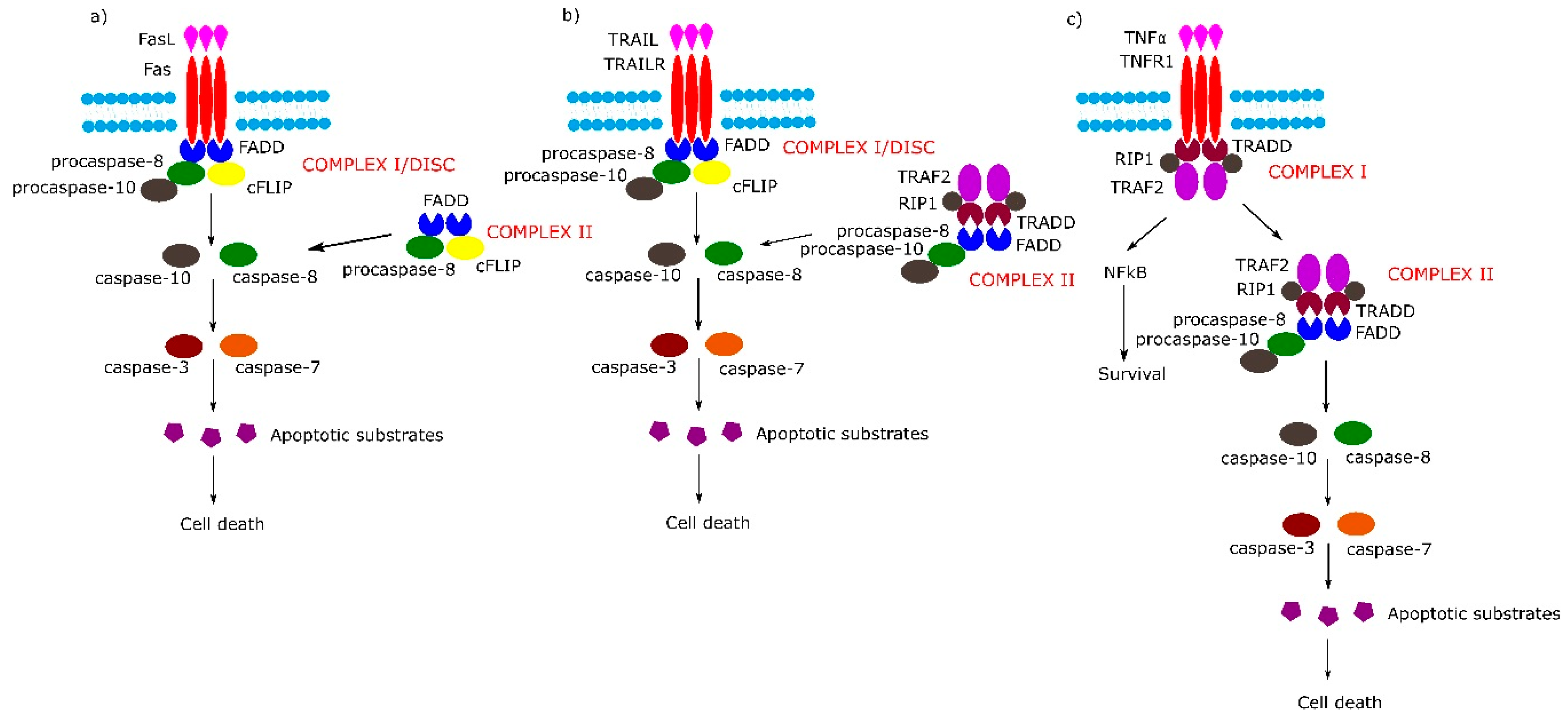
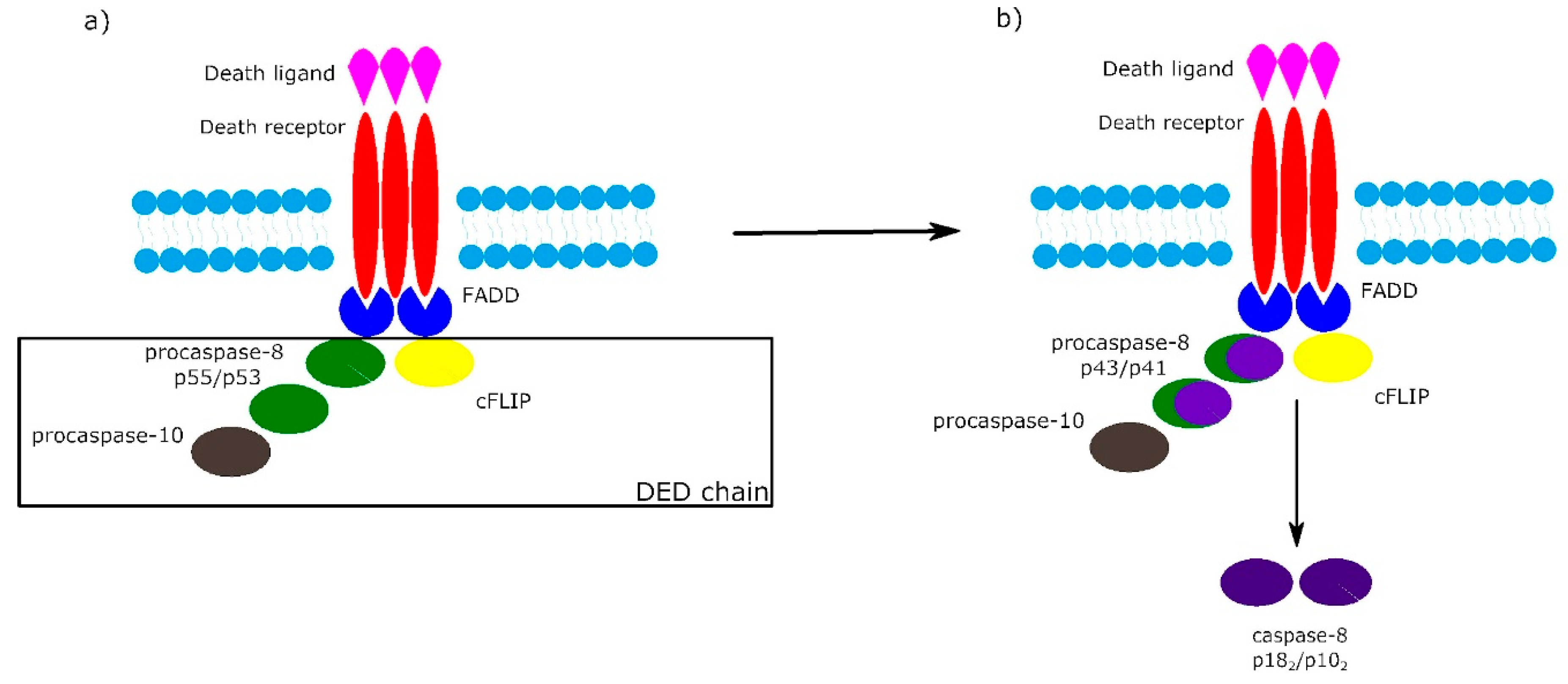
2. Receptor-Dependent Apoptotic Pathway

The TNFRSF consists of TNF receptor-associated factor (TRAF)-interacting receptors, decoy receptors (DcRs) and death receptors (DRs). The DRs can further be classified according to the molecular mechanisms of apoptosis and necroptosis activation: TNF receptor superfamily member 6 (Fas), TNF-related apoptosis-inducing ligand receptor 1 (TRAILR1) and TNF-related apoptosis-inducing ligand receptor 2 (TRAILR2) belong to the first subgroup of the DRs, which on the cytosolic plasma membrane surface, directly interact with an adapter protein, Fas-associated protein with death domain (FADD) and tumor necrosis factor α (TNF-α) receptor 1 (TNFR1/p55), which belongs to the second subgroup of DRs that act directly with TNFR1-associated protein with death domain (TRADD) [26]. Schemes of mentioned above pathways are shown in Figure 1.
The DRs are characterized by an extracellular N-terminal domain with cysteine-rich domains (CRDs), a membrane-spanning region and a C-terminal, an intracellular domain which harbors a death domain (DD). While DRs share the CRDs with all members of the TNFRSF, the DD is only found in DRs. The CRDs contain the N-terminal pre-ligand assembly domain (PLAD) which is involved in receptor–receptor interactions and is involved in binding ligands of the TNF superfamily (TNFSF) [27,28]. The PLAD domains’ interactions cause spontaneous receptor dimerization and/or trimerization. In the case of CD95/Fas and TRAILR death receptors, PLAD–PLAD interaction has a high affinity, in contrast to the TNF1 receptor [29].
Death ligands exist in the cell membrane as trimeric type II transmembrane proteins belonging to the TNFSF. They contain a conserved C-terminal TNF homology domain (THD), an N-terminal intracellular proline-rich domain (PRD) and a stalk region. THD binds to CRDs of the TNFRSF. Membrane-bound death ligands can be processed in the stalk region by metalloproteases, which leads to the release of soluble molecules. The soluble death ligands also own THD and, in consequence, have the capacity to react with TNFRSF receptors [27].
According to the sequential model of TNFRSF receptor activation, a single TNFRSF receptor interacts with a TNFSF ligand trimer and forms a cell surface-bound TNFRSF receptor-TNFSF ligand3 complex. Next, in two subsequent stages, two additional monomeric TNFRSF receptors are added to create an active TNFSF ligand3–TNFRSF receptor3 cluster which is necessary for induction of death-inducing signaling complexes [29].
The binding of Fas with tumor necrosis factor ligand superfamily member 6 (FasL) leads to formation of the DISC, also termed complex I, consisting of FADD, procaspase-8, procaspase-10 and cellular FLICE (FADD-like IL-1β-converting enzyme) inhibitory proteins (c-FLIPs). The formation of DISC leads to activation of caspase-8, caspase-10 and in consequence to cleavage and activation of effector caspase-3 and caspase-7. Additionally, in some cell lines, a second cytosolic complex is formed upon ligand stimulation. This complex, called complex II, composed of FADD, procaspase-8 and c-FLIPs, might amplify caspase activation by processing caspase-3 (Figure 1a) [30].
Upon tumor necrosis factor-related apoptosis-inducing (TRAIL) stimulation, the TRAIL receptor forms membrane-associated DISC/complex I in a similar way to the Fas receptor and secondary complex, similarly to above-mentioned TNFR1 complex II (Figure 1b) [30].
Signaling through TNFR1 leads to the formation of complex I and then complex II. Complex I consists of TRADD, TNFR1, TNF receptor-associated factor 2 (TRAF2) and tumor necrosis factor α (TNFα)-related receptor interacting protein (RIP). Complex I assembles rapidly following TNF-α stimulation, and activates the nuclear factor kappa-light-chain-enhancer of activated B cells (NF-κB) signal transduction pathway, which activates survival signals. In the next step, complex I dissociates from the death receptor and binds FADD and procaspase-8 (complex II), the activation of which leads to an induction of apoptosis (Figure 1c) (reviewed in [30]).
It is worth noting that the activation of procaspase-8 at DISC is driven by death effector domain (DED) “chains”, which include procaspase-8, procaspase-10 and c-FLIP. The number of procaspase-8 molecules in DED chains is 10 times higher than the number of procaspase-10 and c-FLIP molecules. The local increase of procaspase-8 concentration in DISC causes dimerization of procaspase-8. Then, procaspase-8 dimers (also called p55/p53) are converted by autocatalytic two-step transprocessing into mature heterotetrameric caspase-8 (p182/p102) while the DISC-bound caspase-8 prodomain can be replaced by a new procaspase-8 molecule. The termination of the DED chain’s elongation depends on its stability and association/dissociation rates of procaspase-8 to the chain (Figure 2) [26,31,32].
Among the most important regulators of the receptor-dependent apoptosis pathway are c-FLIP isoforms, which are major antiapoptotic proteins. Three isoforms can be distinguished as: short c-FLIPS, Raji c-FLIPR and long c-FLIPL. All c-FLIP isoforms possess two N-terminal DED domains. c-FLIPS and c-FLIPR are truncated version of procaspase-8, whereas c-FLIPL retains a full length procaspase-8 chain in which the catalytic cysteine residue within the active site is missing and thus has no proteolytic activity [32]. The isoforms of c-FLIP can heterodimerize with procaspase-8 via a co-operative and hierarchical binding mechanism. The result of c-FLIPS or c-FLIPR homo- or heterodimerization with procaspase-8 is prevention of DED-mediated caspase-8 oligomerization and inhibition of its activation. The ratio of c-FLIPL to procaspase-8 in c-FLIPL-procaspase-8 heterodimers determines whether procaspase-8 will be activated or inhibited. At physiologically-low levels, c-FLIPL-procaspase-8 heterodimer activates procaspase-8. In contrast, high levels of c-FLIPL block procaspase-8 oligomerization, which results in inhibition of caspase cascade activation (Figure 3) [32,33].
Other regulators of apoptosis are inhibitors of apoptosis (IAPs). The mammalian IAPs, X-linked IAP (XIAP), cellular inhibitor of apoptosis protein 1 (cIAP1) and cellular inhibitor of apoptosis protein 2 (cIAP2) contain three baculovirus IAP repeat (BIR) domains which mediate protein–protein interactions, UB-associated domain (UBA), which is responsible for interaction with ubiquitylated proteins, and the “really interesting new gene” (RING) domain conferring E3-ubiquitin ligase activity. Additionally, cIAP1 and cIAP2 have the caspase recruitment domain (CARD), which has the ability to inhibit their E3 ligase activity [34]. The BIR1 domain of XIAP binds the TGFβ-activated kinase 1 (TAK1)-binding protein 1 (TAB1) [35], whereas cIAPs’ BIR1 domain binds TRAF1 and TRAF2 [36]. In turn, the XIAP BIR2 domain is involved in inhibition of effector caspase-3 and caspase-7, while BIR3 binds to caspase-9 and prevents dimerization of this enzyme [37]. cIAP1 and cIAP2 can bind caspases through BIR2 and BIR3 domains, but do not inhibit them [38]. XIAP is able to induce ubiquitination of active caspases at the K48 residue, causing their (proteasomal) degradation. Additionally, XIAP is involved in covalent tagging of caspase-7 with ubiquitin-like protein NEDD8 leading to inactivation of this caspase [39]. cIAP1 and cIAP2 are able to ubiquinate caspase-3 and caspase-7 [40,41] but only cIAP1 targets them for proteasomal degradation [40].
The linear ubiquitin chain assembly complex (LUBAC) is another regulator of cell death. LUBAC is the only known E3 ligase complex for linear ubiquitination, which consists of ring finger protein 31 (RNF31, also known as HOIP), HOIL-1 and Shank-associated RH domain-interacting protein (sharpin) [42]. Recent studies showed that sharpin has a regulatory function in NF-κB and apoptotic signaling pathways. Absence of sharpin attenuates TNFα-mediated NF-κB activation [43,44,45]. Cleavage of RNF31 by caspase-3 and caspase-6 suppresses activity of LUBAC in the NF-κB signaling pathway. This process weakens the inhibitory role of NF-κB in death signaling, leading to the sensitization of cells to cell death signals [46]. On the other hand, another study showed that RNF31 limits caspase-8 activity in complex I and complex II in TRAIL signaling, which causes inhibition of apoptosis [47,48].

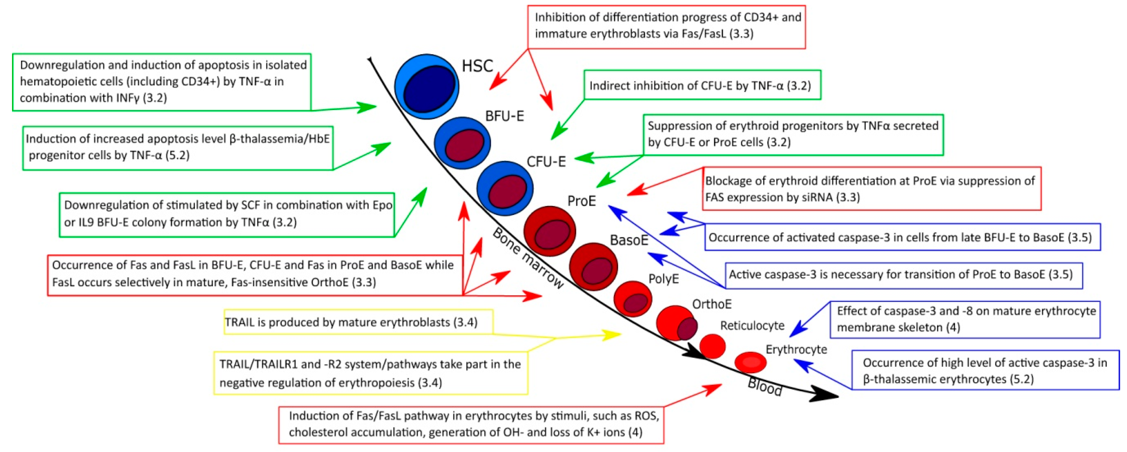
3. Erythropoiesis and Extrinsic Apoptotic Pathway

3.1. Erythropoiesis

In humans, two distinct types of erythroid cell production take place. The first type corresponds to primitive erythropoiesis which occurs in “blood islands” within the yolk sac. The newly formed cells are derived from mesodermal cells. Primitive erythroid cells (EryP) circulating in the early embryo are short-lived (~two days), nucleated, large and express ε-, γ-, α- and ζ -globin genes [10,49]. This process can be regulated by the renal cytokine erythropoietin (Epo). Epo has an influence on the primitive human erythroid precursors’ survival, rate of terminal maturation and proliferation [50].
Definitive erythropoiesis occurs first in the fetal liver and then in postnatal bone marrow. The adult erythroid lineage starts with multipotent hematopoietic stem cells (HSCs), also called long-term repopulating HSCs (LT-HSCs). The asymmetrical division of LT-HSCs gives rise to short-term repopulating HSCs (ST-HSCs), the asymmetrical division of which is the source of the multipotent progenitor cells (MPP-HSCs). The difference among these cells’ characteristics is gradual loss of self-renewal potential, but they sustain the same ability to transform into all blood cell types [51]. Further asymmetrical divisions of MPP-HSCs give slowly proliferating BFU-E and then rapidly dividing CFU-E progenitor cells, which divide three to five times within two to three days. Both BFU-E and CFU-E cells are sensitive to Epo due to the presence of erythropoietin receptor (EpoR) on these cells [52,53].
Continued stimulation with Epo triggers differentiation into erythroblast precursors: ProE, each of which undergoes three to four rounds of mitosis giving sequentially two basophilic erythroblasts (BasoE), four PolyE, and eight orthochromatic erythroblasts (OrthoE). The maturation processes of these cells are characterized by progressive expansion, accumulation of hemoglobin, decrease in RNA content, increase in chromatin density and decrease in cell size [50]. OrthoE transforms into two cell forms: a pyrenocyte (plasma membrane-coated extruded nucleus, which undergoes phagocytosis by macrophage) and a reticulocyte, which is a stage resulting from enucleation [54] which changes into a mature erythrocyte [8]. Reticulocytes and erythrocytes stop expressing EpoR [55].
Erythropoiesis is regulated by multiple factors such as growth factors and cytokines, SCF, G-CSF, and IL3, vitamins (e.g., folic acid and B12), hormones (corticosteroids, thyroid hormones, activin/inhibin, androgens), iron and its regulators (ferritin, transferrin, TfR-1, TfR-2, ferroportin, hepcidin), survival factors (MCL-1, BCL-xL, HSP70), transcription factors (GATA-1, STAT5A, STAT5B), negative growth regulators (TGFβ, BID, DRs, death ligands), differentiation factors (TGFβ, GDF11 and Epo) [56] and non-coding RNAs (e.g., miRNAs) [57]. Epo plays a crucial role in erythroid maturation. The binding of Epo to its receptor EpoR on the surface of erythroid progenitors causes activation of signal transducer and activator of transcription 5 (Stat5), phosphatidylinositol-3-kinase (PI3K)/Akt and Shc/Ras/mitogen-activated kinase (MAPK) pathways. Inactivation of the Stat5 or PI3K/AKT pathway contributes to significant apoptosis of early progenitors. On the other hand, the loss of the Shc/Ras/MAPK pathway exerts rather slight effects on terminal stages of erythroid maturation [52].

3.2. Role of TNF-α in Normal Erythropoiesis

Extensive studies have been devoted to the effects of TNF-α on erythropoiesis. A common feature of these works is that they all show that TNF-α exerted a direct or indirect inhibitory effect on erythroid maturation. One of the first studies showed that the indirect inhibition of human CFU-E colony formation by TNF-α was an effect of interferon β (IFNβ) released by bone marrow accessory cells [58,59]. On the other hand, a direct effect of TNF-α on erythroid progenitors has been described. Namely, TNF-α inhibited erythropoiesis stimulated by Epo alone or in combination with interleukin 3 (IL3) [60,61]. Further study demonstrated that TNF-α can downregulate BFU-E colony formation stimulated by stem cell factor (SCF) with Epo or IL9 with Epo [62]. The same authors showed that TNF-α induced inhibition of BFU-E colony formation is caused by TNFR1/p55. On the other hand, TNF-α receptor 2 (TNFR2/p75) is involved in the inhibition of progenitors’ responses to Epo alone [62,63], which somehow contradicts the fact that TNFR2 does not have a death domain in its cytoplasmic tail [64]. While TNFR1 occurrence was observed only during the initial stages of erythropoiesis, TNFR2 encoding gene (TNFR2) expression could be detected during all stages of erythroid differentiation. The variable occurrence of TNFRs on erythroid progenitors may suggest a different sensitivity of these cells to TNF-α [9,65]
Other studies suggested the existence of negative regulatory feedback of TNF-α within specialized niches-erythroblastic islands. The accumulation of mature erythroblasts that express death ligands may temporarily stop the expansion and differentiation of immature erythroblasts sensitive to death ligands. The interaction between death ligands and death receptors resulted in caspase-mediated degradation of GATA-1 [66,67]. A later study suggested the possible occurrence of negative feedback, in which TNF-α secreted by CFU-Es or ProEs suppresses the maturation of erythroid progenitors [68]. Another study carried out on a heterogeneous cell population constituting a hematopoietic stem and progenitor cells (CD34+ HSPC) culture demonstrated that TNF-α plays an important role in the weakness in balance between GATA-1 and GATA-2, which is important in erythroid lineage development. TNF-α promoted the formation of the GATA-1/PU.1 complex, which blocks the transcriptional activity of GATA-1, consequently inhibiting erythropoiesis in Epo-induced HSPCs [69].
In a dose-dependent manner, TNF-α in combination with interferon γ (INFγ) downregulated growth of purified hematopoietic cells (CD34+/CD38 CD34+/CD38+) in vitro and was also found to induce apoptosis of total bone marrow and CD34+ cells [70]. This effect did not require the presence of accessory cells. It seems that TNF-α and INFγ exert a suppressive effect on erythropoiesis in two ways: 1) inhibition of progenitor CD34+ cell proliferation by TNF-α and INFγ receptors’ activation-triggered phosphorylation of protein kinase R (PKR) [71] and/or 2) induction of apoptosis, which may cause a reduction in the number of stem and progenitor cells [70]. It should be mentioned that the former mechanism has been implicated in the pathophysiology of bone marrow failure and myelodysplastic (MDS) syndromes [71].
To sum up, it seems that TNF-α mainly exerts an inhibitory effect on erythroid maturation. TNF-α can inhibit erythropoiesis induced by Epo alone, Epo with IL3, Epo with SCF or Epo with IL9. The above-mentioned data indicate that TNF-α acts like negative regulator, which could inhibit maturation of erythroid progenitors and immature erythroblasts or regulate their number via the induction of apoptosis.

3.3. Fas/FasL in Normal Erythropoiesis

There is no detectable level of Fas in CD34+ cells freshly isolated from human bone marrow. Treatment with TNF-α and/or INFγ increases the Fas level on CD34+ cells. The inhibition of erythropoiesis by TNF-α and INFγ might be mediated by the Fas/FasL system [72]. The FasL encoding gene (FASLG) expression has not been identified in purified CD34+ cells, but there are experimental results which showed that stroma-derived factor 1α (SDF-1α) induced FasL-level increases in these cells with no effect on Fas production. It seems that SDF-1α inhibited the development of erythroid cells by functional upregulation of FasL [73].
The expression of the Fas encoding gene (FAS) increases from BFU-E to CFU-E stages of erythroid development, to reach a maximal level in ProE and BasoE. In turn, the FASLG gene is expressed in BFU-E, CFU-E and mature OrthoE. The interaction of FasL localized on mature erythroblasts with Fas present on immature erythroblasts causes induction of apoptosis in immature erythroblasts, while mature erythroblasts bearing Fas are insensitive to FasL [74]. Fas–FasL interaction results in apoptotic degradation of the transcription factors Tal-1 and GATA-1. The degradation of both transcription factors is therefore responsible for the Fas-mediated downregulation of maturation of immature erythroblasts [66,75]. This process strongly depends on the concentration of Epo, since Fas-based cytotoxicity against immature erythroblasts could be abrogated by high doses of Epo [74] upregulating GATA-1 expression, which in turn triggers the expression of the BCL2 gene [76]. At an intermediate level of Epo, cell fate depends on the number of mature erythroblasts in the bone marrow, which means that immature erythroblasts can be arrested in their maturation or enter an apoptosis pathway [77].
On the other hand, the suppression of Fas or caspases by siRNA treatment blocked the erythroid differentiation progress at the stage of ProE, i.e., blocking ProE to BasoE transition. This effect was reversed by FasL but not TRAIL treatment and suggests that caspase activation stimulates the erythroid maturation process [78].
SCF inhibits activation of caspase-8 and caspase-3 without decreasing the level of Fas. SCF prevents Fas-mediated apoptosis of human erythroid colony-forming cells, mainly CFU-E. This signal was found to depend on Src-family kinase [79,80]. Further studies showed that the mechanism of SCF action is based on the increase of FLIP expression [81]. The results of experiments indicate that a high cellular level of FLIP protects human HSPCs from Fas-mediated apoptosis [82].
During erythroid development, various long non-coding RNAs (lncRNAs) were found to regulate erythroid gene expression. Fas-AS1, also known as lncRNA Fas-antisense 1 (Saf), is encoded on the antisense strand of the first intron of the human FAS gene on chromosome 10 [83,84]. During erythropoiesis, Fas-AS1 is upregulated by the erythroid transcription factors GATA-1 and Kruppel-like factor 1 (KLF1) and is negatively regulated by NF-κB. Since the level of Fas-AS1 expression increases during erythroid maturation, it is suggested that the role of Fas lncRNA is to protect developing erythroblasts from Fas-mediated apoptosis via reducing the level of Fas [85,86].
The studies cited above on the role of the Fas/FasL pathway in erythropoiesis showed that death ligands or receptors are present up to the stage of basophilic erythroblasts. Both are involved in the inhibition of erythropoiesis in CD34+ cells and immature erythroblasts. The Fas/FasL system plays a significant role in control of the level and rate of immature erythroblast maturation in an Epo-dependent manner.

3.4. Role of TRAIL in Normal Erythropoiesis

Other death receptors expressed by erythroid cells are TRAILR1 and TRAILR2. In immature cells, the expression level of both receptors is higher than in mature erythroblasts [66]. In turn, TRAILR3 and TRAILR4 are not present on the cell surface during erythroid maturation [87]. TRAIL, which binds TRAILR1 and TRAILR2, is produced only by mature erythroblasts [66]. TRAIL, similarly to TNF-α and FasL, negatively regulates adult erythropoiesis [88]. Moreover, TRAIL was found to be involved in INFγ-mediated inhibition of erythropoiesis [89].
There is evidence that TRAIL negatively affects generation of mature erythroblasts by activation of the ERK1/2 pathway [87]. Another study showed that protein kinase Cε (PKCε) protects Epo-responsive mature erythroblasts against TRAIL-mediated apoptosis by up-regulation of BCL-2 [90,91]. In addition, recent research showed that in the absence of Epo, TRAIL behaves like a pro-survival factor by activating the NF-κB/IκBα pathway [92].
In summary, the TRAIL/TRAILR1 and -R2 system/pathways take part mainly in the negative regulation of erythropoiesis in a similar way as the Fas/FasL pathway or TNF-α.

3.5. Effect of Caspases on Normal Erythroid Maturation

The activation of several caspases seems to be essential in earlier stages of erythroblast differentiation [93]. Proenzymes of caspases 1–3 and 5–9 are present in erythroid cells. The level of procaspases-2, -3, -7 and -8 is the highest in immature erythroblasts [66,94]. Caspase-3 is the best known caspase which is involved in erythroid maturation. The occurrence of activated caspase-3 was observed in cells from late BFU-E [95] to BasoE [93,96]. Caspase-3 inhibition resulted in a reduction in erythroid expansion and differentiation [95]. On the other hand, caspase-3 downregulation had no effect on terminal maturation of erythrocytes, such as nuclear condensation and extrusion [97]. As was mentioned above, caspase activation via the Fas/FasL pathway triggers a positive effect on erythropoiesis; in particular, it is necessary for transition of ProE to BasoE [78].
The fate of erythroid precursors was found to depend on the nuclear localization of chaperone HSP70. During erythropoiesis, in the presence of Epo, HSP70 enters the nucleus and protects GATA1 from caspase cleavage. In the absence of Epo, HSP70 leaves the nucleus and allows for the caspase-3-mediated degradation of GATA-1 [98,99].
The above-mentioned data point to the crucial role of caspase-3 in early stages of erythroid maturation and indicate that active caspase-3 is necessary for transition of ProE to BasoE. Moreover, caspase-3 activation plays a major role in apoptosis of erythroid precursors upon Epo starvation.

4. Mature Erythrocytes and Extrinsic Apoptotic Pathway

The absence of an intrinsic apoptotic pathway in mature erythrocyte has an obvious evolutionary connection with the lack of mitochondria. Studies on apoptotic machinery in erythrocytes have clearly indicated that caspase-9, apoptotic protease activating factor 1 (Apaf-1) and CytC were missing, but also showed that red blood cells contain considerable amounts of caspase-3 and caspase-8 [100]. Erythrocytes, except for the above-mentioned caspase, also possess Fas, FasL and FADD, which are vital in the extrinsic apoptotic pathway. All of these components are localized to the detergent-resistant membrane (DRM) fraction of aged erythrocytes and also those which have undergone oxidative stress [101].
Early apoptosis activation study attempts showed that erythrocytes do not enter the path of apoptosis after treatment with staurosporine or when cells were incubated in the absence of serum. Normally these conditions trigger apoptosis in nucleated cells [102]. The next studies confirmed that caspase-8 and caspase-3 cannot be activated by CD95-stimulating antibody [103], etoposide or mitomycin c [100], well known inducers of apoptosis. More recent studies revealed that induction of the Fas-mediated cell death cascade may be a result of reactive oxygen species (ROS) and cholesterol accumulation in the erythrocytes of animals exposed to arsenic derivatives (e.g., sodium arsenite) [104,105]. Another study suggested that chronic lead exposure, which increases generation of OH¯ and loss of K+ ions, may induce Fas translocation into the membrane DRM fraction and in consequence induction of the extrinsic apoptotic pathway in erythrocytes [106].
It is worth noting that activated caspases can take part in degradation of erythrocyte membrane proteins which are involved in maintenance of cell shape and mechanical properties of the erythrocyte membrane. Caspase-8 is involved in β-spectrin degradation. Experiments performed on erythrocyte ghosts showed that spectrin undergoes proteolytic cleavage among other sites at residue 470, which results in generation of an N-terminal fragment containing the actin binding domain (ABD). This process was shown to depend on protein 4.1 [107]. Activated caspase-3 is able to cleave the N-terminal cytoplasmic domain of human erythroid anion exchanger 1 (AE1/band 3) in aged erythrocytes as a consequence of oxidative stress. This cleavage weakens interaction between AE1 and 4.2 protein, which results in weakened interactions of AE1-based complexes with spectrin [108,109,110,111]. Other studies provided evidence that upon storage of erythrocytes in saline-adenine-glucose-mannitol (SAGM) preservation medium caspase-3 mediates formation of a 24 kDa fragment which is a result of degradation of AE1 protein [112]. Data of other authors showed that caspase-3 is involved in clustering of AE1 protein and recognition of erythrocytes by macrophages [113,114]. Additionally, in erythrocytes, externalization of phosphatidylserine (PS) under oxidative stress is dependent on caspase-3, which seems to be connected to the impairment of flippase activity [115].
The observations that mature erythrocytes contain all components of the extrinsic apoptotic pathway raised many questions; however, common inducers of apoptosis do not activate this pathway. Studies based on an animal model suggested that other stimuli, such as ROS or cholesterol accumulation, could activate the extrinsic apoptotic pathway. The above data also demonstrated important effects of caspases-8 and caspase-3 on red cell membrane and membrane skeleton proteins, which are crucial in determining cell shape and erythrocyte deformability and therefore erythrocyte survival in the circulation.

5. β-Thalassemia and Extrinsic Apoptotic Pathway

5.1. β-Thalassemia

β-thalassemia is an autosomal recessive, inherited blood disorder characterized by reduced (β+) or absent (β0) beta-globin chain, which is the main component of adult hemoglobin. The beta-globin chain is encoded by the HBB gene located on chromosome 11. β-thalassemia is characterized by a decrease in hemoglobin synthesis and reduction of erythrocyte production leading to anemia [116]. β-thalassemia is widespread in the Mediterranean region, Middle East, southeast Asia, and North and Central Africa [117]. Every year, about 68,000 children are born with various β-thalassemia syndromes [118]. The heterogeneity of β-thalassemia is a result of different mutations in the HBB gene [119].
β-thalassemia minor is caused by heterozygous mutation that affects only one allele of the β-globin gene. This type of thalassemia can be inherited by the β+ gene and the β0 gene. Carriers of β-thalassemia minor are usually asymptomatic, but sometimes suffer from mild anemia [120]. In turn, β-thalassemia intermedia results from homozygous or compound heterozygous mutations in HBB gene, i.e., both alleles are affected. Patients with β-thalassemia intermedia show mild to moderate anemia. The most severe type of β-thalassemia is β-thalassemia major, which occurs when both alleles of the β-globin gene have mutations. Among above types of β-thalassemia, only patients with β-thalassemia major need regular blood transfusions [120,121].
Another type of β-thalassemia is β-thalassemia/hemoglobin E, which globally makes up about 50% of all cases of severe β-thalassemia [122]. Hemoglobin E (HbE) is an abnormal hemoglobin which is a result of substitution of a glutamic acid to lysine residue at codon 26 in the HBB gene. This mutation contributes to activation of a splice site, which leads to abnormal messenger RNA processing. HbE is produced at a reduced rate and the patient’s symptoms resemble mild β-thalassemia [123]. Patients with β-thalassemia/HbE inherited the β-thalassemia allele from one parent and the structural variant HbE from the other parent [122].
In general, the most important problems in thalassemic patients are iron overload, cardiac arrhythmia, hepatitis, osteoporosis, endocrine disorder and typical symptoms of anemia [119,120,121,122,123,124].

5.2. Role of Extrinsic Apoptotic Pathway in Different Types of β-Thalassemia

In 1970, ferrokinetic studies showed that 60–80% of thalassemic erythroid precursors die in the bone marrow. For comparison, in normal cells, this value reaches 10–20% [125]. The β-thalassemic bone marrow contains about six times more erythroid precursors than normal [126]. The cause of ineffective erythropoiesis in a β-thalassemia major patient was shown in 1993. The authors indicated that α-globin chain accumulation may result in accelerated apoptosis of thalassemic erythroid precursors in bone marrow [127]. Quantitative analysis showed that apoptotic cell death in the β-thalassemia major patient was four times higher than in a healthy patient [126]. Analyses carried out on patients with moderate to severe forms of β-thalassemia revealed a correlation between ineffective erythropoiesis and apoptosis. Patients with β-thalassemia/HbE had the most ineffective erythroid maturation and the most accelerated programmed cell death, for which α-globin accumulation was responsible [128]. β-thalassemic major bone marrow contains two times more PolyE and one third fewer OrthoE than normal. In vitro studies identified PolyE as a stage in which apoptosis occurs [129]. The β-thalassemic erythroid precursors are phagocytosed with about twice as high intensity as normal precursors. The enhanced phagocytosis is the result of: 1) an increased level of apoptosis-connected externalization of PS, 2) an increased number of macrophages and their activation [130].
Flow cytometry measurements showed that thalassemic erythroid precursors expose on their surface significantly higher levels of Fas or FasL [131] than in those of normal erythroid cells. As in typical cells, in β-thalassemia binding of FasL to Fas leads to activation of procaspase-8 [132]. Phosphoproteome analysis of β-thalassemic HSCs pointed to very high abundance of FasL, tumor necrosis factor receptor superfamily member 12A (TNFRSF12A) [133] and TRAF2 [123]. The presence and the level of the mentioned proteins may explain why freshly isolated β-thalassemic HSCs were characterized by shorter survival periods than those from normal donors [123,133].
It was observed that in β-thalassemia patients serum levels of TNF-α [134,135,136], IL-1β and IFNγ are higher than in healthy patients [134]. However, recent research suggested that serum levels of TNF-α in thalassemia patients were decreased before and increased after blood transfusion [137]. IL-1β [138,139], TNF-α and IFNγ were found to induce an increased apoptosis level of β-thalassemia/HbE progenitor cells compared to normal progenitors [138]. TNF-α was found to downregulate EpoR encoding gene (EPOR) expression and consequently Epo-induced cell proliferation of erythroid progenitor cells in β-thalassemia/HbE patients [136].
In contrast to normal erythrocytes, β-thalassemic red blood cells contain a higher level of activated caspase-3, which, similarly to normal cells in a condition of oxidative stress, cleaves AE1 proteins [108,140]. Another example of the results of caspase activation is the cleavage of GATA-1, which becomes unprotected by HSP70 due to its sequestration in the cytoplasm of β-thalassemic erythroblasts by an excess of free α globin chains [141]. Recent data from the research on exportin-1 (XPO1), which is a regulator of HSP70 localization in normal erythroid progenitors, showed the impact of XPO1 activity on β−thalassemic erythroblasts. Namely, XPO1 inhibitor, KPT-251 recovered GATA-1 expression and improved terminal differentiation of β−thalassemic erythroblasts through an increase in the amount of nuclear HSP-70, indicating the possibility of therapeutic treatment of β-thalassemia [142].
It was hypothesized that the excess of ROS could be a reason for increased apoptosis of β-thalassemic erythroid cells. The ROS generation in β-thalassemic precursor cells is probably a result of accumulation of unmatched α-globin, hemichromes, nonheme iron and free iron, the latter being able to generate ROS in a similar way as a Fenton reagent. However, there is no evidence indicating a direct connection between ROS and apoptosis in β-thalassemic erythroid cells [131,143].
The above-mentioned data strongly suggest that ineffective erythropoiesis in β-thalassemia is caused by an increase in the level of apoptosis. In comparison to normal cells, thalassemic cells are characterized by: (1) very high expression of FasL, TNFRSF12A and TRAF2, (2) PolyE-OrthoE arrest, (3) a higher serum level of TNF-α, IL-1β and IFNγ, (4) a higher level of activated caspase-3, and (5) enhanced phagocytosis.

6. Conclusions

Erythropoiesis, and mechanisms regulating this process, have attracted the attention of many scientists in the last two decades. One of the key pathways which is involved in the regulation of erythrocyte production is the extrinsic apoptotic pathway, which plays an important role at various stages of erythropoiesis. Moreover, it is interesting to learn the role, if any, of components of the receptor-dependent apoptotic pathway which are present in mature erythrocytes.
Understanding all of these processes may help to explain the pathophysiology of anemia associated, among other things, with β-thalassemia. Despite extensive research, it is still unclear what the direct mechanism of apoptosis of erythroid cells in β-thalassemic patients is. It seems necessary to specify the mechanism of accelerated apoptosis induced by α-globin chain accumulation. Additionally, it would be worthwhile to focus on the role of ROS overabundance in the increased apoptosis of thalassemic cells, probably existing as a result of unmatched α-globin accumulation. Furthermore, it would be particularly important to examine antiapoptotic processes occurring in thalassemia patients. Moreover, we should ask the following question: at what erythropoietic stage it is possible to inhibit increased apoptosis in β-thalassemic cells? The answer to this question should facilitate understanding of the main mechanism of accelerated apoptosis in β-thalassemia patients and perhaps aid the development of new therapeutic strategies. The summary of apoptotic processes maintaining regulatory function is presented in Figure 4. It seems that available data [144,145,146] coming from detailed Next Generation Sequencing (NGS) and mass spectrometry-based studies, facilitating transcriptome and proteome analyses at the single cell level, may provide a research background for studies seeking near definitive answers concerning the interplay between regulatory events resulting from the sequential regulation of expression of particular genes and apoptotic signals from other cells and the cellular environment.

Author Contributions

O.R.-J. Conceptualization and writing a draft version, preparation of figures, D.M.B. and N.J. discussions and substantial corrections, A.F.S. conceptualization and preparation of the final version. All authors have read and agreed to the published version of the manuscript.

Funding

Supported by National Science Centre, Poland, Project No. 2015/19/B/NZ5/03469.

Conflicts of Interest

The authors declare no conflict of interest. The funder had no role in the writing of the manuscript, or in the decision to publish the results.

Abbreviations

BasoEbasophilic erythroblasts
IAPinhibitors of apoptosis
AMPKAMP-activated kinase
Apaf-1apoptotic protease activating factor 1
BADBCL2 associated death promoter
BAKBCL2 antagonist/killer
BAXBCL2-associated X protein
BCL2B-cell lymphoma 2
BCLBBCL-2 like protein 10
BCLWBCL2-like protein 2
BFL1BCL2-related protein A1
BFU-Eburst-forming unit-erythroid
BHBCL2 homology
BIDBH3 interacting-domain death agonist
BIKBCL2 interacting killer
BIMBCL2 interacting mediator of cell death
BIRbaculovirus IAP repeat domains
BMFBCL2 modifying factor
CARDcaspase recruitment domain
c-FLIPscellular FLICE (FADD-like IL-1β-converting enzyme) inhibitory proteins
CFU-Ecolony-forming unit-erythroid
cIAP1 and cIAP2cellular inhibitor of apoptosis protein 1 and 2
CRDcysteine-rich domain
DcRsdecoy receptors
DDdeath domain
DEDdeath effector domain
DISCdeath-inducing signaling complex
DRMdetergent-resistant membrane
DRsdeath receptors
Epoerythropoietin
FADDFas-associated protein with death domain
FasTNF receptor superfamily member 6
FasLtumor necrosis factor ligand superfamily member 6
HRKactivator of apoptosis harakiri
IAPsinhibitors of apoptosis
INFγinterferon γ
JAK3Janus kinase 3
KLF1Kruppel-like factor 1
lncRNAslong non-coding RNAs
LT-HSCslong-term repopulating HSCs
LUBAClinear ubiquitin chain assembly complex
MAPKShc/Ras/mitogen-activated kinase
MCL1myeloid cell leukemia
MPCsmultipotent progenitor cells
NGSNext Generation Sequencing
NF-κBnuclear factor kappa-light-chain-enhancer of activated B cells
NOXAPhorbol-12-myristate-13-acetate-induced protein 1
OrthoEorthochromatic erythroblasts
PAK2p21-activated kinase 2
PI3Kphosphatidylinositol-3-kinase
PKC protein kinase C
PKCεprotein kinase Cε
PKRprotein kinase R
PLADpre-ligand assembly domain
PolyEpolychromatophilic erythroblasts
ProEProerythroblasts
ProEsProerythroblasts
PUMAp53 upregulated modulator of apoptosis
RING“really interesting new gene” domain
RNF31ring finger protein 31, also known as HOIP
ROSreactive oxygen species
SCFstem cell factor
SDF-1αstroma-derived factor 1α
Stat5signal transducer and activator of transcription 5
ST-HSCsshort-term repopulating HSCs
TAB1TGFβ-activated kinase 1 (TAK1)-binding protein 1
THDTNF homology domain
TNFR1TNF-α receptor 1
TNFR2TNF-α receptor 2
TNFRSFtumor necrosis factor receptor superfamily
TNFSFTNF superfamily
TNFαtumor necrosis factor α
TRADDTNFR1-associated protein with death domain
TRAFTNF receptor-associated factor
TRAILR1/TRAILR2TNF-related apoptosis-inducing ligand receptor 1 and 2
UBAUB-associated domain
XIAPX-linked IAP

References

  1. Ghashghaeinia, M.; Cluitmans, J.C.A.; Akel, A.; Dreischer, P.; Toulany, M.; Köberle, M.; Skabytska, Y.; Saki, M.; Biedermann, T.; Duszenko, M.; et al. The impact of erythrocyte age on eryptosis. Br. J. Haematol. 2012, 157, 606–614. [Google Scholar] [CrossRef]
  2. Repsold, L.; Joubert, A.M. Eryptosis: An Erythrocyte’s Suicidal Type of Cell Death. BioMed Res. Int. 2018, 2018, 9405617. [Google Scholar] [CrossRef]
  3. Lang, P.A.; Kempe, D.S.; Myssina, S.; Tanneur, V.; Birka, C.; Laufer, S.; Lang, F.; Wieder, T.; Huber, S.M. PGE2 in the regulation of programmed erythrocyte death. Cell Death Differ. 2005, 12, 415–428. [Google Scholar] [CrossRef]
  4. Myssina, S.; Huber, S.M.; Birka, C.; Lang, P.A.; Lang, K.S.; Friedrich, B.; Risler, T.; Wieder, T.; Lang, F. Inhibition of Erythrocyte Cation Channels by Erythropoietin. J. Am. Soc. Nephrol. 2003, 14, 2750–2757. [Google Scholar] [CrossRef] [Green Version]
  5. Qadri, S.M.; Bissinger, R.; Solh, Z.; Oldenborg, P.A. Eryptosis in health and disease: A paradigm shift towards understanding the (patho)physiological implications of programmed cell death of erythrocytes. Blood Rev. 2017, 31, 349–361. [Google Scholar] [CrossRef]
  6. Lang, F.; Qadri, S.M. Mechanisms and significance of eryptosis, the suicidal death of erythrocytes. Blood Purif. 2012, 33, 125–130. [Google Scholar] [CrossRef]
  7. Lang, E.; Lang, F. Triggers, Inhibitors, Mechanisms, and Significance of Eryptosis: The Suicidal Erythrocyte Death. BioMed Res. Int. 2015, 2015, 513518. [Google Scholar] [CrossRef] [Green Version]
  8. Nandakumar, S.K.; Ulirsch, J.C.; Sankaran, V.G. Advances in understanding erythropoiesis: Evolving perspectives. Br. J. Haematol. 2016, 173, 206–218. [Google Scholar] [CrossRef] [Green Version]
  9. Testa, U. Apoptotic mechanisms in the control of erythropoiesis. Leukemia 2004, 18, 1176–1199. [Google Scholar] [CrossRef] [Green Version]
  10. Dzierzak, E.; Philipsen, S. Erythropoiesis: Development and Differentiation. Cold Spring Harb. Perspect. Med. 2013, 3, a011601. [Google Scholar] [CrossRef]
  11. Dai, H.; Meng, W.; Kaufmann, S.H. BCL2 Family, Mitochondrial Apoptosis, and Beyond. Cancer Transl. Med. 2016, 2, 7. [Google Scholar] [CrossRef]
  12. Kalkavan, H.; Green, D.R. MOMP, cell suicide as a BCL-2 family business. Cell Death Differ. 2018, 25, 46–55. [Google Scholar] [CrossRef]
  13. Kale, J.; Osterlund, E.J.; Andrews, D.W. BCL-2 family proteins: Changing partners in the dance towards death. Cell Death Differ. 2018, 25, 65–80. [Google Scholar] [CrossRef] [Green Version]
  14. Motoyama, N.; Kimura, T.; Takahashi, T.; Watanabe, T.; Nakano, T. bcl-x Prevents apoptotic cell death of both primitive and definitive erythrocytes at the end of maturation. J. Exp. Med. 1999, 189, 1691–1698. [Google Scholar] [CrossRef] [Green Version]
  15. Dolznig, H.; Habermann, B.; Stangl, K.; Deiner, E.M.; Moriggl, R.; Beug, H.; Müllner, E.W. Apoptosis protection by the Epo target Bcl-XL allows factor-independent differentiation of primary erythroblasts. Curr. Biol. 2002, 12, 1076–1085. [Google Scholar] [CrossRef] [Green Version]
  16. Mason, K.D.; Lin, A.; Robb, L.; Josefsson, E.C.; Henley, K.J.; Gray, D.H.D.; Kile, B.T.; Roberts, A.W.; Strasser, A.; Huang, D.C.S.; et al. Proapoptotic Bak and Bax guard against fatal systemic and organ-specific autoimmune disease. Proc. Natl. Acad. Sci. USA 2013, 110, 2599–2604. [Google Scholar] [CrossRef] [Green Version]
  17. Delbridge, A.R.D.; Opferman, J.T.; Grabow, S.; Strasser, A. Antagonism between MCL-1 and PUMA governs stem/progenitor cell survival during hematopoietic recovery from stress. Blood 2015, 125, 3273–3280. [Google Scholar] [CrossRef] [Green Version]
  18. Opferman, J.T.; Iwasaki, H.; Ong, C.C.; Suh, H.; Mizuno, S.I.; Akashi, K.; Korsmeyer, S.J. Obligate role of anti-apoptotic MCL-1 in the survival of hematopoietic stem cells. Science 2005, 307, 1101–1104. [Google Scholar] [CrossRef]
  19. Delbridge, A.R.D.; Aubrey, B.J.; Hyland, C.; Bernardini, J.P.; Di Rago, L.; Garnier, J.M.; Lessene, G.; Strasser, A.; Alexander, W.S.; Grabow, S. The BH3-only proteins BIM and PUMA are not critical for the reticulocyte apoptosis caused by loss of the pro-survival protein BCL-XL. Cell Death Dis. 2017, 8, e2914-9. [Google Scholar] [CrossRef] [Green Version]
  20. Abutin, R.M.; Chen, J.; Lung, T.K.; Lloyd, J.A.; Sawyer, S.T.; Harada, H. Erythropoietin-Induced Phosphorylation/Degradation of BIM Contributes to Survival of Erythroid Cells. Exp. Hematol. 2009, 37, 151–158. [Google Scholar] [CrossRef] [Green Version]
  21. Wensveen, F.M.; Geest, C.R.; Libregts, S.F.W.M.; Derks, I.A.M.; Ekert, P.G.; Labi, V.; Villunger, A.; Nolte, M.A.; Eldering, E. BH3-only protein Noxa contributes to apoptotic control of stress-erythropoiesis. Apoptosis 2013, 18, 1306–1318. [Google Scholar] [CrossRef] [Green Version]
  22. Schug, Z.T.; Gonzalvez, F.; Houtkooper, R.H.; Vaz, F.M.; Gottlieb, E. BID is cleaved by caspase-8 within a native complex on the mitochondrial membrane. Cell Death Differ. 2011, 18, 538–548. [Google Scholar] [CrossRef]
  23. Milhas, D.; Cuvillier, O.; Therville, N.; Clavé, P.; Thomsen, M.; Levade, T.; Benoist, H.; Ségui, B. Caspase-10 triggers bid cleavage and caspase cascade activation in fasL-induced apoptosis. J. Biol. Chem. 2005, 280, 19836–19842. [Google Scholar] [CrossRef] [Green Version]
  24. Peslak, S.A.; Wenger, J.; Bemis, J.C.; Kingsley, P.D.; Frame, J.M.; Koniski, A.D.; Chen, Y.; Williams, J.P.; Mcgrath, K.E.; Dertinger, S.D.; et al. Sublethal radiation injury uncovers a functional transition during erythroid. Exp. Hematol. 2011, 39, 434–445. [Google Scholar] [CrossRef] [Green Version]
  25. Lamarque, M.; Guillerm, F.; Belaid, Z.; Hermine, O.; Courtois, G. BID Cleavage Pattern Is a Novel Check-Point That Determines the Fate of Human Erythroid Progenitors (Differentiation or Apoptosis). Blood 2016. [Google Scholar] [CrossRef]
  26. Siegmund, D.; Lang, I.; Wajant, H. Cell death-independent activities of the death receptors CD95, TRAILR1, and TRAILR2. FEBS J. 2017, 284, 1131–1159. [Google Scholar] [CrossRef]
  27. Bodmer, J.L.; Schneider, P.; Tschopp, J. The molecular architecture of the TNF superfamily. Trends Biochem. Sci. 2002, 27, 19–26. [Google Scholar] [CrossRef] [Green Version]
  28. Guicciardi, M.E.; Gores, G.J. Life and death by death receptors. FASEB J. 2009, 23, 1625–1637. [Google Scholar] [CrossRef] [Green Version]
  29. Wajant, H. Principles of antibody-mediated TNF receptor activation. Cell Death Differ. 2015, 22, 1727–1741. [Google Scholar] [CrossRef] [Green Version]
  30. Tourneur, L.; Chiocchia, G. FADD: A regulator of life and death. Trends Immunol. 2010, 31, 260–269. [Google Scholar] [CrossRef]
  31. Schleich, K.; Warnken, U.; Fricker, N.; Öztürk, S.; Richter, P.; Kammerer, K.; Schnölzer, M.; Krammer, P.H.; Lavrik, I.N. Stoichiometry of the CD95 Death-Inducing Signaling Complex: Experimental and Modeling Evidence for a Death Effector Domain Chain Model. Mol. Cell 2012, 47, 306–319. [Google Scholar] [CrossRef] [Green Version]
  32. Schleich, K.; Buchbinder, J.H.; Pietkiewicz, S.; Kähne, T.; Warnken, U.; Öztürk, S.; Schnölzer, M.; Naumann, M.; Krammer, P.H.; Lavrik, I.N. Molecular architecture of the DED chains at the DISC: Regulation of procaspase-8 activation by short DED proteins c-FLIP and procaspase-8 prodomain. Cell Death Differ. 2016, 23, 681–694. [Google Scholar] [CrossRef]
  33. Hughes, M.A.; Powley, I.R.; Jukes-Jones, R.; Horn, S.; Feoktistova, M.; Fairall, L.; Schwabe, J.W.R.; Leverkus, M.; Cain, K.; MacFarlane, M. Co-operative and Hierarchical Binding of c-FLIP and Caspase-8: A Unified Model Defines How c-FLIP Isoforms Differentially Control Cell Fate. Mol. Cell 2016, 61, 834–849. [Google Scholar] [CrossRef] [Green Version]
  34. Silke, J.; Meier, P. Inhibitor o fApoptosis (IAP) Proteins-Modulators of Cell Death and Inflammation. Cold Spring Harb. Perspect. Biol. 2013, 5, 1–19. [Google Scholar] [CrossRef]
  35. Lu, M.; Lin, S.C.; Huang, Y.; Kang, Y.J.; Rich, R.; Lo, Y.C.; Myszka, D.; Han, J.; Wu, H. XIAP Induces NF-κB Activation via the BIR1/TAB1 Interaction and BIR1 Dimerization. Mol. Cell 2007, 26, 689–702. [Google Scholar] [CrossRef] [Green Version]
  36. Vince, J.E.; Wong, W.W.L.; Khan, N.; Feltham, R.; Chau, D.; Ahmed, A.U.; Benetatos, C.A.; Chunduru, S.K.; Condon, S.M.; McKinlay, M.; et al. IAP Antagonists Target cIAP1 to Induce TNFα-Dependent Apoptosis. Cell 2007, 131, 682–693. [Google Scholar] [CrossRef] [Green Version]
  37. Gyrd-Hansen, M.; Meier, P. IAPs: From caspase inhibitors to modulators of NF-κB, inflammation and cancer. Nat. Rev. Cancer 2010, 10, 561–574. [Google Scholar] [CrossRef]
  38. Eckelman, B.P.; Salvesen, G.S. The human anti-apoptotic proteins cIAP1 and cIAP2 bind but do not inhibit caspases. J. Biol. Chem. 2006, 281, 3254–3260. [Google Scholar] [CrossRef] [Green Version]
  39. Berthelet, J.; Dubrez, L. Regulation of Apoptosis by Inhibitors of Apoptosis (IAPs). Cells 2013, 2, 163–187. [Google Scholar] [CrossRef] [Green Version]
  40. Choi, Y.E.; Butterworth, M.; Malladi, S.; Duckett, C.S.; Cohen, G.M.; Bratton, S.B. The E3 ubiquitin ligase cIAP1 binds and ubiquitinates caspase-3 and -7 via unique mechanisms at distinct steps in their processing. J. Biol. Chem. 2009, 284, 12772–12782. [Google Scholar] [CrossRef] [Green Version]
  41. Huang, H.K.; Joazeiro, C.A.P.; Bonfoco, E.; Kamada, S.; Leverson, J.D.; Hunter, T. The inhibitor of apoptosis, cIAP2, functions as a ubiquitin-protein ligase and promotes in vitro monoubiquitination of caspases 3 and 7. J. Biol. Chem. 2000, 275, 26661–26664. [Google Scholar]
  42. Kirisako, T.; Kamei, K.; Murata, S.; Kato, M.; Fukumoto, H.; Kanie, M.; Sano, S.; Tokunaga, F.; Tanaka, K.; Iwai, K. A ubiquitin ligase complex assembles linear polyubiquitin chains. EMBO J. 2006, 25, 4877–4887. [Google Scholar] [CrossRef]
  43. Gerlach, B.; Cordier, S.M.; Schmukle, A.C.; Emmerich, C.H.; Rieser, E.; Haas, T.L.; Webb, A.I.; Rickard, J.A.; Anderton, H.; Wong, W.W.L.; et al. Linear ubiquitination prevents inflammation and regulates immune signalling. Nature 2011, 471, 591–596. [Google Scholar] [CrossRef]
  44. Ikeda, F.; Deribe, Y.L.; Skånland, S.S.; Stieglitz, B.; Grabbe, C.; Franz-Wachtel, M.; Van Wijk, S.J.L.; Goswami, P.; Nagy, V.; Terzic, J.; et al. SHARPIN forms a linear ubiquitin ligase complex regulating NF-κB activity and apoptosis. Nature 2011, 471, 637–641. [Google Scholar] [CrossRef]
  45. Tokunaga, F.; Nakagawa, T.; Nakahara, M.; Saeki, Y.; Taniguchi, M.; Sakata, S.I.; Tanaka, K.; Nakano, H.; Iwai, K. SHARPIN is a component of the NF-κB-activating linear ubiquitin chain assembly complex. Nature 2011, 471, 633–636. [Google Scholar] [CrossRef]
  46. Joo, D.; Tang, Y.; Blonska, M.; Jin, J.; Zhao, X. Regulation of linear ubiquitin chain assembly complex by Caspase-mediated cleveage of RNF31. Mol. Cell. Biol. 2016, 36, 3010–3018. [Google Scholar] [CrossRef] [Green Version]
  47. Lafont, E.; Kantari-Mimoun, C.; Draber, P.; De Miguel, D.; Hartwig, T.; Reichert, M.; Kupka, S.; Shimizu, Y.; Taraborrelli, L.; Spit, M.; et al. The linear ubiquitin chain assembly complex regulates TRAIL-induced gene activation and cell death. EMBO J. 2017, 36, 1147–1166. [Google Scholar] [CrossRef]
  48. Lafont, E.; Hartwig, T.; Walczak, H. Paving TRAIL’s Path with Ubiquitin. Trends Biochem. Sci. 2018, 43, 44–60. [Google Scholar] [CrossRef]
  49. Lawson, K.A.; Meneses, J.J.; Pedersen, R.A. Clonal analysis of epiblast fate during germ layer formation in the mouse embryo. Development 1991, 113, 891–911. [Google Scholar]
  50. Palis, J. Primitive and definitive erythropoiesis in mammals. Front. Physiol. 2014, 5, 1–9. [Google Scholar] [CrossRef] [Green Version]
  51. Herrera-Merchan, A.; Hidalgo, I.; Arranz, L.; Gonzalez, S. Insights into Stem Cell Aging. In Advances in Hematopoietic Stem Cell Research; IntechOpen: London, UK, 2012. [Google Scholar] [CrossRef] [Green Version]
  52. Hattangadi, S.M.; Wong, P.; Zhang, L.; Flygare, J.; Lodish, H.F. From stem cell to red cell: Regulation of erythropoiesis at multiple levels by multiple proteins, RNAs, and chromatin modifications. Blood 2011, 118, 6258–6268. [Google Scholar] [CrossRef] [PubMed] [Green Version]
  53. Bunn, H.F. Erythropoietin. Cold Spring Harb. Perspect. Med. 2013, 3, a011619. [Google Scholar] [CrossRef] [PubMed] [Green Version]
  54. Nigra, A.D.; Casale, C.H.; Santander, V.S. Human erythrocytes: Cytoskeleton and its origin. Cell. Mol. Life Sci. 2019. [Google Scholar] [CrossRef] [PubMed]
  55. Elliott, S.; Pham, E.; Macdougall, I.C. Erythropoietins: A common mechanism of action. Exp. Hematol. 2008, 36, 1573–1584. [Google Scholar] [CrossRef]
  56. Valent, P.; Büsche, G.; Theurl, I.; Uras, I.Z.; Germing, U.; Stauder, R.; Sotlar, K.; Füreder, W.; Bettelheim, P.; Pfeilstöcker, M.; et al. Normal and pathological erythropoiesis in adults: From gene regulation to targeted treatment concepts. Haematologica 2018, 103, 1593–1603. [Google Scholar] [CrossRef]
  57. Listowski, M.A.; Heger, E.; Bogusławska, D.M.; Machnicka, B.; Kuliczkowski, K.; Leluk, J.; Sikorski, A.F. MicroRNAs: Fine tuning of erythropoiesis. Cell. Mol. Biol. Lett. 2012, 18, 34–46. [Google Scholar] [CrossRef]
  58. Means, R.T.; Dessypris, E.N.; Krantz, S.B. Inhibition of human colony-forming-unit erythroid by tumor necrosis factor requires accessory cells. J. Clin. Investig. 1990, 86, 538–541. [Google Scholar] [CrossRef]
  59. Means, R.T.; Krantz, S.B. Inhibition of human erythroid colony-forming units by tumor necrosis factor requires beta interferon. J. Clin. Investig. 1993, 91, 416–419. [Google Scholar] [CrossRef] [Green Version]
  60. Backx, B.; Broeders, L.; Bot, F.J.; Lowenberg, B. Positive and negative effects of tumor necrosis factor on colony growth from highly purified normal marrow progenitors. Leukemia 1991, 5, 66. [Google Scholar]
  61. Caux, C.; Saeland, S.; Favre, C.; Duvert, V.; Mannoni, P.; Banchereau, J. Tumor Necrosis Factor-alpha Strongly Potentiates Interleukin-3 and Granulocyte-Macrophage Colony-Stimulating Factor-Induced Proliferation of Human CD34+ Hematopoietic Progenitor Cells. Blood 1990, 75, 2292–2298. [Google Scholar] [CrossRef]
  62. Rusten, L.S.; Jacobsen, S.E.W. Tumor Necrosis Factor (TNF)-a Directly Inhibits Human Erythropoiesis In Vitro: Role. J. Biol. Chem. 1995, 85, 989–996. [Google Scholar]
  63. Dicato, M.; Morceau, F.; Diederich, M. Pro-inflammatory cytokine-mediated anemia: Regarding molecular mechanisms of erythropoiesis. Mediators Inflamm. 2009, 2009, 405016. [Google Scholar]
  64. Li, J.; Yin, Q.; Wu, H. Structural Basis of Signal Transduction in the TNF Receptor Superfamily. Adv. Immunol. 2013, 119, 135–153. [Google Scholar]
  65. Xiao, W.; Koizumi, K.; Nishio, M.; Endo, T.; Osawa, M.; Fujimoto, K.; Sato, I.; Sakai, T.; Koike, T.; Sawada, K.I. Tumor necrosis factor-α inhibits generation of glycophorin A+ cells by CD34+ cells. Exp. Hematol. 2002, 30, 1238–1247. [Google Scholar] [CrossRef]
  66. De Maria, R.; Zeuner, A.; Eramo, A.; Domenichelli, C.; Bonci, D.; Grignani, F.; Srinivasula, S.M.; Alnemri, E.S.; Testa, U.; Peschle, C. Negative regulation of erythropoiesis by caspase-mediated cleavage of GATA-1. Nature 1999, 401, 489–493. [Google Scholar] [CrossRef]
  67. Chasis, J.A.; Mohandas, N. Erythroblastic islands: Niches for erythropoiesis. Blood 2008, 112, 470–478. [Google Scholar] [CrossRef] [Green Version]
  68. Jacobs-Helber, S.M.; Roh, K.; Bailey, D.; Dessypris, E.N.; Ryan, J.J.; Chen, J.; Wickrema, A.; Barber, D.L.; Dent, P.; Sawyer, S.T. Tumor necrosis factor-alpha expressed constitutively in erythroid cells or induced by erythropoietin has negative and stimulatory roles in normal erythropoiesis and erythroleukemia. Blood 2003, 101, 524–531. [Google Scholar] [CrossRef]
  69. Grigorakaki, C.; Morceau, F.; Chateauvieux, S.; Dicato, M.; Diederich, M. Tumor necrosis factor alpha-mediated inhibition of erythropoiesis involves GATA-1/GATA-2 balance impairment and PU.1 over-expression. Biochem. Pharmacol. 2011, 82, 156–166. [Google Scholar] [CrossRef] [Green Version]
  70. Selleri, C.; Sato, T.; Anderson, S.; Young, N.S.; Maciejewski, J.P. Interferon-gamma and tumor necrosis factor-alpha suppress both early and late stages of hematopoiesis and induce programmed cell death. J. Cell. Physiol. 1995, 165, 538–546. [Google Scholar] [CrossRef]
  71. Sharma, B.; Altman, J.K.; Goussetis, D.J.; Verma, A.K.; Platanias, L.C. Protein kinase R as mediator of the effects of interferon (IFN) γ and tumor necrosis factor (TNF) α on normal and dysplastic hematopoiesis. J. Biol. Chem. 2011, 286, 27506–27514. [Google Scholar] [CrossRef] [Green Version]
  72. Maciejewski, B.J.; Selleri, C.; Anderson, S.; Young, N.S. Fas antigen expression on CD34+ human marrow cells is induced by interferon gamma and tumor necrosis factor alpha and potentiates cytokine-mediated hematopoietic suppression in vitro. Blood 1995, 85, 3183–3190. [Google Scholar] [CrossRef]
  73. Gibellini, D.; Bassini, A.; Re, M.C.; Ponti, C.; Miscia, S.; Gonelli, A.; La Placa, M.; Zauli, G. Stroma-derived factor 1alpha induces a selective inhibition of human erythroid development via the functional upregulation of Fas/CD95 ligand. Br. J. Haematol. 2000, 111, 432–440. [Google Scholar] [CrossRef]
  74. De Maria, R.; Testa, U.; Luchetti, L.; Zeuner, A.; Stassi, G.; Pelosi, E.; Riccioni, R.; Felli, N.; Samoggia, P.; Peschle, C. Apoptotic role of Fas/Fas ligand system in the regulation of erythropoiesis. Blood 1999, 93, 796–803. [Google Scholar] [CrossRef]
  75. Zeuner, A.; Eramo, A.; Testa, U.; Felli, N.; Pelosi, E.; Mariani, G.; Srinivasula, S.M.; Alnemri, E.S.; Condorelli, G.; Peschle, C.; et al. Control of erythroid cell production via caspase-mediated cleavage of transcription factor SCL/Tal-1. Cell Death Differ. 2003, 10, 905–913. [Google Scholar] [CrossRef] [Green Version]
  76. Gregory, T.; Yu, C.; Ma, A.; Orkin, S.H.; Blobel, G.; Weiss, M.J. GATA-1 and erythropoietin cooperate to promote erythroid cell survival by regulating bcl-xL expression. Blood 1999, 94, 87–96. [Google Scholar] [CrossRef]
  77. Vandekerkhove, J.; Courtois, G.; Coulon, S.; Ribeli, J.A.; Hermine, O. Regulation of erythropoesis. ESH Handb. Disord. Iron Metab. Eur. Sch. Hematol. 2009, 2, 44–87. [Google Scholar]
  78. Carlile, G.W.; Smith, D.H.; Wiedmann, M. A non-apoptotic role for Fas/FasL in erythropoiesis. FEBS Lett. 2009, 583, 848–854. [Google Scholar] [CrossRef] [Green Version]
  79. Nishio, M.; Oda, A.; Koizumi, K.; Satoh, I.; Sato, Y.; Endoh, T.; Tsutsumi, A.; Fujihara, M.; Ikebuchi, K.; Ikeda, H.; et al. Stem cell factor prevents Fas-mediated apoptosis of human erythroid precursor cells with Src-family kinase dependency. Exp. Hematol. 2001, 29, 19–29. [Google Scholar] [CrossRef]
  80. Oda, A.; Nishio, M.; Sawada, K.I. Stem Cell Factor Regulation of Fas-Mediated Apoptosis of human erythroid precursor cell. J. Hematother. 2001, 10, 595–600. [Google Scholar] [CrossRef]
  81. Chung, I.J.; Dai, C.; Krantz, S.B. Stem cell factor increases the expression of FLIP that inhibits IFNgamma -induced apoptosis in human erythroid progenitor cells. Blood 2003, 101, 1324–1328. [Google Scholar] [CrossRef] [Green Version]
  82. Kim, H.; Whartenby, K.A.; Georgantas, R.W.; Wingard, J.; Civin, C.I. Human CD34+ Hematopoietic Stem/Progenitor Cells Express High Levels of FLIP and Are Resistant to Fas-Mediated Apoptosis. Stem Cells 2002, 20, 174–182. [Google Scholar] [CrossRef] [PubMed]
  83. De Yan, M.; Hong, C.C.; Lai, G.M.; Cheng, A.L.; Lin, Y.W.; Chuang, S.E. Identification and characterization of a novel gene Saf transcribed from the opposite strand of Fas. Hum. Mol. Genet. 2005, 14, 1465–1474. [Google Scholar] [CrossRef] [PubMed] [Green Version]
  84. Kulczyńska, K.; Siatecka, M. A regulatory function of long non-coding RNAs in red blood cell development. Acta Biochim. Pol. 2016, 63, 675–680. [Google Scholar] [CrossRef] [PubMed] [Green Version]
  85. Villamizar, O.; Chambers, C.B.; Mo, Y.Y.; Torry, D.S.; Hofstrand, R.; Riberdy, J.M.; Persons, D.A.; Wilber, A. Fas-antisense long noncoding RNA is differentially expressed during maturation of human erythrocytes and confers resistance to Fas-mediated cell death. Blood Cells Mol. Dis. 2016, 58, 57–66. [Google Scholar] [CrossRef] [PubMed]
  86. Villamizar, O.; Chambers, C.B.; Mo, Y.Y.; Torry, D.S.; Hofstrand, R.; Riberdy, J.M.; Persons, D.A.; Wilber, A. Data in support of transcriptional regulation and function of Fas-antisense long noncoding RNA during human erythropoiesis. Data Br. 2016, 7, 1288–1295. [Google Scholar] [CrossRef] [PubMed] [Green Version]
  87. Secchiero, P.; Melloni, E.; Heikinheimo, M.; Mannisto, S.; Di Pietro, R.; Iacone, A.; Zauli, G. TRAIL regulates normal erythroid maturation through an ERK-dependent pathway. Blood 2004, 103, 517–522. [Google Scholar] [CrossRef] [PubMed] [Green Version]
  88. Zamai, L.; Secchiero, P.; Pierpaoli, S.; Bassini, A.; Papa, S.; Alnemri, E.S.; Guidotti, L.; Vitale, M.; Zauli, G. TNF-related apoptosis-inducing ligand (TRAIL) as a negative regulator of normal human erythropoiesis. Blood 2000, 95, 3716–3724. [Google Scholar]
  89. Felli, N.; Pedini, F.; Zeuner, A.; Petrucci, E.; Testa, U.; Conticello, C.; Biffoni, M.; Di Cataldo, A.; Winkles, J.A.; Peschle, C.; et al. Multiple Members of the TNF Superfamily Contribute to IFN-γ-Mediated Inhibition of Erythropoiesis. J. Immunol. 2005, 175, 1464–1472. [Google Scholar] [CrossRef] [Green Version]
  90. Mirandola, P.; Gobbi, G.; Ponti, C.; Sponzilli, I.; Cocco, L.; Vitale, M. PKCε controls protection against TRAIL in erythroid progenitors. Blood 2006, 107, 508–513. [Google Scholar] [CrossRef] [Green Version]
  91. Vitale, M.; Gobbi, G.; Mirandola, P.; Ponti, C.; Sponzilli, I.; Rinaldi, L.; Manzoli, F.A. TNF-related apoptosis-inducing ligand (TRAIL) and erythropoiesis: A role for PKCε. Eur. J. Histochem. 2006, 50, 15–18. [Google Scholar]
  92. Sancilio, S.; Giacomo, V.D.; Quaglietta, A.M.; Iacone, A.; Angelucci, D.; Tatasciore, U.; Rana, R.A.; Cataldi, A.; Zauli, G.; Pietro, R. TRAIL promotes a pro-survival signal in erythropoietin-deprived human erythroblasts throught the activation of an NF-κB/IκBα pathway. J. Biol. Regul. Homeost. Agents 2011, 25, 375–386. [Google Scholar] [PubMed]
  93. Zermati, Y.; Garrido, C.; Amsellem, S.; Fishelson, S.; Bouscary, D.; Valensi, F.; Varet, B.; Solary, E.; Hermine, O. Caspase activation is required for terminal erythroid differentiation. J. Exp. Med. 2001, 193, 247–254. [Google Scholar] [CrossRef] [PubMed] [Green Version]
  94. Gregoli, P.A.; Bondurant, M.C. Function of caspases in regulating apoptosis caused by erythropoietin deprivation in erythroid progenitors. J. Cell. Physiol. 1999, 178, 133–143. [Google Scholar] [CrossRef]
  95. Boehm, D.; Mazurier, C.; Giarratana, M.C.; Darghouth, D.; Faussat, A.M.; Harmand, L.; Douay, L. Caspase-3 Is Involved in the Signalling in Erythroid Differentiation by Targeting Late Progenitors. PLoS ONE 2013, 8, e62303. [Google Scholar] [CrossRef] [PubMed] [Green Version]
  96. Carlile, G.W.; Smith, D.H.; Wiedmann, M. Caspase-3 has a nonapoptotic function in erythroid maturation. Blood 2004, 103, 4310–4316. [Google Scholar] [CrossRef] [Green Version]
  97. Krauss, S.W.; Lo, A.J.; Short, S.A.; Koury, M.J.; Mohandas, N.; Chasis, J.A. Nuclear substructure reorganization during late-stage erythropoiesis is selective and does not involve caspase cleavage of major nuclear substructural proteins. Blood 2005, 106, 2200–2206. [Google Scholar] [CrossRef]
  98. Ribeil, J.-A.; Zermati, Y.; Vandekerckhove, J.; Cathelin, S.; Kersual, J.; Dussiot, M.; Coulon, S.; Moura, I.C.; Zeuner, A.; Kirkegaard-Sørensen, T.; et al. Hsp70 regulates erythropoiesis by preventing caspase-3-mediated cleavage of GATA-1. Nature 2007, 445, 102–105. [Google Scholar] [CrossRef]
  99. Hermine, O.; Arlet, J.B.; Ribeil, J.A.; Guillerm, F.; Vandekerkhove, J.; Courtois, G. HSP70, an erythropoiesis regulator that determines the fate of erythroblasts between death and differenciation. Transfus. Clin. Biol. 2013, 20, 144–147. [Google Scholar] [CrossRef]
  100. Berg, C.P.; Engels, I.H.; Rothbart, A.; Lauber, K.; Renz, A.; Schlosser, S.F.; Schulze-Osthoff, K.; Wesselborg, S. Human mature red blood cells express caspase-3 and caspase-8, but are devoid of mitochondrial regulators of apoptosis. Cell Death Differ. 2001, 8, 1197–1206. [Google Scholar] [CrossRef]
  101. Mandal, D.; Mazumder, A.; Das, P.; Kundu, M.; Basu, J. Fas-, caspase 8-, and caspase 3-dependent signaling regulates the activity of the aminophospholipid translocase and phosphatidylserine externalization in human erythrocytes. J. Biol. Chem. 2005, 280, 39460–39467. [Google Scholar] [CrossRef] [Green Version]
  102. Weil, M.; Jacobson, M.D.; Davies, T.J.; Gardner, R.L.; Raft, K.D.; Raft, M.C. Constitutive Expression of the Machinery for Programmed Cell Death. J. Cell Biol. 1996, 133, 1–7. [Google Scholar] [CrossRef] [PubMed]
  103. Sagan, D.; Jermnim, N.; Tangvarasittichai, O. CD95 is not functional in human erythrocytes. Int. J. Lab. Hematol. 2010, 32, 244–247. [Google Scholar] [CrossRef] [PubMed]
  104. Biswas, D.; Banerjee, M.; Sen, G.; Das, J.K.; Banerjee, A.; Sau, T.J.; Pandit, S.; Giri, A.K.; Biswas, T. Mechanism of erythrocyte death in human population exposed to arsenic through drinking water. Toxicol. Appl. Pharmacol. 2008, 230, 57–66. [Google Scholar] [CrossRef] [PubMed]
  105. Biswas, D.; Sen, G.; Sarkar, A.; Biswas, T. Atorvastatin acts synergistically with N-acetyl cysteine to provide therapeutic advantage against Fas-activated erythrocyte apoptosis during chronic arsenic exposure in rats. Toxicol. Appl. Pharmacol. 2011, 250, 39–53. [Google Scholar] [CrossRef]
  106. Mandal, S.; Mukherjee, S.; Chowdhury, K.D.; Sarkar, A.; Basu, K.; Paul, S.; Karmakar, D.; Chatterjee, M.; Biswas, T.; Sadhukhan, G.C.; et al. S-allyl cysteine in combination with clotrimazole downregulates Fas induced apoptotic events in erythrocytes of mice exposed to lead. Biochim. Biophys. Acta-Gen. Subj. 2012, 1820, 9–23. [Google Scholar] [CrossRef]
  107. Toporkiewicz, M.; Grzybek, M.; Meissner, J.; Michalczyk, I.; Dubielecka, P.M.; Korycka, J.; Seweryn, E.; Sikorski, A.F. Release of an ~55kDa fragment containing the actin-binding domain of β-spectrin by caspase-8 during FND-induced apoptosis depends on the presence of protein 4.1. Arch. Biochem. Biophys. 2013, 535, 205–213. [Google Scholar] [CrossRef]
  108. Mandal, D.; Baudin-Creuza, V.; Bhattacharyya, A.; Pathak, S.; Delaunay, J.; Kundu, M.; Basu, J. Caspase 3-mediated Proteolysis of the N-terminal Cytoplasmic Domain of the Human Erythroid Anion Exchanger 1 (Band 3). J. Biol. Chem. 2003, 278, 52551–52558. [Google Scholar] [CrossRef] [Green Version]
  109. Machnicka, B.; Grochowalska, R.; Bogusławska, D.M.; Sikorski, F.; Lecomte, M.C. Spectrin-based skeleton as an actor in cell signaling. Cell. Mol. Life Sci. 2012, 69, 191–201. [Google Scholar] [CrossRef] [Green Version]
  110. Machnicka, B.; Czogalla, A.; Hryniewicz-Jankowska, A.; Bogusławska, D.M.; Grochowalska, R.; Heger, E.; Sikorski, A.F. Spectrins: A structural platform for stabilization and activation of membrane channels, receptors and transporters. Biochim. Biophys. Acta 2014, 1838, 620–634. [Google Scholar] [CrossRef] [Green Version]
  111. Machnicka, B.; Grochowalska, R.; Bogusławska, D.M.; Sikorski, A.F. The role of spectrin in cell adhesion and cell–cell contact. Exp. Biol. Med. 2019, 1303–1312. [Google Scholar] [CrossRef]
  112. Rinalducci, S.; Ferru, E.; Blasi, B.; Turrini, F.; Zolla, L. Oxidative stress and caspase-mediated fragmentation of cytoplasmic domain of erythrocyte band 3 during blood storage. Blood Transfus. 2012, 10, 55–62. [Google Scholar]
  113. Pietraforte, D.; Matarrese, P.; Straface, E.; Gambardella, L.; Metere, A.; Scorza, G.; Leto, T.L.; Malorni, W.; Minetti, M. Two different pathways are involved in peroxynitrite-induced senescence and apoptosis of human erythrocytes. Free Radic. Biol. Med. 2007, 42, 202–214. [Google Scholar] [CrossRef] [PubMed]
  114. Miki, Y.; Tazawa, T.; Hirano, K.; Matsushima, H.; Kumamoto, S.; Hamasaki, N.; Yamaguchi, T.; Beppu, M. Clearance of oxidized erythrocytes by macrophages: Involvement of caspases in the generation of clearance signal at band 3 glycoprotein. Biochem. Biophys. Res. Commun. 2007, 363, 57–62. [Google Scholar] [CrossRef] [PubMed]
  115. Mandal, D.; Moitra, P.K.; Saha, S.; Basu, J. Caspase 3 regulates phosphatidylserine externalization and phagocytosis of oxidatively stressed erythrocytes. FEBS Lett. 2002, 513, 184–188. [Google Scholar] [CrossRef] [Green Version]
  116. Ingram, V.M.; Stretton, A.O.W. Genetic basis of the thalassemia diseases. Nature 1959, 184, 632–633. [Google Scholar] [CrossRef] [PubMed]
  117. Weatherall, D.J.; Williams, T.N.; Allen, S.J.; O’Donnell, A. The population genetics and dynamics of the thalassemias. Hematol. Oncol. Clin. N. Am. 2010, 24, 1021–1031. [Google Scholar] [CrossRef] [PubMed]
  118. Modell, B.; Darlison, M. Global epidemiology of haemoglobin disorders and derived service indicators. Bull. World Health Organ. 2008, 86, 480–487. [Google Scholar] [CrossRef]
  119. Origa, R. β-Thalassemia. Genet. Med. 2017, 19, 609–619. [Google Scholar] [CrossRef] [Green Version]
  120. Tari, K.; Valizadeh Ardalan, P.; Abbaszadehdibavar, M.; Atashi, A.; Jalili, A.; Gheidishahran, M. Thalassemia an update: Molecular basis, clinical features and treatment. Int. J. Biomed. Public Heal. 2018, 1, 48–58. [Google Scholar] [CrossRef] [Green Version]
  121. Musallam, K.M.; Rivella, S.; Vichinsky, E.; Rachmilewitz, E.A. Non-transfusion-dependent thalassemias. Haematologica 2013, 98, 833–844. [Google Scholar] [CrossRef] [Green Version]
  122. Olivieri, N.; Pakbaz, Z.; Vichinsky, E. Hb E/beta-thalassaemia: A common & clinically diverse disorder. Indian J. Med. Res. 2011, 134, 522–531. [Google Scholar] [PubMed]
  123. Ponnikorn, S.; Kong, S.P.; Thitivirachawat, S.; Tanjasiri, C.; Tungpradabkul, S.; Hongeng, S. Proteomic Analysis of β Thalassemia/HbE: A perspective from Hematopoietic Stem Cells (HSCs). In Proteomics Technologies and Applications; IntechOpen: London, UK, 2019. [Google Scholar] [CrossRef] [Green Version]
  124. Fucharoen, S.; Weatherall, D.J. The hemoglobin E thalassemias. Cold Spring Harb. Perspect. Med. 2012, 2, a011734. [Google Scholar] [CrossRef] [PubMed]
  125. Finch, C.A.; Deubelbeiss, K.; Cook, J.D.; Eschbach, J.W.; Harker, L.A.; Funk, D.D.; Mrsaglia, G.; Hillman, R.S.; Slichter, S.; Adamson, J.W.; et al. Ferrokinetics in man. Medicine 1970, 49, 17–53. [Google Scholar] [CrossRef] [PubMed]
  126. Centis, F.; Tabellini, L.; Lucarelli, G.; Buffi, O.; Tonucci, P.; Persini, B.; Annibali, M.; Emiliani, R.; Iliescu, A.; Rapa, S.; et al. The importance of erythroid expansion in determining the extent of apoptosis in erythroid precursors in patients with beta-thalassemia major. Blood 2000, 96, 3624–3629. [Google Scholar] [CrossRef]
  127. Yuan, J.; Angelluci, E.; Lucarelli, G.; Aljurf, M.; Sznder, L.M.; Kiefer, C.R.; Ma, L.; Schrier, S.L. Accelerated Programmed Cell Death (Apoptosis) in Erythroid Precursors of Patients With Severe β-Thalassemia (Cooley’s Anemia). Blood 1993, 82, 374–377. [Google Scholar] [CrossRef] [Green Version]
  128. Pootrakul, P.; Sirankapracha, P.; Hemsorach, S.; Moungsub, W.; Kumbunlue, R.; Piangitjagum, A.; Wasi, P.; Ma, L.; Schrier, S.L. A correlation of erythrokinetics, ineffective erythropoiesis, and erythroid precursor apoptosis in thai patients with thalassemia. Blood 2000, 96, 2606–2612. [Google Scholar] [CrossRef]
  129. Mathias, L.A.; Fisher, T.C.; Zeng, L.; Meiselman, H.J.; Weinberg, K.I.; Hiti, A.L.; Malik, P. Ineffective erythropoiesis in β-thalassemia major is due to apoptosis at the polychromatophilic normoblast stage. Exp. Hematol. 2000, 28, 1343–1353. [Google Scholar] [CrossRef]
  130. Angelucci, E.; Bai, H.; Centis, F.; Bafti, M.S.; Lucarelli, G.; Ma, L.; Schrier, S. Enhanced macrophagic attack on β-thalassemia major erythroid precursors. Haematologica 2002, 87, 578–583. [Google Scholar]
  131. Schrier, S.L. Pathophysiology of thalassemia. Curr. Opin. Hematol. 2002, 9, 123–126. [Google Scholar] [CrossRef]
  132. Schrier, S.L.; Centis, F.; Verneris, M.; Ma, L.; Angelucci, E. The role of oxidant injury in the pathophysiology of human thalassemias. Redox Rep. 2003, 8, 241–245. [Google Scholar] [CrossRef]
  133. Ponnikorn, S.; Panichakul, T.; Sresanga, K.; Wongborisuth, C.; Roztrakul, S.; Hongeng, S.; Tungpradabkul, S. Phosphoproteomic analysis of apoptotic hematopoietic stem cells from hemoglobin E/β-thalassemia. J. Transl. Med. 2011, 9, 1–10. [Google Scholar] [CrossRef] [PubMed] [Green Version]
  134. Wickramasinghe, S.N.; Wiener, E.; Siripanyaphinyo, U.; Chinprasertsuk, S.; Mawas, F.; Fucharoen, S.; Wickramasinghe, S.N.; Pootrakul, P.; Visudhiphan, S. Serum Levels of Tumor Necrosis Factor alpha, Interleukin-1, and Interferon gamma in Betta-Thalassemia/HbE and Their Clinical Significance. Viral Immunol. 1999, 111, 75–79. [Google Scholar]
  135. Abo Shanab, A.M.; El-Desouky, M.A.; Kholoussi, N.; El-Kamah, G.; Fahmi, A.A. Evaluation of neopterin as a prognostic factor in patients with beta-thalassemia, in comparison with cytokines and immunoglobulins. Arch. Hell. Med. 2015, 32, 60–65. [Google Scholar]
  136. Tanyong, D.I.; Panichob, P.; Kheansaard, W.; Fucharoen, S. Effect of Tumor Necrosis Factor-Alpha on Erythropoietin and Erythropoietin Receptor-Induced Erythroid Progenitor Cell Proliferation in β-Thalassemia/Hemoglobin E Patients. Turkish J. Haematol. Off. J. Turkish Soc. Haematol. 2015, 32, 304–310. [Google Scholar] [CrossRef]
  137. Fadhil, R.; Mohammed, H.; Faraj, S. Evaluation of cellular immunity for β-thalassemia major patients in waist thalassemia center. Int. J. Microbiol. Genet. Mol. Biol. Res. 2017, 3, 1–8. [Google Scholar]
  138. Kheansaard, W.; Panichob, P.; Fucharoen, S.; Tanyong, D.I. Cytokine-induced apoptosis of beta-thalassemia/hemoglobin e erythroid progenitor cells via nitric oxide-mediated process in vitro. Acta Haematol. 2011, 126, 224–230. [Google Scholar] [CrossRef]
  139. Gupta, U.; Fucharoen, P.; Tanyong, D.; Al, E. Interleukin-1 involved in apoptosis of Beta-thalassemia/Hemoglobin E erythroid progenitor cells. Asian J. Med. Sci. 2012, 3, 8–14. [Google Scholar] [CrossRef] [Green Version]
  140. Ficarra, S.; Tellone, E.; Giardina, B.; Scatena, R.; Russo, A.; Misiti, F.; Clementi, M.E.; Colucci, D.; Bellocco, E.; Laganà, G.; et al. Derangement of erythrocytic AE1 in beta-thalassemia by caspase 3: Pathogenic mechanisms and implications in red blood cell senescence. J. Membr. Biol. 2009, 228, 43–49. [Google Scholar] [CrossRef]
  141. Arlet, J.B.; Ribeil, J.A.; Guillem, F.; Negre, O.; Hazoume, A.; Marcion, G.; Beuzard, Y.; Dussiot, M.; Moura, I.C.; Demarest, S.; et al. HSP70 sequestration by free α-globin promotes ineffective erythropoiesis in β-thalassaemia. Nature 2014, 514, 242–246. [Google Scholar] [CrossRef]
  142. Guillem, F.; Dussiot, M.; Colin, E.; Suriyun, T.; Arlet, J.B.; Goudin, N.; Marcion, G.; Seigneuric, R.; Causse, S.; Gonin, P.; et al. XPO1 regulates erythroid differentiation and is a new target for the treatment of β-thalassemia. Haematologica 2018, 210054. [Google Scholar] [CrossRef] [Green Version]
  143. Ribeil, J.; Arlet, J.; Dussiot, M.; Al, E. Ineffective Erythropoiesis in β-Thalassemia. Hindawi Publ. Corp. 2013, 2013, 1–11. [Google Scholar]
  144. Gautier, E.F.; Ducamp, S.; Leduc, M.; Salnot, V.; Guillonneau, F.; Dussiot, M.; Hale, J.; Giarratana, M.C.; Raimbault, A.; Douay, L.; et al. Comprehensive Proteomic Analysis of Human Erythropoiesis. Cell Rep. 2016, 16, 1470–1484. [Google Scholar] [CrossRef] [PubMed] [Green Version]
  145. An, X.; Schulz, V.P.; Li, J.; Wu, K.; Liu, J.; Xue, F.; Hu, J.; Mohandas, N.; Gallagher, P.G. Global transcriptome analyses of human and murine terminal erythroid differentiation. Blood 2014, 123, 3466–3477. [Google Scholar] [CrossRef] [PubMed] [Green Version]
  146. Hennrich, M.L.; Romanov, N.; Horn, P.; Jaeger, S.; Eckstein, V.; Steeples, V.; Ye, F.; Ding, X.; Poisa-Beiro, L.; Lai, M.C.; et al. Cell-specific proteome analyses of human bone marrow reveal molecular features of age-dependent functional decline. Nat. Commun. 2018, 9, 4004. [Google Scholar] [CrossRef] [PubMed]
Figure 1. Extrinsic apoptotic signaling pathway. (a,b) Tumor necrosis factor (TNF) receptor superfamily member 6/tumor necrosis factor-related apoptosis-inducing (Fas/TRAIL) mediated apoptotic pathway leads to the formation of Complex I/death-inducing signaling complex (DISC) which consists of Fas-associated protein with death domain (FADD), procaspase 8/10 and cellular FLICE (FADD-like IL-1β-converting enzyme) inhibitory proteins (cFLIP). (c) Binding of tumor necrosis factor α (TNFα) to TNF-α receptor 1 (TNFR1) receptor causes the formation of Complex I (TNFR1-associated protein with death domain (TRADD), TNFR1, TNF receptor-associated factor 2 (TRAF2), TNFα-related receptor interacting protein (RIP), which in the next stage either activates the nuclear factor kappa-light-chain-enhancer of activated B cells (NF-κB) pathway or transforms into Complex II (TRADD, TNFR1, TRAF2, RIP, FADD, procaspase-8, procaspase-10).
Ijms 21 03325 g001
Figure 2. Death effector domain (DED) chains and procaspase-8 transprocessing. (a) Activation of procaspase-8 begins with DED chains, which consist of procaspase-8 p55/p53, procaspase-10 and c-FLIP. (b) Procaspase-8 homodimers are converted into active caspase-8 heterotetramers p182/p102 via the intermediate procaspase-8 p43/p41.
Ijms 21 03325 g002
Figure 3. Role of different isoforms of cFLIP in extrinsic apoptotic pathway. The ratio of cFLIP to procaspase-8 in cFLIP-procaspases-8 heterodimer determines cell fate. (a) High levels of cFLIPS/R/L block formation of procaspase-8 oligomer and in consequence inhibit cell death. (b) At low level of cFLIPL, cFLIPL - procaspase-8 heterodimer activates cell death.
Ijms 21 03325 g003
Figure 4. Erythropoiesis and chosen regulatory events connected with apoptotic signals and effectors mentioned in the text. Color code: green: TNFα-, red: Fas/tumor necrosis factor ligand superfamily member 6 (FasL), yellow TRAIL/TNF-related apoptosis-inducing ligand receptor (TRAILR)-, and blue caspase-3/-8-related events.
Ijms 21 03325 g004

