- Research article
- Open access
- Published:
Molecular characterisation of cell line models for triple-negative breast cancers
BMC Genomics volume 13, Article number: 619 (2012)
Abstract
Background
Triple-negative breast cancers (BC) represent a heterogeneous subtype of BCs, generally associated with an aggressive clinical course and where targeted therapies are currently limited. Target validation studies for all BC subtypes have largely employed established BC cell lines, which have proven to be effective tools for drug discovery.
Results
Given the lines of evidence suggesting that BC cell lines are effective tools for drug discovery, we assessed the similarities between triple-negative BCs and cell lines, to identify in vitro representatives, modelling the diversity within this BC subtype. 25 BC cell lines, enriched for those lacking ER, PR and HER2 expression, were subjected to transcriptomic, genomic and epigenomic profiling analyses and comparisons were made to existing knowledge of corresponding perturbations in triple-negative BCs. Transcriptional analysis segregated ER-negative BC cell lines into three groups, displaying distinctive abundances for genes involved in epithelial-mesenchymal transition, apocrine and high-grade carcinomas. DNA copy number aberrations of triple-negative BCs were well represented in cell lines and genes with coordinately altered gene expression showed similar patterns in tumours and cell lines. Methylation events in triple-negative BCs were mostly retained in epigenomes of cell lines. Combined methylation and gene expression analyses revealed a subset of genes characteristic of the Claudin-low BC subtype, exhibiting epigenetic-regulated gene expression in BC cell lines and tumours, suggesting that methylation patterns are likely to underpin subtype-specificity.
Conclusion
Here, we provide a comprehensive analysis of triple-negative BC features on several molecular levels in BC cell lines, thereby creating an in-depth resource to access the suitability of individual lines as experimental models for studying BC tumour biology, biomarkers and possible therapeutic targets in the context of preclinical target validation.
Background
Oestrogen-receptor (ER) negative breast cancer (BC) accounts for approximately 20% of all newly diagnosed breast malignancies [1–3]. Clinically, however, this group of BCs contains different subtypes and can be subdivided into either HER2-positive or triple-negative BCs, defined by very low or absent immunohistochemical expression of ER and progesterone receptor (PR), and low expression and lack of amplification of HER2 [4]. Triple-negative BCs account for 10-15% of all breast tumours and are mostly of high grade, have a high incidence of TP53 mutations, and show proliferative characteristics with a higher propensity to spread to visceral organs [4]. Sharing many of these phenotypic features with triple-negative BCs are breast tumours of the ‘intrinsic’ basal-like subtype. These tumours generally lack ER and HER2 expression and are molecularly characterised by the expression of genes associated with both basal epithelium and myoepithelium of the normal mammary gland (e.g. KRT5/6, KRT14, VIM, CDH3, CRYAB, CAV1 and CAV2, as well as EGFR) [2, 5]. Approximately, 80% of triple-negative BCs show features of basal-like BCs [4, 6, 7]. While most triple-negative BCs show aggressive clinical behaviour and have very limited targeted therapies, they also encompass subgroups of cancers sensitive to chemotherapy and having a good prognosis [4]. Hence, continuous efforts to characterise this BC population have already identified several subgroups. One of the proposed groups comprise “Claudin-low” tumours, which are characterised by gene expression profiles similar to those found in the so-called breast ‘cancer stem cell’ populations [8], while other subgroups were classified as having higher expression of the interferon-related or apocrine genes [9–12]. BC cell lines are essential tools in BC research and have been widely used to elucidate BC biology and new therapies [13, 14]. Since cell lines are easily propagated and genetically manipulated, extensive information about their transcriptome, genome and to a lesser extent epigenome has been produced [11, 15–19]. Several studies have compared and integrated gene expression profiles and genomic alterations between primary breast tumours and BC cell lines, demonstrating that the heterogeneity found in primary BCs is to a certain extent recapitulated in the panel of commonly used BC cell lines [15, 16, 18]. Given the increasing knowledge of the diversity and complexity among BC subtypes it has also become evident that no individual cell line will recapitulate all aspects of the disease. Here we interrogated genome-wide transcriptional profiles with genomic and epigenetic profiling in a collection of 25 BC cell lines enriched for those of triple-negative phenotype. We have focused on gene signatures, underlying DNA copy number aberrations (CNAs) and epigenetic events specifically associated with triple-negative BCs. By cataloguing these perturbations on a gene-centric basis we have extended the characterisation of these BC cell lines and offer valuable insights on their suitability in modelling certain features of this heterogeneous disease.
Results
BC cell lines segregate into three groups based on their transcriptional profiles
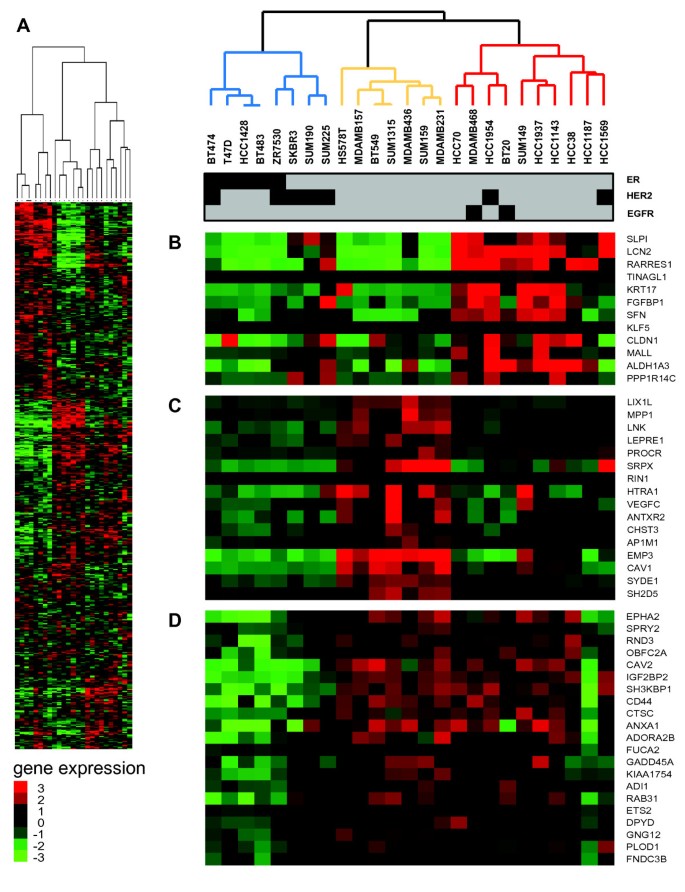
To investigate the molecular heterogeneity of triple-negative BC cell lines and their representativeness of triple-negative breast cancers, we used Illumina HumanWG-6v2.0 to survey the phenotypic and genotypic characteristics of seven ER-negative mesenchymal BC cell lines (Hs578T, BT549, MDAMD157, MDAMD231, MDAMD436 and SUM159) and compared them with 13 ER-negative epithelial-like BC cell lines (BT20, HCC38, HCC70, HCC1143, HCC1937, MDAMD468, SUM149, SKBR3, SUM190, SUM225). Five ER-positive BC cell lines (T47D, BT474, ZR7530, BT483 and HCC1428) were also included in our dataset, to evaluate ER-responsive transcriptional signatures in ER-negative BC cell lines. In addition, two ER-negative/ HER2-positive epithelial BC cell lines were included (i.e. HCC1954 and HCC1569), and were employed as comparators with other triple-negative BC cell lines. Molecular pathological features of all BC cell lines are provided in Additional file 1 Table S1. Unsupervised hierarchical clustering of 5,693 highly variable Ensembl genes separated ER-negative BC cell lines into three groups (Figure 1). One group, designated “Cluster 1” (blue lines, Figure 1) included three ER-negative/HER2-positive BC cell lines, namely SKBR3, SUM190 and SUM225, which clustered with ER-positive/HER2-negative (T47D, HCC1428 and BT483) and ER-positive/HER2-positive (BT474, ZRF7530) cell lines. “Cluster 2” was uniformly composed of ER-negative/HER2-negative BC cell lines (orange lines, Figure 1) and was in complete concordance with the Basal “B” BC cell line subtype described in previous BC cell line studies [16–18]. “Cluster 3” consisted of cell lines (red lines, Figure 1) either having a triple-negative phenotype or showing amplification and higher abundance of HER2 (HCC1954, HCC1569) and EGFR gene amplification (BT20, MDAMB468). Investigating the expression levels of known triple-negative BC-related genes demonstrated subtle differences within these three groups. While “Cluster 3” cell lines expressed genes commonly found in the intrinsic gene list preferentially expressed in basal-like primary BC (e.g. LCN, RARRES1, CLDN1, KRT17; Figure 1B) [20], “Cluster 2” was characterised by a higher abundance of CAV1, a marker for the basal-like phenotype of sporadic and hereditary breast cancers [21], and the lymphangiogenic factor VEGFC, a potential therapeutic target for triple-negative BCs [22] (Figure 1C). Other genes previously associated with triple-negative BCs such as c-MET, CD44 and CAV2[23, 24] exhibited higher expression in both groups in comparison to “Cluster 1” cell lines (Figure 1D).
Subtype-specific gene expression and molecular characteristics of breast cancer cell lines, using the Illumina HumanWG-6v2.0 microarray platform. (A) “Two-way” hierarchical clustering of 25 BC cell lines and 5,693 variably expressed genes segregate into three groups, “Cluster 1, 2 and 3” indicated as blue, orange and red dendrogram branches. (B) Selected cluster commonalities in gene expression for ”Cluster 2”; (C) “Cluster 3”; and (D) both cell lines clusters are shown. Bar below dendrogram indicates phenotypic features of cell lines for ER, HER2 and EGFR.
Representation of ER-negative breast tumour-related gene signatures in BC cell lines
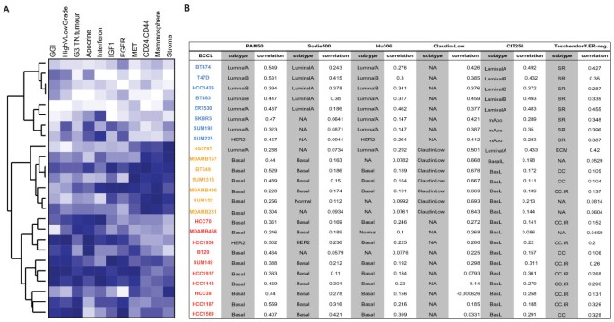
Given that the combined expression pattern of certain genes can uniquely characterise different BC subtypes or can be used as surrogate markers for pathway activation, we selected a compendium of gene signatures representing various features of triple-negative BCs. Firstly, we investigated the representation of eleven gene signatures based on a “relative similarity score” and ranked the BC cell lines accordingly (Additional file 2 Table S2). Overall “Cluster 3” cell lines (red names, Figure 2) showed a good representation of gene signatures identified for “Apocrine”, G3.TN.Tumour” and high-grade breast carcinomas, while “Cluster 2” cell lines exhibited high similarities with the “Stroma”, “Mammosphere” and “CD24.CD44” expression patterns (orange names, Figure 2). The resemblance to the remaining four gene signatures showed a less striking concordance with the gene expression defined clusters, and identified SUM1315, HCC1569, SUM149 and HCC1143 cell lines as the best representatives for the “MET”, “EGFR”, “IGF1” and “Interferon” gene signatures, respectively. Secondly, we examined the 5 different ER-negative BC subgroups associated with a prognostic outcome [25] in the BC cell line transcriptomes based on centroid correlation classification. As shown in Figure 2B, “Cluster 3” cell lines had activation of the “cell cycle and cell proliferation pathway/immune response genes” and the “cell cycle and cell proliferation pathway” groups (Figure 2B), both of which showed association to basal-like BCs in their study [25]. In contrast, “Cluster 1” cell lines displayed highest correlation to ER-negative tumours of the steroid response group, supporting the outcome of our hierarchical clustering in which these cell lines group with the ER-positive cell lines (Figure 1). Lastly, to explore the representation of the molecular basal-like BC subtype within this BC cell line set, we performed nearest centroid classification using the class centroids from Parker et al. [26], Sorlie et al. [27], Hu et al. [28], Prat et al. [8] and Guedj et al., [10] (referred to as PAM50, Sorlie500, Hu306, Claudin.Low and CIT256 respectively, in Figure 2B). As expected given the manner in which the “Claudin.Low” gene signature was originally established [8], “Cluster 2” cell lines (Hs578T, MDAMB157, MDAMB231, MDAMB436, BT549, SUM159 and SUM1315) are representatives of the described “Claudin.Low” subtype. The recently published CIT classification [10] assigned all “Cluster 2” and “Cluster 3” cell lines to the basalL group (Figure 2B) with the exception of Hs578T which was assigned to the “LuminalA” subtype. Interestingly, in “Cluster 1”, ER-negative/HER2-positive cell lines SKBR3, SUM190 and SUM225 were labelled as mApo (‘molecular Apocrine’) groups, in agreement with ER-negative/HER2-positive breast carcinomas in this molecular subtype [9]. PAM50 classification assigned all “Cluster 2 and 3” BC cell lines except Hs578T and HCC1954 to the basal-like BC subtype. Assignments of cell lines into the basal-like subtype with class centroids obtained from Sorlie500 and Hu306 were in good agreement with those obtained with PAM50, however only 2 out 10 cell lines were consistently classified as of luminal A, luminal B or HER2 by all methods. To determine the agreement of these three centroid classifications, we used the free-marginal Kappa statistics of Brennan and Prediger [29] and saw substantial agreement in the classification of the basal-like (Kappa score = 0.78), luminal A (Kappa score = 0.7) and HER2 subtypes (Kappa=0.63), in agreement with the results of previous studies [7, 30].
Representation of gene signatures across BC cell lines. BC cell lines are ranked based on their gene expression clustering. (A) Expression levels of eleven gene signatures previously associated with ER-negative BC. Similarities between gene signature and each BC cell line transcriptome was established as described in Material and Methods, and used for ranking lines. Each coloured square represents the rank (between 1 and 25) of the cell line to the specific gene signature, whereby blue indicates the highest rank (best resemblance to the gene signature), while white being the lowest rank. Gene signatures used were: G3.TN.Tumour [43]; Apocrine.Basal [9]; Interferon [28]; IGF1 [39]; EGFR [41]; c-MET [42]; Mammosphere [40]; CD24.CD44 [44]; HighGradeVLowGrade [47]; GGI [68]; Stroma [45]. (B) Classification of BC cell lines to BC subtypes by nearest centroid correlation based on gene expression signatures: PAM50 [26]; Sorlie500 [27]; Hu306 [69]; Claudin.Low [8], Teschendorff.ER.neg [25]; CIT256 [10]; both subtype assignment and Spearman correlation are presented.
Copy number aberrations and associated gene expression changes in BC cell lines represent those observed in triple-negative BCs
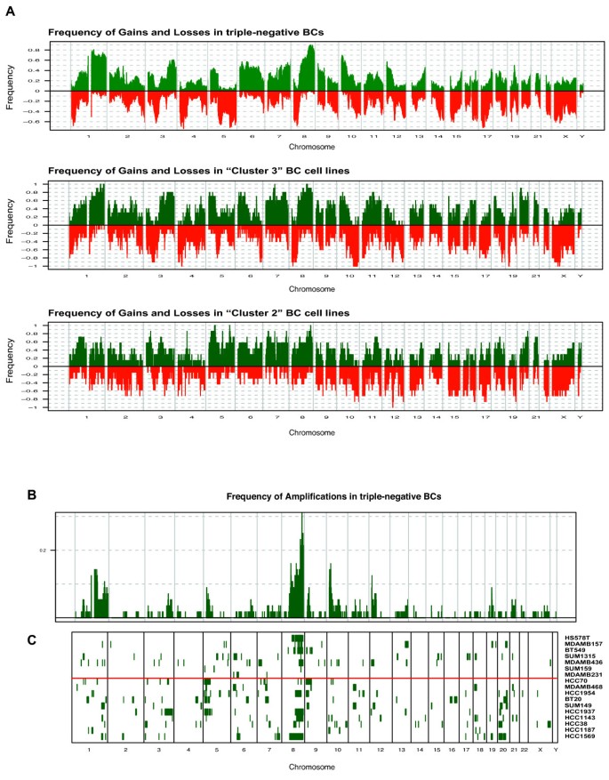
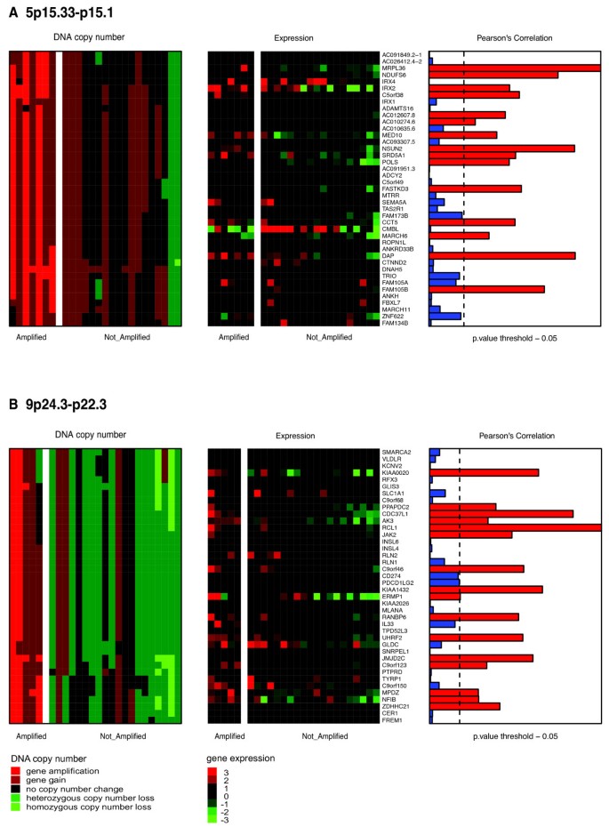
To identify BC cell lines harbouring copy number aberrations specific for triple-negative BC, we performed aCGH using a 32k tiling path array platform and surveyed their genomic changes (individual aCGH-profiles are provided as Additional file 3 Figure S1). By grouping the BC cell lines based on the three expression clusters, “Cluster 2” cell lines displayed significantly less high-level amplifications and deletions compared with the other two clusters (Additional file 4 Figure S2). Next, we retrieved CNAs identified in our previous study on 56 triple-negative BCs [31], analysed on the same genomic platform. Overall the frequency of gains and losses seen in triple-negative BCs was more similar to “Cluster 3” than to “Cluster 2” and “Cluster 1” cell lines (Figure 3A). Recurrent amplification seen in primary triple-negative BCs were recapitulated in at least one BC cell line (Additional file 5 Table S3). The most highly recurrent triple-negative BC-specific amplicons in BC cell lines were on 5p15.33-p15.1 (HCC1143, HCC1937, HCC1954, HCC70, MDAMB468 and BT20), followed by 9p24.3-22.3 and 7q11.1 found in 6 “Cluster 3” cell lines and 4 “Cluster 3” cell lines, respectively (Figure 3B). Given that these regions are also characterised by common polymorphisms, genes such as JAK2 (9p24) [32], NUNS2 (5p15) [33] or LIMK1 (7q11) [34] previously associated with breast cancers, and gained preferentially in basal-like breast cancers [35] might validate that these regions contain genes providing a selective advantage for triple-negative BCs. A comprehensive integration of genes lost or gained in the 2,000 breast cancer study is provided in Additional file 6 Table S4. Amplifications on 3q24-q25.1, 3q25.32-q25.33, 5p14.3-p14.1, 7p11.2 and 9p22.3 were observed only in ”Cluster 3” cell lines, and the 13q32.3-q33.3 amplicon only in “Cluster 2” cell lines, illustrating a good representation of triple-negative BC-specific CNAs in ER-negative cell lines, with some of them having a higher prevalence in one than the other expression defined group. We and others have previously demonstrated that the expression levels of certain genes located in triple-negative BCs specific CNAs is copy number dependent [31, 36]. To investigate if these dependencies are also recapitulated in BC cell lines, we integrated expression data with cbs-smoothed aCGH profiles of each BC cell lines. Using Pearson’s correlation (fdr adjusted P_value <0.05), 4,571 genes showed significantly correlation between their expression and DNA copy number levels (Additional file 6 Table S4). This set encompassed 1,158/2,064 triple-negative BC copy-number dependent genes as determined by Turner [31], and included genes such as transcriptional regulators (n=98), kinases (n=47), phosphatases (n=24) and transmembrane receptors (n=5), as well as biomarkers for diagnosis (n=42), prognosis (n=14), disease progression (n=7) and known drug targets (n=20) (Additional file 5 Table S4). Triple-negative BC–specific amplicons, recurrently amplified in our BC cell lines (e.g. 5p15.33-p15.1 and 9p24.3-22.3) and harbouring genes with DNA copy number-dependent expression levels are shown in Figure 4. Among those were genes, such as JAK2, NSUN2 and NFIB previously shown to have pathogenic roles, but also novel potential drivers e.g. PPAPDC or RANBP6, which could be selectively required for the survival of cells harbouring those amplification (Figure 4).
Genomic alteration of triple-negative BCs and ER-negative BC cell lines. (A) Genomic profiles of 56 triple-negative BCs were obtained from Turner [31]. The proportion of samples for each group (triple-negative BCs, ”Cluster 2 and 3” BC cell lines) is plotted in which each genomic area is gained (green) or lost (red) according to their genomic position. (B) The frequency of triple-negative BCs in which the smoothed log2ratios of each BAC clone above 0.45 is plotted (y-axis) according to its genomic location (x-axis). (C) Using the same criteria for the aCGH data of BC cell lines, amplified BAC clones for each BC cell line (row) are represented as green lines along the genome. The separation between ”Cluster 2 and 3” BC cell lines are indicated with a red line.
BC cell lines recapitulate recurrent triple-negative BC-specific amplicons and their possible drivers. Matched heatmaps of gene expression and aCGH within regions of recurrent amplification in BC cell lines (A) and (B). Cell lines were split into those with amplification (AMP) and those without (NA). Heatmap of aCGH (left) shows amplification in red, deletion in green and no change in black for each corresponding chromosomal location of the respective gene. Expression heatmap (middle) indicates if expression values for the gene are above (red) or below (green) the median value. Barplots (right) illustrate of a Pearson’s correlation analysis between expression and DNA copy number levels. Black dotted lines show adjusted P_value threshold of 0.05.
Epigenetic influence on sub-type specific genes in ER-negative BC cell lines
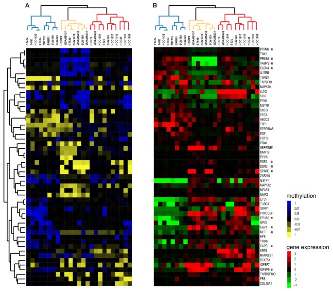
The functional validity of the methylation pattern found on CpG islands in cultured cancer cell lines has been the matter of controversy [37]. To study epigenetic patterns in BC cell lines, we produced genome-wide methylation profiles on Illumina GoldenGate bead arrays. Unsupervised hierarchical clustering using 1,223 CpG probes, corresponding to 707 genes, revealed a different grouping (Additional file 7 Figure S3) as it was observed when their expression profiles were clustered (Figure 1). To validate that the BC cell line specific methylation pattern can be also found in triple-negative BC, we retrieved methylation data of 189 fresh-frozen BCs performed on the same microarray [38]. Due to the lack of HER2 status information in their study, 43 basal-like BCs were used as surrogates for triple-negative tumours and 165 CpG islands with variable methylation levels were observed. Of those 165, 128 CpG probes showed also changes in their methylation status in our BC cell lines (Additional file 6 Table S4). Investigating the methylation state of these CpG islands in each BC cell line individually, demonstrated an overall good representation of ≥70% of hypo- and hyper- basal-like BC-specific methylation events specifically in ER-negative BC cell lines (Additional file 8 Figure S4). Given that BC subtype specific expression has been suggested to be under epigenetic influence [38], we surveyed the methylation effect on gene expression in BC cell lines. Integration of methylation and gene expression data resulted in 1,129 CpG gene pairs (corresponding to 652 genes) and identified an inverse correlation between methylation and gene expression levels for 93 pairs (correlation < 0.55; adjusted P_value 0.05). Performing bootstrap analysis by randomly sampling the BC cell lines, we showed that the number of significant association between gene expression and methylation was more than 90 fold higher than expected. Using these 93 CpG gene pairs in a multiclass SAM analysis revealed 73 with specific methylation patterns over the three expression clusters, particularly distinguishing “Cluster 2” cell lines from the others (Figure 5). As described previously, “Cluster 2” cell lines exhibited among others, expression patterns similar to the “Claudin.low” gene signature [8]. Fifteen genes of the “Claudin.low” gene signature had CpG sites with varying methylation pattern over these BC cell lines which was significantly higher than expected by chance (hypergeometric testing P_values < 0.001), whereby genes downregulated in “Claudin.low” cancers according to “Claudin.low” signature were methylated and vice-versa, such as “Claudin.low” signature genes such as PRSS8, CLDN4 and VAMP8 were downregulated in “Cluster 2” and their CpG islands were methylated, while genes like SPARC or DDR2 had unmethylated CpG islands and showed higher abundance in these BC cell lines. By comparing those with epigenetic-regulated genes in breast cancers [38], we identified 12 genes with the same concordant pattern (asterisks, Figure 5). Taken together, our analyses demonstrate that BC cell lines retain methylation–dependent gene expression patterns observed in basal-like BCs, and strengthen an epigenetic influence on some BC phenotypes that are retained in their equivalent model systems.
Matched heatmaps of gene expression and methylation profiling. 51 genes whose epigenetic-regulated expression varied between the three expression clusters. (A) Methylation heatmap (left) illustrates unmethylated probes in yellow, methylated in blue and partially methylated in black. (B) Expression heatmap displays the mean centred expression level for the corresponding gene. BC cell lines are ordered based on the gene expression clustering, and groups are illustrated as blue, orange and red dendrogram branches for “Cluster 1, 2 and 3” BC cell lines, respectively. Genes of the Claudin-low signature are indicated with asterisks.
Discussion
Triple-negative BCs represent a heterogeneous group with diverse deregulation of biological pathways. Here, we extended the molecular characterisation of ER-negative BC cell lines based on their genetic, epigenetic and transcriptional profiles, and correlated these with a comprehensive compendium of gene signatures reflecting different features of ER-negative BCs [8, 9, 28, 39–45]. Initial cluster analysis of BC cell lines’ expression profiles resulted in three groups, two clusters encompassing purely ER-negative BC cell lines (“Cluster 3” and “Cluster 2”), while one consisted of three ER-negative and all ER-positive BC cell lines. The first two cell line clusters were in good agreement with recent BC cell line studies [6, 11, 15–18, 31, 46]. While “Cluster 2” encompassed cell lines that were all represented in the Basal “B” cluster of Neve et al.[18] and were assigned to the triple-negative mesenchymal phenotype by Lehmann et al. [11], most of the “Cluster 3” cell lines were part of Neve’s Basal “A” cluster [18] and part of the basal-like subtype according to Lehmann et al., [11]. Our “Cluster 3” cell lines exhibited expression patterns found in transcriptional profiles of microdissected grade 3 triple-negative breast tumours [43] as well as grade 3 versus grade 1 breast carcinomas [9, 47]. HCC1143, an ER-negative/HER2-negative cell line, was the top in vitro representative “Cluster 3” cell line for the triple-negative phenotype of microdissected grade 3 triple-negative breast tumours [43]. The transcriptional profile of HCC1143 also seemed very suitable in modelling the Interferon, IGF1 and MET signalling pathways. BC cell lines with expression patterns most closely associated with the Apocrine.Basal subtype [9] were not defined to one or the other cluster and HCC1954, an ER-negative/HER2-positive cell line of “Cluster 3” displayed the highest representation. These BCs were originally defined on the basis of their androgen receptor level and many of them harboured ERBB2 amplifications [9]. This is in agreement with our findings, whereby using a recently published BC classifier, named CIT, three ER-negative/HER2-positive cell lines SKBR3, SUM190 and SUM225 were classified to the mApo (molecular Apocrine) breast cancer subtype [10]. In a study, MDAMB453, SUM185, CAL148 and MFM223 showed expression patterns associated with androgen receptor signalling and were more sensitive to androgen receptor antagonist bicalutamide and an Hsp90 inhibitor [11]. While none of those cell lines were part of our study, BT549 and HCC1937, BC cell lines used in our study and good representatives of the Apocrine.Basal subtype showed high sensitivity to Hsp90 inhibitors in Lehmann’s work [11]. The Claudin-low subtype has been described as BC entity [8, 48], which is enriched for ER-negative invasive ductal carcinomas, while displaying low levels of luminal differentiation markers and activation of pathways involved in epithelial-to-mesenchymal transition, stem cell-like features and the immune response [8]. Integration of gene expression with methylation data over BC cell lines revealed a group of CpG islands corresponding to genes within the Claudin-low signature, showing an inverse correlation between their methylation and the genes expression in BC cell lines and BCs [38]. Our findings are in agreement with those from a recent report that led to the identification of a set of genes whose expression was epigenetically regulated and when used as a gene signature identified mesenchymal features in Claudin-Low breast tumours [19]. Furthermore, they postulated that a deviant methylation might reflect cell lineage commitment in agreement with our hypothesis of a contribution of an epigenetic regulation to the Claudin-Low subtype. Aberrant DNA methylation events have initially been thought to accumulate in a random fashion within cells in pre-malignant tissues, however, lately it has also been shown that de novo methylation has a predictable pattern, creating plasticity followed by commitment to alternative cell lineages [49]. Holm and colleagues proposed that BC subtypes might be driven by different epigenetic events and could reflect their different cellular origins [38]. Nevertheless, an alternative hypothesis might also be that the methylation patterns are a result from mutations in genes controlling the epigenetic landscape in breast cancer [50]; thus further investigation is warranted to determine whether these distinctive methylation patterns are results of genetic aberration in epigenetic regulator genes and/or contribute to delineation of the differentiation hierarchy of Claudin-Low and other BC subtypes.
We and others have recently shown that basal-like BCs are most likely derived from luminal progenitor cells [51, 52]. Identifying in vitro models would enhance our understanding of these cell populations. Interestingly, our cluster and gene signature analysis revealed ER-responsive features for SKBR3, SUM190 and SUM225, three ER-negative/HER2-positive cell lines. SKBR3 cells are well known to have luminal BC characteristics [53]. In contrast, the classification of SUM190 and SUM225 is controversial. While some BC cell line studies assigned them to basal-like cell lines [15, 18], others supported our finding of SUM190 within the ER-positive cluster [16]. SUM225, although not included in this study, was classified as of luminal phenotype in other studies [54]. Common to both is the expression of luminal cytokeratins 8, 18 and 19 [55] as well as genes found in luminal progenitor cell population (data not shown) [51], more consistent with a luminal classification. Although SUM225 was found to highly express ALDH1, a marker for the so-called BC stem cells [56], further investigations are necessary to ascertain whether SUM190 and SUM225 represent appropriate in vitro models for luminal intermediate progenitor populations.
High-level amplifications are less likely to represent random aberrations and often encompass genes driving the development or maintenance of tumour growth. Three-quarters of triple-negative BCs harbour at least one amplicon [31], however, their recurrence rates are lower than those of high-level CNAs found in ER-positive/ HER2-negative and HER2-positive BC subtypes (e.g. ERBB2-amplicon in HER2, and CCND1 and FGFR1 in luminal breast tumours [57]). Here, we demonstrated that triple-negative BC-specific amplicons are recapitulated in ER-negative BC cell lines and that some of them are associated with higher frequencies either to ”Cluster 2” or “Cluster 3” expression clusters. For example, the region on 5p15.33-p15.1 was found to be recurrently amplified in 5/56 and 10/28 triple-negative BCs [31, 36], was present in six ”Cluster 3” but only in one ”Cluster 2” cell lines. Notably, these genomic sites map to regions of common germline copy number polymorphism and the functional consequences of their increased DNA levels require further validation. Nevertheless, several genes located within these amplified regions were found gained with a higher frequency in basal-like BCs in a recent study investigating 2,000 breast tumours [35] and expression levels significantly correlated with their DNA copy number in triple-negative BC cell lines and tumours for several of these genes [31]. A recent study investigated genes on 5p15.33-p15.1 in more detail and showed that silencing of the overexpressed and amplified NUNS2, a MYC target gene, reduced cell number in some BC cell lines [33]. NUNS 2 expression has been found significantly increased in malignant tissues whereas it could only be found in testis in normal tissues, furthermore its role in stabilising the mitotic spindle and phosphorylation by Aurora-B make it an interesting target for cancer diagnostics and molecular therapeutics.
Conclusion
Taken together, transcriptional, genomic and epigenetic profiles of 25 BC cell lines, enriched for those representing triple-negative features, help to define cell lines that most closely capture individual examples of the heterogeneous characteristics within triple-negative BCs. By cross-referencing different high-resolution datasets, we provide useful resources to further study transcriptional, as well as genetic and epigenetic modulation and inform the best selection of available in vitro models for the identification and validation of potential novel therapeutic targets relevant to triple-negative BCs.
Methods
BC cell lines
BT20, BT474, BT483, BT549, Hs578T, MDAMB157, MDAMB231, MDAMB436, MDAMB468, T47D, SKBR3, ZR75-30, HCC1937, HCC70, HCC1428, HCC1143, HCC38, HCC1187, HCC1569, HCC1954 were obtained from ATCC (Manassas, VA, USA). SUM159, SUM149, SUM1315, SUM225, SUM190 were purchased from Asterand plc (Detroit, MI, USA) (Additional file 1 Table S1). All lines were grown according to the supplier’s recommendation and authenticated by means of Short Tandem Repeat (STR) analysis (PowerPlex® 1.2 System, Promega, WI, US) as previously described [58]. STR profiles were matched to the German Collection of Microorganisms and Cell Cultures (DSMZ)–database (http://www.dsmz.com). BC cell lines were stratified into mesenchymal and epithelial-like morphological groups based on previous studies [11, 16, 18].
RNA and DNA isolation
Cells were grown to ~70% confluence before harvesting nucleic acids. DNA was prepared using the Qiagen DNeasy tissue kit (Qiagen, Valencia, CA) and RNA was isolated using Trizol (Invitrogen, Carlsbad, CA) according to the manufacturers’ protocol. DNA concentration was measured with Picogreen (Invitrogen, Paisley, UK). Integrity of RNA was quantified using the Agilent 2100 Bioanalyser with RNA Nano LabChip Kits (Agilent Biosystems, Foster City, CA).
Microarray analyses
Analyses of microarray data were performed in the R environment 2.12.0 (http://www.r-project.org/) making use of several Bioconductor packages (http://www.bioconductor.org/). All Microarray probes and external gene signatures were mapped to the Ensembl 55 (human genome build 37) to ensure uniform annotation. Microarray data have been deposited in Array Express (E-TABM-928; http://www.ebi.ac.uk/arrayexpress/). A Sweave document describing the statistical analysis is provided as Supplemental Methods (Addition file 9).
Gene expression profiling
Using the Illumina Totalprep RNA amplification kit (Ambion, UK), 200ng total BC cell line RNA was amplified and hybridised to Illumina HumanWG-6v2.0 arrays gene expression bead-chips at Genizon BioSiences Inc (Quebec, CA). Raw data obtained from Illumina BeadStudio (Illumina, San Diego, CA) were preprocessed using the “lumi” -Bioconductor package [59]. Microarray probes absent in more than 80% of samples based on an Illumina BeadStudio detection P_value >0.01 were removed from further analysis. For unsupervised hierarchical clustering of gene expression, 5,693 unique Ensembl genes with a median absolute deviation (MAD) of ≥0.4 across all BC cell lines were selected. Ward clustering was applied to genes and arrays after median centring using Pearson’s correlation as a distance measurement and 10,000 bootstrap iterations were performed to assess the significance of the observed the stability of the clusters using the pvclust package for R [60]. Resulting clusters were visualised with Java TreeView [61]. Two strategies were applied for gene expression signature analysis: (1) When centroids for specific classes (e.g. BC subtypes or groups of ER-negative breast tumours [25]) were publicly available, assignment of BC cell lines to these classes was based on their highest Spearman rank correlation. Classification included class centroids defined by Sorlie[27], Hu [28], Parker [26], Prat [8], CIT256 [10] and Teschendorff [25]. (2) To monitor specific ER-related features, 11 gene signatures were retrieved from publication (see Additional file 3 Table S2 for a detailed description). For the “G3.TN.Tumour” signature, we used our previously published expression data of microdissected breast tumours [43]. Significance Analysis of Microarrays (SAM) [62] with 1,000 permutations and 0% fdr was used to identify significant genes for triple-negative BCs, using a two-class comparisons between tumours belonging to the triple-negative subtype and all other subtypes. For each BC cell line, a weighted mean expression of genes present in the respective signature was determined, and cell lines were ranked based on their concordance.
Array-based comparative genomic hybridisation (aCGH)
Labelling, hybridisation, image and initial data analysis of the 32k BAC tiling path aCGH platform, produced at the Breakthrough Breast Cancer Research Centre, London, UK [63] was carried out as previously described [43]. Breakpoint analysis was performed using the circular binary segmentation (cbs) algorithm [64] and rescaled such that the genome MAD was the same in each sample. Only segments of ≥ 3 BAC clones were used in further analyses. Thresholds for cbs-smoothed data were estimated as described previously [65]. Briefly, cbs-smoothed aCGH Log2 values <−0.08 were classified as losses, >0.08 but ≤0.45 were categorised as gains, and >0.45 were referred to as high-level gains/ amplifications. To determine genomic instability, the fraction of amplified, deleted or total BACs over the whole data set was calculated and presented as a proportion. Gene expression values were compared with median cbs-smoothed aCGH data for all BACs encompassing the genomic position using Pearson’s correlation adjusted for multiple testing [66]. Matched heatmaps between gene expression and genomic data were created as described in [31] showing the minus log10 Pearson’s P_value of each gene-aCGH pair correlation. The raw and cbs-smoothed aCGH data are deposited at http://rock.icr.ac.uk/collaborations/GrigoriadisA/.
Methylation array analysis
Hybridisation and image analysis of the Illumina GoldenGate methylation beadarrays were performed at the Genome Centre (Barts and the London School of Medicine and Dentistry, London, UK). Methylation profiles of the BC cell lines, obtained through the BeadStudio Methylation Module (Illumina, San Diego, CA), was normalised by dichotomising the un- /methylated CpG islands separately before equalising their median according to the “methylumi” package (http://www.bioconductor.org/). CpG sites located on the X chromosomes were removed, as well as constitutively un-/ methylated probes, resulting in 1,223 CpG sites (data are available at http://rock.icr.ac.uk/collaborations/GrigoriadisA/). The methylation state of CpG islands given as a ß-value [67] was stratified into three categories: ß-values ≤ 0.25, ≥ 0.75 and between ≥ 0.25 and ≤ 0.75; and interpreted as un-/, methylated and partially methylated CpG sites, respectively. These cut-offs are slightly more stringent than Holm et al. has used them for the analysis of breast carcinomas using the same methylation array platform [38] to increase the chances of true-positive events. Initial analysis revealed a similar methylation frequency in all BC cell lines, determined as the fraction of methylated CpG sites, affecting on average 31% of all CpG islands. Using a total of 10,000 permutations to obtain reasonable estimates of dependencies, sample labels were permuted and correlation analyses between gene expression and methylation values were carried out on the resampled data set.
Abbreviations
- BC:
-
Breast cancer
- ER:
-
Estrogen-receptor
- PR:
-
Progesterone receptor
- CNAs:
-
Copy Number Aberrations
- MAD:
-
Median Absolute Deviation
- SAM:
-
Significance Analysis of Microarrays
- ACGH:
-
Array-based comparative genomic hybridisation
- CBS:
-
Circular binary segmentation
- BAC:
-
Bacterial Artificial Chromosome
- AMP:
-
Amplification.
References
Rakha EA, Ellis IO: Triple-negative/basal-like breast cancer: review. Pathology. 2009, 41 (1): 40-47. 10.1080/00313020802563510.
Reis-Filho JS, Tutt AN: Triple negative tumours: a critical review. Histopathology. 2008, 52 (1): 108-118.
Voduc D, Nielsen TO: Basal and triple-negative breast cancers: impact on clinical decision-making and novel therapeutic options. Clin Breast Cancer. 2008, 8 (Suppl 4): S171-178.
Foulkes WD, Smith IE, Reis-Filho JS: Triple-negative breast cancer. N Engl J Med. 2010, 363 (20): 1938-1948. 10.1056/NEJMra1001389.
Cheang MC, Voduc D, Bajdik C, Leung S, McKinney S, Chia SK, Perou CM, Nielsen TO: Basal-like breast cancer defined by five biomarkers has superior prognostic value than triple-negative phenotype. Clin Cancer Res. 2008, 14 (5): 1368-1376. 10.1158/1078-0432.CCR-07-1658.
Geyer FC, Weigelt B, Natrajan R, Lambros MB, de Biase D, Vatcheva R, Savage K, Mackay A, Ashworth A, Reis-Filho JS: Molecular analysis reveals a genetic basis for the phenotypic diversity of metaplastic breast carcinomas. J Pathol. 2010, 220 (5): 562-573. 10.1002/path.2675.
Weigelt B, Mackay A, A'Hern R, Natrajan R, Tan DS, Dowsett M, Ashworth A, Reis-Filho JS: Breast cancer molecular profiling with single sample predictors: a retrospective analysis. Lancet Oncol. 2010, 11 (4): 339-349. 10.1016/S1470-2045(10)70008-5.
Prat A, Parker JS, Karginova O, Fan C, Livasy C, Herschkowitz JI, He X, Perou CM: Phenotypic and molecular characterization of the claudin-low intrinsic subtype of breast cancer. Breast Cancer Res. 2010, 12 (5): R68-10.1186/bcr2635.
Farmer P, Bonnefoi H, Becette V, Tubiana-Hulin M, Fumoleau P, Larsimont D, Macgrogan G, Bergh J, Cameron D, Goldstein D, et al: Identification of molecular apocrine breast tumours by microarray analysis. Oncogene. 2005, 24 (29): 4660-4671. 10.1038/sj.onc.1208561.
Guedj M, Marisa L, de Reynies A, Orsetti B, Schiappa R, Bibeau F, MacGrogan G, Lerebours F, Finetti P, Longy M, et al: A refined molecular taxonomy of breast cancer. Oncogene. 2012, 31 (9): 1196-1206. 10.1038/onc.2011.301.
Lehmann BD, Bauer JA, Chen X, Sanders ME, Chakravarthy AB, Shyr Y, Pietenpol JA: Identification of human triple-negative breast cancer subtypes and preclinical models for selection of targeted therapies. J Clin Invest. 2011, 121 (7): 2750-2767. 10.1172/JCI45014.
Sotiriou C, Pusztai L: Gene-expression signatures in breast cancer. N Engl J Med. 2009, 360 (8): 790-800. 10.1056/NEJMra0801289.
Lacroix M, Leclercq G: Relevance of breast cancer cell lines as models for breast tumours: an update. Breast Cancer Res Treat. 2004, 83 (3): 249-289. 10.1023/B:BREA.0000014042.54925.cc.
Barretina J, Caponigro G, Stransky N, Venkatesan K, Margolin AA, Kim S, Wilson CJ, Lehar J, Kryukov GV, Sonkin D, et al: The Cancer Cell Line Encyclopedia enables predictive modelling of anticancer drug sensitivity. Nature. 2012, 483 (7391): 603-607. 10.1038/nature11003.
Chin K, DeVries S, Fridlyand J, Spellman PT, Roydasgupta R, Kuo WL, Lapuk A, Neve RM, Qian Z, Ryder T, et al: Genomic and transcriptional aberrations linked to breast cancer pathophysiologies. Cancer Cell. 2006, 10 (6): 529-541. 10.1016/j.ccr.2006.10.009.
Kao J, Salari K, Bocanegra M, Choi YL, Girard L, Gandhi J, Kwei KA, Hernandez-Boussard T, Wang P, Gazdar AF, et al: Molecular profiling of breast cancer cell lines defines relevant tumor models and provides a resource for cancer gene discovery. PLoS One. 2009, 4 (7): e6146-10.1371/journal.pone.0006146.
Mackay A, Tamber N, Fenwick K, Iravani M, Grigoriadis A, Dexter T, Lord CJ, Reis-Filho JS, Ashworth A: A high-resolution integrated analysis of genetic and expression profiles of breast cancer cell lines. Breast Cancer Res Treat. 2009, 118 (3): 481-498. 10.1007/s10549-008-0296-7.
Neve RM, Chin K, Fridlyand J, Yeh J, Baehner FL, Fevr T, Clark L, Bayani N, Coppe JP, Tong F, et al: A collection of breast cancer cell lines for the study of functionally distinct cancer subtypes. Cancer Cell. 2006, 10 (6): 515-527. 10.1016/j.ccr.2006.10.008.
Sproul D, Nestor C, Culley J, Dickson JH, Dixon JM, Harrison DJ, Meehan RR, Sims AH, Ramsahoye BH: Transcriptionally repressed genes become aberrantly methylated and distinguish tumors of different lineages in breast cancer. Proc Natl Acad Sci USA. 2011, 108 (11): 4364-4369. 10.1073/pnas.1013224108.
Perou CM, Sorlie T, Eisen MB, van de Rijn M, Jeffrey SS, Rees CA, Pollack JR, Ross DT, Johnsen H, Akslen LA, et al: Molecular portraits of human breast tumours. Nature. 2000, 406 (6797): 747-752. 10.1038/35021093.
Savage K, Lambros MB, Robertson D, Jones RL, Jones C, Mackay A, James M, Hornick JL, Pereira EM, Milanezi F, et al: Caveolin 1 is overexpressed and amplified in a subset of basal-like and metaplastic breast carcinomas: a morphologic, ultrastructural, immunohistochemical, and in situ hybridization analysis. Clin Cancer Res. 2007, 13 (1): 90-101. 10.1158/1078-0432.CCR-06-1371.
Linderholm BK, Hellborg H, Johansson U, Elmberger G, Skoog L, Lehtio J, Lewensohn R: Significantly higher levels of vascular endothelial growth factor (VEGF) and shorter survival times for patients with primary operable triple-negative breast cancer. Ann Oncol. 2009, 20 (10): 1639-1646. 10.1093/annonc/mdp062.
Klingbeil P, Natrajan R, Everitt G, Vatcheva R, Marchio C, Palacios J, Buerger H, Reis-Filho JS, Isacke CM: CD44 is overexpressed in basal-like breast cancers but is not a driver of 11p13 amplification. Breast Cancer Res Treat. 2010, 120 (1): 95-109. 10.1007/s10549-009-0380-7.
Savage K, Leung S, Todd SK, Brown LA, Jones RL, Robertson D, James M, Parry S, Rodrigues Pinilla SM, Huntsman D, et al: Distribution and significance of caveolin 2 expression in normal breast and invasive breast cancer: an immunofluorescence and immunohistochemical analysis. Breast Cancer Res Treat. 2008, 110 (2): 245-256. 10.1007/s10549-007-9718-1.
Teschendorff AE, Miremadi A, Pinder SE, Ellis IO, Caldas C: An immune response gene expression module identifies a good prognosis subtype in estrogen receptor negative breast cancer. Genome Biol. 2007, 8 (8): R157-10.1186/gb-2007-8-8-r157.
Parker JS, Mullins M, Cheang MC, Leung S, Voduc D, Vickery T, Davies S, Fauron C, He X, Hu Z, et al: Supervised risk predictor of breast cancer based on intrinsic subtypes. J Clin Oncol. 2009, 27 (8): 1160-1167. 10.1200/JCO.2008.18.1370.
Sorlie T, Tibshirani R, Parker J, Hastie T, Marron JS, Nobel A, Deng S, Johnsen H, Pesich R, Geisler S, et al: Repeated observation of breast tumor subtypes in independent gene expression data sets. Proc Natl Acad Sci USA. 2003, 100 (14): 8418-8423. 10.1073/pnas.0932692100.
Hu Z, Fan C, Oh DS, Marron JS, He X, Qaqish BF, Livasy C, Carey LA, Reynolds E, Dressler L, et al: The molecular portraits of breast tumors are conserved across microarray platforms. BMC Genomics. 2006, 7: 96-10.1186/1471-2164-7-96.
Brennan RL, Prediger DJ: Coeffecient kappa: some uses, misuses, and alternatives. Educ Psychol Meas. 1981, 41: 687-699. 10.1177/001316448104100307.
Haibe-Kains B, Desmedt C, Loi S, Culhane AC, Bontempi G, Quackenbush J, Sotiriou C: A three-gene model to robustly identify breast cancer molecular subtypes. J Natl Cancer Inst. 2012, 104 (4): 311-325. 10.1093/jnci/djr545.
Turner N, Lambros MB, Horlings HM, Pearson A, Sharpe R, Natrajan R, Geyer FC, van Kouwenhove M, Kreike B, Mackay A, et al: Integrative molecular profiling of triple negative breast cancers identifies amplicon drivers and potential therapeutic targets. Oncogene. 2010, 29 (14): 2013-2023. 10.1038/onc.2009.489.
Ding L, Ellis MJ, Li S, Larson DE, Chen K, Wallis JW, Harris CC, McLellan MD, Fulton RS, Fulton LL, et al: Genome remodelling in a basal-like breast cancer metastasis and xenograft. Nature. 2010, 464 (7291): 999-1005. 10.1038/nature08989.
Frye M, Dragoni I, Chin SF, Spiteri I, Kurowski A, Provenzano E, Green A, Ellis IO, Grimmer D, Teschendorff A, et al: Genomic gain of 5p15 leads to over-expression of Misu (NSUN2) in breast cancer. Cancer Lett. 2010, 289 (1): 71-80. 10.1016/j.canlet.2009.08.004.
Wang W, Mouneimne G, Sidani M, Wyckoff J, Chen X, Makris A, Goswami S, Bresnick AR, Condeelis JS: The activity status of cofilin is directly related to invasion, intravasation, and metastasis of mammary tumors. J Cell Biol. 2006, 173 (3): 395-404. 10.1083/jcb.200510115.
Curtis C, Shah SP, Chin SF, Turashvili G, Rueda OM, Dunning MJ, Speed D, Lynch AG, Samarajiwa S, Yuan Y, et al: The genomic and transcriptomic architecture of 2,000 breast tumours reveals novel subgroups. Nature. 2012, 486 (7403): 346-352.
Han W, Jung EM, Cho J, Lee JW, Hwang KT, Yang SJ, Kang JJ, Bae JY, Jeon YK, Park IA, et al: DNA copy number alterations and expression of relevant genes in triple-negative breast cancer. Gene Chromosome Canc. 2008, 47 (6): 490-499. 10.1002/gcc.20550.
Hinshelwood RA, Clark SJ: Breast cancer epigenetics: normal human mammary epithelial cells as a model system. J Mol Med. 2008, 86 (12): 1315-1328. 10.1007/s00109-008-0386-3.
Holm K, Hegardt C, Staaf J, Vallon-Christersson J, Jonsson G, Olsson H, Borg A, Ringner M: Molecular subtypes of breast cancer are associated with characteristic DNA methylation patterns. Breast Cancer Res. 2010, 12 (3): R36-10.1186/bcr2590.
Creighton CJ, Casa A, Lazard Z, Huang S, Tsimelzon A, Hilsenbeck SG, Osborne CK, Lee AV: Insulin-like growth factor-I activates gene transcription programs strongly associated with poor breast cancer prognosis. J Clin Oncol. 2008, 26 (25): 4078-4085. 10.1200/JCO.2007.13.4429.
Creighton CJ, Li X, Landis M, Dixon JM, Neumeister VM, Sjolund A, Rimm DL, Wong H, Rodriguez A, Herschkowitz JI, et al: Residual breast cancers after conventional therapy display mesenchymal as well as tumor-initiating features. Proc Natl Acad Sci USA. 2009, 106 (33): 13820-13825. 10.1073/pnas.0905718106.
Hoadley KA, Weigman VJ, Fan C, Sawyer LR, He X, Troester MA, Sartor CI, Rieger-House T, Bernard PS, Carey LA, et al: EGFR associated expression profiles vary with breast tumor subtype. BMC Genomics. 2007, 8: 258-10.1186/1471-2164-8-258.
Kaposi-Novak P, Lee JS, Gomez-Quiroz L, Coulouarn C, Factor VM, Thorgeirsson SS: Met-regulated expression signature defines a subset of human hepatocellular carcinomas with poor prognosis and aggressive phenotype. J Clin Invest. 2006, 116 (6): 1582-1595. 10.1172/JCI27236.
Natrajan R, Lambros MB, Rodriguez-Pinilla SM, Moreno-Bueno G, Tan DS, Marchio C, Vatcheva R, Rayter S, Mahler-Araujo B, Fulford LG, et al: Tiling path genomic profiling of grade 3 invasive ductal breast cancers. Clin Cancer Res. 2009, 15 (8): 2711-2722. 10.1158/1078-0432.CCR-08-1878.
Shipitsin M, Campbell LL, Argani P, Weremowicz S, Bloushtain-Qimron N, Yao J, Nikolskaya T, Serebryiskaya T, Beroukhim R, Hu M, et al: Molecular definition of breast tumor heterogeneity. Cancer Cell. 2007, 11 (3): 259-273. 10.1016/j.ccr.2007.01.013.
West RB, Nuyten DS, Subramanian S, Nielsen TO, Corless CL, Rubin BP, Montgomery K, Zhu S, Patel R, Hernandez-Boussard T, et al: Determination of stromal signatures in breast carcinoma. PLoS Biol. 2005, 3 (6): e187-10.1371/journal.pbio.0030187.
Natrajan R, Weigelt B, Mackay A, Geyer FC, Grigoriadis A, Tan DS, Jones C, Lord CJ, Vatcheva R, Rodriguez-Pinilla SM, et al: An integrative genomic and transcriptomic analysis reveals molecular pathways and networks regulated by copy number aberrations in basal-like, HER2 and luminal cancers. Breast Cancer Res Treat. 2010, 121 (3): 575-589. 10.1007/s10549-009-0501-3.
Cordenonsi M, Zanconato F, Azzolin L, Forcato M, Rosato A, Frasson C, Inui M, Montagner M, Parenti AR, Poletti A, et al: The Hippo transducer TAZ confers cancer stem cell-related traits on breast cancer cells. Cell. 2011, 147 (4): 759-772. 10.1016/j.cell.2011.09.048.
Herschkowitz JI, Simin K, Weigman VJ, Mikaelian I, Usary J, Hu Z, Rasmussen KE, Jones LP, Assefnia S, Chandrasekharan S, et al: Identification of conserved gene expression features between murine mammary carcinoma models and human breast tumors. Genome Biol. 2007, 8 (5): R76-10.1186/gb-2007-8-5-r76.
Reynolds PA, Sigaroudinia M, Zardo G, Wilson MB, Benton GM, Miller CJ, Hong C, Fridlyand J, Costello JF, Tlsty TD: Tumor suppressor p16INK4A regulates polycomb-mediated DNA hypermethylation in human mammary epithelial cells. J Biol Chem. 2006, 281 (34): 24790-24802. 10.1074/jbc.M604175200.
Garber K: Genetic discoveries propagate new epigenetic drugs. J Natl Cancer Inst. 2012, 104 (3): 174-176. 10.1093/jnci/djs021.
Lim E, Vaillant F, Wu D, Forrest NC, Pal B, Hart AH, Asselin-Labat ML, Gyorki DE, Ward T, Partanen A, et al: Aberrant luminal progenitors as the candidate target population for basal tumor development in BRCA1 mutation carriers. Nat Med. 2009, 15 (8): 907-913. 10.1038/nm.2000.
Molyneux G, Geyer FC, Magnay FA, McCarthy A, Kendrick H, Natrajan R, Mackay A, Grigoriadis A, Tutt A, Ashworth A, et al: BRCA1 basal-like breast cancers originate from luminal epithelial progenitors and not from basal stem cells. Cell Stem Cell. 2010, 7 (3): 403-417. 10.1016/j.stem.2010.07.010.
Charafe-Jauffret E, Ginestier C, Monville F, Finetti P, Adelaide J, Cervera N, Fekairi S, Xerri L, Jacquemier J, Birnbaum D, et al: Gene expression profiling of breast cell lines identifies potential new basal markers. Oncogene. 2006, 25 (15): 2273-2284. 10.1038/sj.onc.1209254.
Behbod F, Kittrell FS, LaMarca H, Edwards D, Kerbawy S, Heestand JC, Young E, Mukhopadhyay P, Yeh HW, Allred DC, et al: An intraductal human-in-mouse transplantation model mimics the subtypes of ductal carcinoma in situ. Breast Cancer Res. 2009, 11 (5): R66-10.1186/bcr2358.
Forozan F, Veldman R, Ammerman CA, Parsa NZ, Kallioniemi A, Kallioniemi OP, Ethier SP: Molecular cytogenetic analysis of 11 new breast cancer cell lines. Br J Cancer. 1999, 81 (8): 1328-1334. 10.1038/sj.bjc.6695007.
Charafe-Jauffret E, Ginestier C, Birnbaum D: Breast cancer stem cells: tools and models to rely on. BMC Cancer. 2009, 9: 202-10.1186/1471-2407-9-202.
Reis-Filho JS, Savage K, Lambros MB, James M, Steele D, Jones RL, Dowsett M: Cyclin D1 protein overexpression and CCND1 amplification in breast carcinomas: an immunohistochemical and chromogenic in situ hybridisation analysis. Mod Pathol. 2006, 19 (7): 999-1009. 10.1038/modpathol.3800621.
Brough R, Frankum JR, Sims D, Mackay A, Mendes-Pereira AM, Bajrami I, Costa-Cabral S, Rafiq R, Ahmad AS, Cerone MA, et al: Functional viability profiles of breast cancer. Cancer Discov. 2011, 1 (3): 260-273. 10.1158/2159-8290.CD-11-0107.
Du P, Kibbe WA, Lin SM: lumi: a pipeline for processing Illumina microarray. Bioinformatics. 2008, 24 (13): 1547-1548. 10.1093/bioinformatics/btn224.
Suzuki R, Shimodaira H: Pvclust: an R package for assessing the uncertainty in hierarchical clustering. Bioinformatics. 2006, 22 (12): 1540-1542. 10.1093/bioinformatics/btl117.
Saldanha AJ: Java Treeview–extensible visualization of microarray data. Bioinformatics. 2004, 20 (17): 3246-3248. 10.1093/bioinformatics/bth349.
Tusher VG, Tibshirani R, Chu G: Significance analysis of microarrays applied to the ionizing radiation response. Proc Natl Acad Sci USA. 2001, 98 (9): 5116-5121. 10.1073/pnas.091062498.
Lacroix-Triki M, Suarez PH, MacKay A, Lambros MB, Natrajan R, Savage K, Geyer FC, Weigelt B, Ashworth A, Reis-Filho JS: Mucinous carcinoma of the breast is genomically distinct from invasive ductal carcinomas of no special type. J Pathol. 2010, 222 (3): 282-298. 10.1002/path.2763.
Lai WR, Johnson MD, Kucherlapati R, Park PJ: Comparative analysis of algorithms for identifying amplifications and deletions in array CGH data. Bioinformatics. 2005, 21 (19): 3763-3770. 10.1093/bioinformatics/bti611.
Duprez R, Wilkerson PM, Lacroix-Triki M, Lambros MB, Mackay A, Hern RA, Gauthier A, Pawar V, Colombo PE, Daley F, et al: Immunophenotypic and genomic characterization of papillary carcinomas of the breast. J Pathol. 2012, 226 (3): 427-441. 10.1002/path.3032.
Benjamini Y, Hochberg Y: Controlling the false discovery rate: a pratical and powerful approach to multiple testing. J Royal Stat Soc B. 1995, 57: 289-300.
Bibikova M, Lin Z, Zhou L, Chudin E, Garcia EW, Wu B, Doucet D, Thomas NJ, Wang Y, Vollmer E, et al: High-throughput DNA methylation profiling using universal bead arrays. Genome Res. 2006, 16 (3): 383-393. 10.1101/gr.4410706.
Sotiriou C, Wirapati P, Loi S, Harris A, Fox S, Smeds J, Nordgren H, Farmer P, Praz V, Haibe-Kains B, et al: Gene expression profiling in breast cancer: understanding the molecular basis of histologic grade to improve prognosis. J Natl Cancer Inst. 2006, 98 (4): 262-272. 10.1093/jnci/djj052.
Hu X, Stern HM, Ge L, O'Brien C, Haydu L, Honchell CD, Haverty PM, Peters BA, Wu TD, Amler LC, et al: Genetic alterations and oncogenic pathways associated with breast cancer subtypes. Mol Cancer Res. 2009, 7 (4): 511-522. 10.1158/1541-7786.MCR-08-0107.
Acknowledgments
This study was funded by Breakthrough Breast Cancer. This research was supported by the Experimental Cancer Medicine Centre at King’s College London and also by the National Institute for Health Research (NIHR) Biomedical Research Centre based at Guy’s and St Thomas’ NHS Foundation Trust and King’s College. RN, JT and JSR-F acknowledge NHS funding for the NIHR Biomedical Research Centre. The views expressed are those of the author(s) and not necessarily those of the NHS, the NIHR or the Department of Health. We would like to thank Dr. Alice Gao and Prof. Marketa Zevlebil for providing access to the ROCK database. We are also grateful to Dr Olorunsola Agbaje for advise in statistical analysis.
Author information
Authors and Affiliations
Corresponding author
Additional information
Competing interests
The authors have declared no conflict of interests.
Authors' contributions
Conceived and designed the experiments: AG, AM, EN, RN, JSR-F and AT. Performed the experiments: PJW, RN and JF. Analysed the data: AG, AM. Wrote the paper: AG, EN, JSR-F and AT. All authors read and approved the final manuscript.
Electronic supplementary material
12864_2012_4470_MOESM1_ESM.pdf
Additional file 1: Table S1. Clinicopathological features of breast cell lines. Clinicopathological characteristics of BC cell lines. (PDF 52 KB)
12864_2012_4470_MOESM2_ESM.xlsx
Additional file 2: Table S2. Gene signatures with relevance in ER-negative breast tumours. Compendium of gene signatures, listing their genes, their citation and their relevance for triple-negative BCs. (XLSX 202 KB)
12864_2012_4470_MOESM3_ESM.pdf
Additional file 3: Figure S1. aCGH profiles Of BCCLs.zip. Folder provided aCGH-profiles for each BCCL individually. Gains are coloured in green, while copy number loss is shown in red. (PDF 46 KB)
12864_2012_4470_MOESM4_ESM.pdf
Additional file 4: Figure S2. Distribution of CNAs over 3 gene expression clusters. Genomic instability varies between different BC cell lines expression clusters. For each BC cell line the genomic instability was determined, defined as the fraction of altered genome, and compared between the three expression clusters. Total genomic aberrations, amplifications and deletions were investigated separately. P_values (Welch t-test) for pairwise comparison are shown in red. (PDF 241 KB)
12864_2012_4470_MOESM5_ESM.xlsx
Additional file 5: Table S3. Recurrent amplicons of 56 Grade 3 TNBC in BCCLs. Recurrent amplicons of TNBC found in BC cell lines of “Cluster 1, 2 and 3”. (XLSX 39 KB)
12864_2012_4470_MOESM6_ESM.xls
Additional file 6: Table S4. Gene centric analysis in 25 BCCL. Gene centric table of BC cell lines, showing the copy number state of each gene in each BC cell line, their un/adjusted Pearson’s correlation between gene expression and copy number; their correlation between gene expression and copy number in triple-negative BCs (both taken from Turner [31] their methylation states in BC cell lines, their Pearson’s correlation between methylated state and their gene expression in BC cell lines and basal-like BCs [38]. (XLS 23 MB)
12864_2012_4470_MOESM7_ESM.pdf
Additional file 7: Figure S3. Hierarchical clustering of BCCL methylation data. Unsupervised hierarchical clustering of BC cell lines based CpG islands. BC cell lines of ”Cluster 1, 2, 3” are shown in blue, orange and red, respectively. (PDF 181 KB)
12864_2012_4470_MOESM8_ESM.pdf
Additional file 8: Figure S4. Distribution of methylated and unmethylated CpG islands in each BCCL. representation of BC specific un/methylated CpG sites in BC cell lines. Methylation marks for triple-negative BC were retrieved from Holm’s methylation profiling analysis [38]. Barplots represent the number of un/methylated CpG islands in each BC cell lines as identified of being un/methylated in BCs. BC cell lines of ”Cluster 1, 2, 3” are shown in blue, orange and red, respectively. The order of the BC cell lines is based on their gene expression clustering. (PDF 479 KB)
Authors’ original submitted files for images
Below are the links to the authors’ original submitted files for images.
Rights and permissions
This article is published under license to BioMed Central Ltd. This is an Open Access article distributed under the terms of the Creative Commons Attribution License (http://creativecommons.org/licenses/by/2.0), which permits unrestricted use, distribution, and reproduction in any medium, provided the original work is properly cited.
About this article
Cite this article
Grigoriadis, A., Mackay, A., Noel, E. et al. Molecular characterisation of cell line models for triple-negative breast cancers. BMC Genomics 13, 619 (2012). https://doi.org/10.1186/1471-2164-13-619
Received:
Accepted:
Published:
DOI: https://doi.org/10.1186/1471-2164-13-619
