

Abstract
Background/Aim: Malignant pleural mesothelioma (MPM) is a therapy-resistant neoplasm of the pleura. Standard chemotherapy consists of a combination of cisplatin (CPDD) and pemetrexed (PEM). The aim of this study was to assess whether inhibition of aerobic glycolysis by 2-deoxy-glucose (2DG) would enhance the effects of standard chemotherapy. Materials and Methods: MeT-5A, M14K, MSTO and ZL34 cell lines were used. Cell viability with 2DG and cell proliferation and spheroid formation with CPDD+PEM alone and with 2-DG were tested. Results: Viability with 2-DG was dose-dependent. Cell proliferation with CPDD+PEM on 2D surface was reduced in all cell types, 2-DG inclusion demonstrated a synergistic effect in MSTO and ZL34 cells. Spheroid growth in 3D with CPDD+PEM or CPDD+PEM+2-DG lowered spheroid growth in all cell types. Conclusion: 2-DG synergizes with CPDD+PEM in lowering MPM cell proliferation in 2D to <20%. In 3D MPM spheroid growth 2-DG synergism with CPDD+PEM treatment is not maintained.
- 2-deoxy-glucose
- 2D cell cultures
- 3D cell cultures
- cell proliferation
- chemotherapy
- malignant pleural mesothelioma
- Warburg effect
Malignant pleural mesothelioma (MPM) is a therapy-resistant neoplasm of the pleura arising from asbestos exposure with a median survival after diagnosis of around one year (1). In a cohort of 19,000 MPM cases it was shown that with no treatment, the overall survival was 4.8 months, while with chemotherapy the overall survival was reported to be 11.3 months (2). The most frequent histotype of MPM is epithelioid followed by biphasic and sarcomatoid. The sarcomatoid histotype is considered to be the most aggressive followed by the biphasic and epithelioid type (3). Usually due to late diagnosis the tumor is unresectable, therefore the main treatment is chemotherapy by the combination of cisplatin (CPDD) and pemetrexed (PEM) (1).
A characteristic feature of MPM compared to other solid tumors, is that it develops as a sheet in the pleural membrane (in a 2-dimentional like fashion) wrapping and congesting the lung parenchyma (4). On the other hand, the spontaneous formation of spheroid structures in explant culture indicates an inherent property of the primary cells to recapitulate a morphological 3-dimentional character, that may be associated with local dissemination of MPM in the pleural cavity. Previously, pleural fluid cytology demonstrated the presence of 3-dimentional spheroid like cell aggregates with coiled cords of cells, small papillae with a central core and at times tissue fragments with pseudoacinar formation (5). Therefore, when assessing the efficacy of chemotherapeutic agents in vitro, it is reasonable to do so both in 2-D and 3-D cultures.
CPDD and PEM treatment have been shown to directly induce the expression of multidrug resistance ABC transporter proteins like ABCG2 in biphasic and sarcomatoid mesothelioma cell models. The induction of such transporters results in the expulsion of chemotherapeutic agents that renders MPM chemo-resistant (6). Furthermore, cell–extracellular matrix (ECM) interactions apart from determining tumor cell implantation to novel sites, also induce the expression of multidrug resistance ABC transporter proteins like ABCC1, contributing to chemo-resistance of the tumor (7). Thus, ECM, an acellular component of the tumor structure influences the MPM pathophysiology.
In order to counteract chemo-resistance, some investigators have attempted to manipulate the tendency of cancer cells to depend on aerobic glycolysis for their energy production (Warburg effect), which is also the case in MPM cells. It was shown that citrate in combination with cisplatin was effective in enhancing the cisplatin cytotoxic effects in vitro in MSTO cells, a chemo-resistant biphasic MPM cell line (8). Furthermore, the same group tested this approach in vivo in a model of intraperitoneal carcinomatosis using MSTO cells and 3-bromopyruvate (3-BrPA), as the Warburg effects inhibiting factor in combination to cisplatin (9). In this setting it was shown that although cisplatin alone had mild effects in improving mice survival, the addition of 3-BrPA significantly enhanced the survival of mice. This points to the importance of glycolysis for the viability of MPM cells and hence tumor growth.
In line with the above, the aim of the current study was to administer 2-DG in combination to first-line chemotherapy (CPDD+PEM) in benign mesothelial cells and in epithelioid, biphasic and sarcomatoid cell lines in order to assess their effects on 2-D and 3-D cell growth. This approach would provide information regarding the potential of this treatment in reducing the 2D tumor growth encountered in MPM development and the 3D spheroid mediated local metastasis within the pleural cavity.
Materials and Methods
Cell culture and drugs. Cells used in this study were MeT-5A (benign mesothelial cells), M14K (epithelioid MPM), MSTO-211H (MSTO, biphasic MPM) and ZL34 (sarcomatoid MPM). Cells were cultured in 10% FBS in RPMI with L-Glutamine and antibiotics (complete media; Sigma, Darmstadt, Germany). For synchronization purposes, cells were maintained in 0.5% FBS-RPMI for 24 h.
Drugs. 2-Deoxy-glucose, (2-DG, D8375-1G, Sigma), cisplatin (CPDD, PHR1624-200MG, Sigma) and pemetrexed (PEM, CDS024404-50MG, Sigma) stock solutions were diluted into complete media and used as indicated.
ECM treatment of culture surface. ECM treatments were performed as described previously (10). Cells were seeded with complete media in 96-well plates (104 cells/well), and cultured for 48 h. The cells were then dissolved with sterile ECM extraction buffer (0.5% Triton ×100, 1% sodium deoxycholate in PBS) followed by 2× washes with sterile PBS and a treatment with DNaseI for 30 min. The wells were then washed again once with sterile PBS and immediately used for experiments.
Colorimetric assay with crystal violet stain. Cells were washed with PBS followed by fixation with 4% para-formaldehyde followed by staining with 0.5% crystal violet (CV) stain. The cells were then gently washed 3 times with tap water to remove unbound stain, followed by drying overnight. The CV stain was released with 10% acetic acid and measured at 540 nm using a spectrophotometer.
Cell viability assay. Cells (5×10³/well) were seeded on ECM treated plates and maintained in 0.5% FBS RPMI media for 24 h. Viability of cells in 2-dexoy-glucose at concentrations of 0.5 mM, 1.0 mM, 2.0 mM and 3.0 mM were tested for 48h in complete media. At the end of the assay, each plate was subject to colorimetric assay. All experiments were repeated twice with an n=8 in each experiment.
Cell proliferation assay. Serum starved, synchronized cells (5×10³/well) were seeded on ECM treated plates with complete media (Control), with complete media containing CPDD (10 μM) + PEM (200 μM), or the combination of 2DG (3.0 mM) with CPDD+PEM. Media were changed after 48 h. At the end of the 72 h, plates were subjected to colorimetric assay. Each experiment was repeated twice with an n=4-6 in each experiment.
Spheroid formation assay. Serum-starved, synchronized cells (50 cells in 25 μl) were cultured in a hanging drop configuration for 72 h in complete media (Control) or along with CPDD+PEM or 2DG+CPDD+PEM. All treatments received 250 ng/ml homologous ECM. The spheroids were imaged for perimeter measurement with ImageJ. All experiments were repeated twice with an n=8-12 in each experiment.
Statistical analysis. Analyses were performed using Prism 8 for Mac. Data were tested for normality with D'Agostino & Pearson omnibus normality test and comparisons were performed with One-way ANOVA and Dunnett or Tukey multiple-comparisons post-hoc test where appropriate for parametric data, while for non-parametric data comparisons were made with Kruskal-Wallis and Dunn's multiple-comparisons post-hoc test. All data are presented as means±SEM. Values of p<0.05 were deemed significant.
Results
Benign mesothelial and MPM cells demonstrate dose-dependent viability with 2-dexoy-glucose. All cell types demonstrated significant reduction in viability with increasing 2DG doses of 0.5 mM, 1.0 mM, 2.0 mM and 3.0 mM. 2DG at 3.0 mM dose in MeT-5A cells (Figure 1A) significantly reduced viability (Control: 100.0±5.6%, 2DG: 45.0±4.4%, p<0.001). In M14K cells (Figure 1B), viability was significantly reduced compared to control (Control: 100.0±7.4%, 2DG: 47.0±2.5%, p<0.001). In MSTO cells (Figure 1C) the viability was significantly reduced compared to control (Control: 100.0±6.8%, 2DG: 36.0±1.2%, p<0.0010), and in ZL34 cells (Figure 1D) the viability was significantly reduced compared to control (Control: 100.0±8.5%, 2DG:50.9±6.5%, p<0.001).
Biphasic and sarcomatoid MPM cells show higher susceptibility to 2-DG+CPDD+PEM compared to CPDD+ PEM treatment on 2D surface. In MeT-5A cells, mean % cell proliferation was significantly lower in cell media with drugs compared to control (Control: 100±5.0%, CPDD+PEM: 23.0±4.0%, 2DG+CPDD+PEM: 10.6±0.6%; p<0.001 versus Control; Figure 2A). In M14K cells, in media with drugs the mean % cell proliferation was significantly lower compared to control (Control: 100±1.6%, CPDD+ PEM: 13.0±0.8%, 2DG+CPDD+PEM: 11.2±0.5; p<0.001 versus Control; Figure 2B). In MSTO cells, mean % cell proliferation in media with drugs was significantly lower compared to control (Control: 98.7±2.5%, CPDD+PEM: 16.0±0.7%, 2DG+CPDD+PEM: 5.4±0.1%; p<0.001 versus Control; Figure 2C). Additionally, the mean % cell proliferation in 2DG+CPDD+PEM was significantly lower (p<0.001) compared to CPDD+PEM treatment (Figure 2C). In ZL34 cells, mean % cell proliferation in media with drugs was significantly lower compared to control (Control: 100.0±2.8, CPDD+PEM: 29.6±1.0%, 2DG+CPDD+PEM: 12.5±0.7; p<0.001 versus Control; Figure 2D). Also, the mean % cell proliferation in 2DG+CPDD+PEM was significantly lower (p<0.001) compared to CPDD+PEM treatment (Figure 2D).
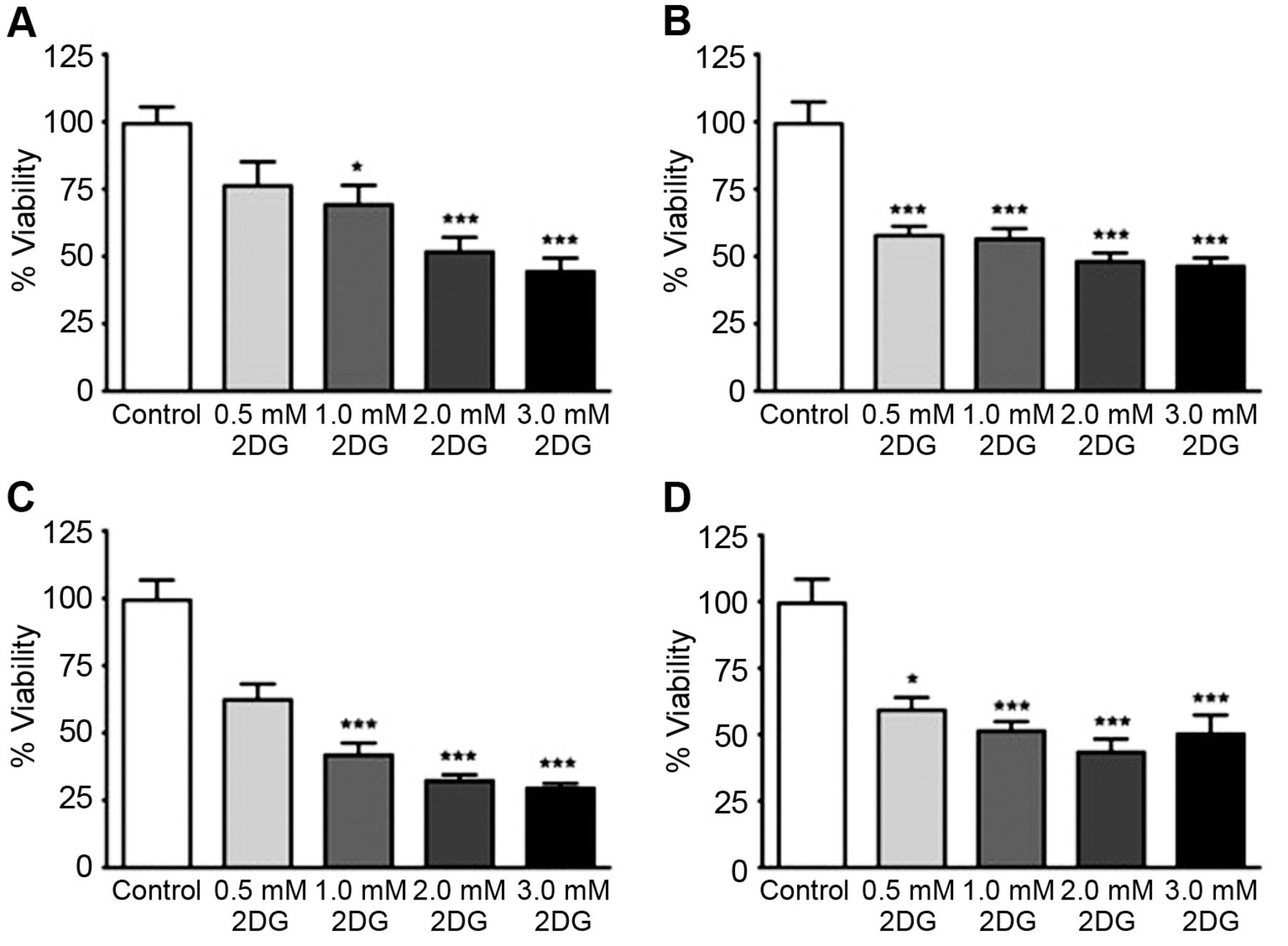
Mean±SEM % viability of (A) MeT-5A cells, (B) M14K cells, (C) MSTO cells and (D) ZL34 cells. *p<0.05 and ***p<0.001 versus Control. Data combined from 2 independent experiments, n=8 in each experiment.
3D spheroid cultures are equally sensitive to CPDD+PEM and 2DG+CPDD+PEM. In 3D spheroids of MeT-5A cells, drug treated spheroids had a significantly lower % mean perimeter compared to control (Control: 100.0±3.6%, CPDD+ PEM: 68.9±1.4%, 2DG+CPDD+ PEM: 69.5±2.3%; p<0.001 versus Control; Figure 3A). In M14K cells (Figure 3B), the treated % mean perimeter of spheroid was significantly lower compared to control (Control: 100.0±2.0%, CPDD+PEM: 67.0±1.6%, 2DG+CPDD+PEM: 69.6±2.3%; p<0.001 versus Control). In MSTO cells (Figure 3C), the treated % mean perimeter of spheroid was significantly lower compared to control (Control: 100.0±2.2%, CPDD+PEM: 78.3±1.7%, 2DG+CPDD+PEM: 77.0±1.8%; p<0.001). Finally, in ZL34 cells (Figure 3D), the % mean perimeter of treated spheroids was significantly lower compared to control (Control: 100.0±3.5%, CPDD+PEM: 74.9±1.7%, 2DG+CPDD+PEM: 72.5±1.1%; p<0.001 versus Control).
Discussion
In this study we have shown that treatment of benign mesothelial and malignant mesothelioma cells renders them susceptible to inhibition of glycolysis by 2-DG treatment. The epithelioid and sarcomatoid histological subtypes (M14K and ZL34 cell lines respectively) were more sensitive to 2-DG treatment since they had significantly decreased viability even with 0.5 mM treatment, while all cell lines had a viability on the range of 50% when treated with 3 mM of 2-DG. Compared to other cell types, mesothelial cells were more susceptible to 2-DG than the NSCLC cell lines A549 and PC-9 as well as the liver cancer cell lines HepF2 and Hep3B, but more resilient than papillary thyroid carcinoma cells CG3 (11-13). Contrary to the above experiments, the experiment we performed in this study used homologous cell derived ECM as a substrate, as described previously (10). This approach provides an additional layer of information given that cell-ECM interaction has been reported to contribute to the development of chemoresistance in tumors (7). Our in vitro system recapitulates well the cell-ECM interactions encountered in vivo.

Mean±SEM % cell proliferation of (A) MeT-5A cells after 72 h, (B) of M14K cells, (C) of MSTO and (D) of ZL34. ***p<0.001 versus Control, ###p<0.001 versus CPDD+PEM. Data combined from 2 independent experiments, n=4-6 in each experiment.

Mean±SEM % spheroid perimeter after 72 h of (A) MeT-5A cells, (B) of M14K cells, (C) of MSTO and (D) of ZL34. ***p<0.001 versus Control. Data combined from 2 independent experiments, n=8-12 in each experiment.
Based on the above findings we also assessed whether 2-DG would enhance the effects of the first-line chemotherapeutics, CPDD and PEM, in decreasing tumor cell growth in 2D and 3D, testing namely cell proliferation and spheroid formation phenotypes. In both cases the homologous cell derived ECM component was maintained. 2DG in combination with CPDD and PEM showed <25% proliferation in all cell types. In MSTO and ZL34 cells, 2DG synergized with CPDD+PEM treatment, which is suggestive of dependence on glucose metabolism. The 3D spheroid formation assay differs from cell proliferation in 2D in that, the cells are subject to ECM without a solid substratum. Spheroid formation and growth limits drug availability in the interior of the spheroid structure. Our data demonstrate 75% growth under cytotoxic challenge that is in contrast to 25-30% growth on 2D surface. In MPM, this is the first study demonstrating experimental data on 2D and 3D cell line models with 2DG as a therapeutic and with homologous cell derived ECM. The spontaneous formation of spheroid structures in explant cultures indicates an inherent property of primary cells to recapitulate a morphological character associated with their neoplastic nature. Previously, pleural fluid cytology demonstrated presence of spheroid like cell aggregates with coiled cords of cells, small papillae with a central core and at times tissue fragments with pseudoacinar formation (5). Cell aggregation behavior confers resistance to anoikis through cell–cell interaction, calcium dependent intercellular junctions (14). Spheroid aggregates from cell cultures on non-adherent surfaces are chemo-resistant through Src family kinase(s) activation that quenches caspase activity (15).
Glycolysis modulation occurs with the end product pyruvic acid. 3-Bromo pyruvic acid (3BrPA) has been experimentally tested in MSTO biphasic MPM cells (9). 3BrPA treatment lowered viability of MSTO cells in a time- and dose-dependent manner, and in vivo combination with CPDD prolonged survival. Similar modulation of glycolysis with the accumulation of citrate regulates glucose catabolism by inhibiting the key step of activating glucose by phosphofructokinase. Citrate mediated lowering of glucose metabolism in MPM has been reported with citrate+CPDD. Citrate in media activated caspase 3 and 9 suggestive of mitochondrial involvement, citrate combined with CPDD demonstrated a significant reduction in viability as opposed to citrate or CPDD (8). Also, 2DG has been reported as a combination therapeutic with docetaxel in patients with solid tumors (breast, thyroid, lung, head and neck). A dose of 63 mg/kg was evaluated to be safe, with low adverse events and hence recommended for progression to phase II trial (13).
In conclusion our results showed that first-line chemotherapeutics in combination with 2-DG in MPM enhances the reduction of 2D cell proliferation, that is pertinent to tumor growth and spread in the thoracic wall and lung parenchyma. On the other hand, 3D spheroid experiments showed that addition of 2-DG to first-line chemotherapeutics does not further decrease the growth of spheroids implying that it would not influence the development of new tumor foci within the pleural cavity. Further experiments are required to determine the potential of 2-DG in negating MPM tumor growth and progression.
Acknowledgements
This work was supported by a Hellenic Thoracic Society Research Award 2017, awarded to Dr. Irini Gerogianni.
Footnotes
Authors' Contributions
Irini Gerogianni, Rajesh M. Jagirdar, Chrissi Hatzoglou, and Sotirios G. Zarogiannis designed the study, analyzed the data, prepared the figures, and wrote the draft manuscript; Eleanna Pitaraki, Olympia Kouliou, Lydia Giannakou, Stefanos Giannopoulos and Eleftherios Papazoglou performed the experiments and revised the manuscript; Irene Gerogianni, Konstantinos I. Gourgoulianis and Sotirios G. Zarogiannis oversaw the study and revised the manuscript.
Conflicts of Interest
The Authors declare no conflicts of interest regarding this study.
- Received April 16, 2019.
- Revision received June 9, 2019.
- Accepted June 10, 2019.
- Copyright© 2019, International Institute of Anticancer Research (Dr. George J. Delinasios), All rights reserved