

Abstract
Aim: We describe the development of a highly-invasive, triple-negative breast cancer (TNBC) variant using serial orthotopic implantation of MDA-MB-231 human breast cancer in nude mice. Materials and Methods: MDA-MB-231 cells expressing red fluorescent protein (RFP) (1×10⁷ cells/site) were initially injected subcutaneously in the flank of nude mice. After the subcutaneous tumors grew, they were harvested and cut into small pieces for orthotopic implantation in the right lower mammary gland. After the orthotopic tumors grew, they were resected and cut into small pieces and orthotopically re-implanted into the mammary gland of nude mice. The tumors grew and metastasized to lymph nodes. The lymph node metastases were harvested and cut into small pieces and orthotopically re-implanted into the mammary gland of nude mice. After the orthotopic tumors grew, the tumor was removed leaving residual cancer cells, which grew and metastasized to lymph nodes. The lymph node metastases were harvested, cut into pieces and orthotopically re-implanted into the mammary gland of nude mice for two cycles and then isolated. Results: The isolated variant is highly invasive in the mammary gland and metastasized to lymph nodes in 10 of 12 mice compared to 2 of 12 of the parental cell line. Conclusion: The availability of a highly invasive variant of TNBC targeting lymph nodes will be very useful for drug discovery of TNBC, a recalcitrant cancer and for mechanistic studies of its aggressiveness.
- Breast cancer
- triple negative
- MDA-MB-231
- red fluorescent protein (RFP)
- nude mice
- orthothopic transplantation
- metastasis
- lymph node
- in vivo selection
- high metastasis variant
Triple-negative breast cancer (TNBC) is defined by a lack of expression of estrogen, progesterone, ERBB2 receptors and is highly aggressive (1, 2). TNBC is usually resistant to therapy (1, 2) and classified as a recalcitrant.
An effective way to study metastasis is to compare high- and low-metastatic variants of the same cancer line. Fidler's group (3) selected metastatic variants from the human Colo-357 pancreatic-cancer cell line by serial orthotopic passage. Cells were injected into the spleen or pancreas of nude mice. Hepatic metastases were harvested and tumor cells were re-injected into the spleen or pancreas. This cycle was repeated several times to yield cell lines L3.6sl (spleen to liver) and L3.6pl (pancreas to liver). The variant cells produced a significantly higher incidence and number of lymph-node and liver metastases than the parental cells (4).
In a previous study from our laboratory, serial passage of BxPC-3 and Panc-1 human pancreatic-cancer cell lines enabled isolation of highly-metastatic variants. Panc-1 contains the KRAS mutation, while BxPC-3 does not. However, both cell lines demonstrated, with in vivo selection, increasingly more aggressive behavior as evident by faster tumor growth and earlier metastasis occurring with each in vivo passage (4).
In another previous study, we developed a highly aggressive variant of human pancreatic cancer cell line XPA-1, which showed more rapid primary tumor growth, faster time to metastasis and more rapid lethality than the parental cell line. The high-metastatic variant developed a much denser tumor vasculature early during growth within the pancreas. Real-time polymerase chain reaction (RT-PCR) evaluation of genes involved in angiogenesis revealed a 24-fold increase in thrombospondin-1 (THBS1) expression in cells derived from the high-metastatic variant when compared with the parental cell line (5).
In another previous study from our laboratory, we demonstrated that in vivo passaging of lung metastasis in nude mice could generate an aggressive variant of human osteosarcoma cells. Experimental metastases were established by injecting 143B human osteosarcoma cells, expressing green fluorescent protein (GFP) in the nucleus and red fluorescent protein (RFP) in the cytoplasm, in the tail vein of nude mice. Lung metastases were harvested under fluorescence microscopy from nude mice to establish cell lines that were then injected via the tail vein of additional nude mice. This procedure was repeated for four passages in order to isolate highly-metastatic variant sublines. When the parental and metastatic variants were transplanted orthotopically into the tibia of nude mice, the 143B-LM4 variant had the highest metastatic rate, approximately 18-fold higher than the parent. αvβ3 integrin expression was increased approximately 5.6-fold in 143B-LM4 compared to parental cells (6).
We have also previously isolated high-metastatic variants of prostate cancer by injection of PC-3 human prostate cancer cells labeled with GFP or RFP into the left ventricle or intratibial bone marrow of nude mice. High-metastatic PC-3-GFP-BM6 targeted and grew in the bone marrow (7).
In another study from our laboratory, high bone-metastatic variants of human breast cancer cells were selected in nude mice by cardiac injection. After cardiac injection of a high bone-metastatic variant of MDA-MB-435 breast cancer, all mice had bone metastases compared to only 20% with parental cells (8).
In the present study, we demonstrate the generation by in vivo passage and initial characterization of a high-invasive variant of TNBC MDA-MB-231.
Materials and Methods
Cell culture. Parental breast cancer cell line, MDA-MB-231 (9, 10), expressing RFP (MDA-MB-231P-RFP) was maintained and cultured in DMEM medium with 10% fetal bovine serum (FBS) and 5% penicillin/streptomycin. Highly metastatic MDA-MB-231-expressing RFP (MDA-MB-231H-RFP), derived from lymph node metastasis after surgery, was also maintained and cultured in DMEM medium with 10% fetal bovine serum (FBS) and 5% penicillin/streptomycin.
Animal experiments. Athymic nude mice were kept in a barrier facility under high-efficiency particulate arrestance (HEPA) filtration. Mice were fed with autoclaved laboratory rodent diet (Tecklad LM-485; Envigo, Indianapolis, IN, USA). All animal studies were conducted in accordance with the principals and procedures outlined in the National Institutes of Health Guide for the Care and Use of Laboratory Animals under assurance A3873–01.
Comparison of local recurrence and lymph-node metastasis of low-invasive and high-invasive triple-negative breast cancer (TNBC) MDA-MB-231-red fluorescent protein (RFP) after primary tumor resection.
Establishment of orthotopic, highly invasive breast cancer variant. MDA-MB-231P-RFP cells (1×10⁷ cells/mouse) were initially injected subcutaneously in the flank of nude mice. Small pieces of harvested subcutaneous tumor were then transplanted orthotopically into the mammary gland of nude mice (11-14). After the tumor grew, the orthotopic tumor was harvested with residual cancer cells remaining. After the residual tumor grew, the tumor was harvested cut into small pieces and re-implanted into the mammary gland. After the tumor metastasized to lymph nodes, the metastatic tumor in the lymph nodes was harvested and cut into pieces and re-implanted into the mammary gland. After tumor growth, the orthotopic tumor was resected with residual cancer cells remaining. After the residual cancer cells grew and metastasized to lymph nodes, the metastatic tumor was harvested and cut into small pieces and re-implanted into the mammary gland of nude mice. After 2 more cycles of orthotopic transplantation, the high-metastatic variant was isolated. The resulting highly metastatic MDA-MB-231-RFP cells were termed MDA-MB-231H-RFP.
In vivo whole-body/whole-tumor imaging. The OV100 small animal imaging system (Olympus Corp., Tokyo, Japan), was used for fluorescence whole-body imaging (15-20). The OV100 contains an MT-20 light source (Olympus Biosystems, Planegg, Germany) and DP70 CCD camera (Olympus) for subcellular imaging in live mice. The optics of the OV100 have been specially developed for macro imaging, as well as micro imaging with high light-gathering capacity. Four individually optimized objective lenses, parcentered and parfocal, provide a 105-fold magnification range. High-resolution images were captured directly on a PC (Fujitsu Siemens, Munich, Germany). Images were processed for contrast and brightness and analyzed with the use of Paint Shop Pro 8 and CellR (21).
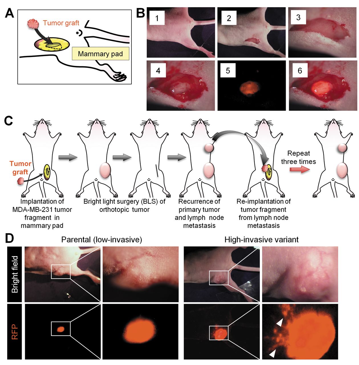
Isolation and characterization of high-invasive variant of triple-negative breast cancer (TNBC). To obtain tumor stock, 1×10⁷ RFP-expressing MDA-MB-231-RFP cells were suspended in Matrigel and inoculated subcutaneously into the right flank of 5-week-old female athymic nude mice. After tumor growth, the tumor was harvested and cut into small pieces (2-3 mm diameter) for subsequent orthotopic implantation. A. Scheme of orthotopic implantation of tumor into the mammary gland. B. Step-by-step procedure of orthotopic implantation. The right-lower mammary gland [1] was incised to make a pocket [2, 3]. A single piece of tumor was put into the pocket [4]. The tumor was readily imaged due to strong RFP fluorescence [5, 6]. The wound was sutured with 6-0 nylon. C. Scheme of serial orthotopic implantation used to isolate the high-metastatic variant. D. Representative images of low-invasive (parental) and high-invasive MDA-MB-231-RFP TNBC.
Results and Discussion
We developed a highly invasive, TNBC variant using serial orthotopic implantation of MDA-MB-231-RFP human breast cancer that targets lymph nodes (Figure 1). The high-invasive variant was termed MDA-MB-231H-RFP. The parental MDA-MB-231P-RFP primary orthotopic tumor has a smooth margin (Figure 1). In contrast, the high-invasive variant MDA-MB-231H-RFP primary tumor shows extensive tumor invasion from multiple parts of the primary tumor. The primary tumor formed by the high-invasive variant, MDA-MB-231H-RFP, was also larger and had more fluorescence intensity than the primary tumor formed from the parental cell line (Figure 1).
The high-invasive MDA-MB-231P-RFP formed lymph node metastasis in 10 of 12 mice after orthotopic implantation after resection of the primary tumor. In contrast, the parental MDA-MB-231-RFP formed lymph node metastasis in only 2 or 12 mice after orthotopic implantation and resection of the primary tumor (Table I).
Conclusion
A high-invasive variant of human TNBC was isolated by serial orthotopic transplantation. The variant, termed MDA-MB-231H-RFP, targeted lymph nodes after orthotopic implantation. The high-invasive variant, labeled with RFP, can serve as a useful model for anti-metastatic drug evaluation and mechanistic studies of this recalcitrant disease.
Footnotes
This article is freely accessible online.
Conflicts of Interest
Y. Urata is President & CEO of Oncolys BioPharma, Inc. H. Tazawa and T. Fujiwara are consultants of Oncolys BioPharma, Inc. S. Yano, K. Takehara, S. Miwa and R.M. Hoffman are unsalaried AntiCancer Inc. associates.
Dedication
This paper is dedicated to the memory of A.R. Moossa, M.D. and Sun Lee, M.D.
Grant Support
This study was supported in part by the National Cancer Institute grant CA 132971 and CA142669. This study was supported in part by grants from the Ministry of Health, Labour, and Welfare, Japan (to T. Fujiwara; No. 10103827, No. 13801426, No. 14525167) and grants from the Ministry of Education, Culture, Sports, Science and Technology, Japan (to T. Fujiwara; No. 25293283).
- Received May 9, 2016.
- Revision received June 7, 2016.
- Accepted June 8, 2016.
- Copyright© 2016 International Institute of Anticancer Research (Dr. John G. Delinassios), All rights reserved