

Abstract
Background: Laboratory and epidemiological studies have indicated that 1α,25-dihydroxyvitamin D3 [1α,25(OH)2D3] and dietary omega 3 (ω3)-polyunsaturated fatty acids (PUFAs) are capable of inhibiting the proliferation of various cancer cells. Materials and Methods: Human hepatoblastoma cells (HepG2) were treated with 1α,25(OH)2D3 and fish oil alone and in combination. Cell proliferation was measured either by the uptake of [3H]-thymidine into DNA or by counting the cell numbers using a hemocytometer. Results: The HepG2 cell proliferation was inhibited by 1α,25(OH)2D3 and fish oil in a dose-dependent manner. The lowest effective concentration of 1α,25(OH)2D3 was 10-7 M and 10-8 M using the [3H]-thymidine incorporation method and the cell counting method, respectively. Fish oil also caused a significant inhibition in HepG2 cell proliferation at 25 μg/mL. When HepG2 cells were treated with 1α,25(OH)2D3 in combination with fish oil, it was found that fish oil increased the antiproliferative effect of 1α,25(OH)2D3 on HepG2 cell growth compared to treatment with 1α,25(OH)2D3 alone. Conclusion: 1α,25(OH)2D3 could be used to treat hepatocellular carcinoma (HCC). However, the major side-effect of hypercalcemia limits its use. An enhanced 1α,25(OH)2D3-induced inhibition of HepG2 cell proliferation in the presence of PUFAs in the form of fish oil suggests that a lower concentration of 1α,25(OH)2D3 could be used to treat hepatocellular carcinoma in the presence of PUFAs to decrease the risk of hypercalcemia caused by high concentrations of 1α,25(OH)2D3.
- Vitamin D
- 1α,25(OH)2D3
- proliferation
- fish oil
- ω-3 PUFAs
- HepG2
- hepatoma
Hepatocellular carcinoma (HCC) is the fifth most common malignancy in the world and the third most deadly cancer worldwide (1). Surgical resection offers the only potentially curative treatment but most patients are not suitable for liver resection when they are diagnosed with HCC. Even after receiving a hepatectomy, postoperative recurrence is high and remains the main cause of late deaths (2-5). Several factors have been suggested to predispose tumor recurrence and Lee et al. proposed prognostic analysis after hepatectomy for HCC (6-8). Therefore, alternative treatments for HCC patients are needed regardless of whether they can or cannot undergo a hepatectomy.
The antiproliferative action of 1α,25(OH)2D3 on cancer cells was first described in 1981 (9). Since that time, abundant reports have shown inhibition of cell growth in many different cell lines derived from either normal or malignant tissues. However, the use of 1α,25(OH)2D3-based therapies is limited by the risk of hypercalcemia (10, 11).
Omega 3 (ω3)-polyunsaturated fatty acids (PUFAs), which are the major constituents of fish oils and certain vegetable oils, have been demonstrated to reduce carcinogenesis in several organs. In contrast, ω6 fatty acids (e.g. linoleic acid), found in corn oil or safflower oil and high fat diets, increase carcinogenesis (12-15).
In this study, the antiproliferative effects of 1α,25(OH)2D3 and ω3 PUFAs in the form of fish oil either alone or in combination were determined in human hepatoblastoma cells (HepG2). The results indicate that fish oil enhances the inhibitory effect of 1α,25(OH)2D3 on HepG2 cell growth.
Materials and Methods
Materials. 1α,25(OH)2D3 was a generous gift from Dr. M. Uskokovic. Fish oil and corn oil were purchased from General Nutrition. Fish oil and corn oil were emulsified with 5% (wt/wt) egg phosphatidycholine (Sigma) and 0.03% phosphate-buffered saline (PBS) at a final oil concentration of 15 mg/mL.
Cell culture. The human hepatoblastoma cell, HepG2, was obtained from ATCC (Manassas, VA, USA). Cells were grown in DMEM (Sigma, St. Louis, MO, USA) supplemented with 5% fetal bovine serum (FBS). Culture medium was changed 3 times per week.
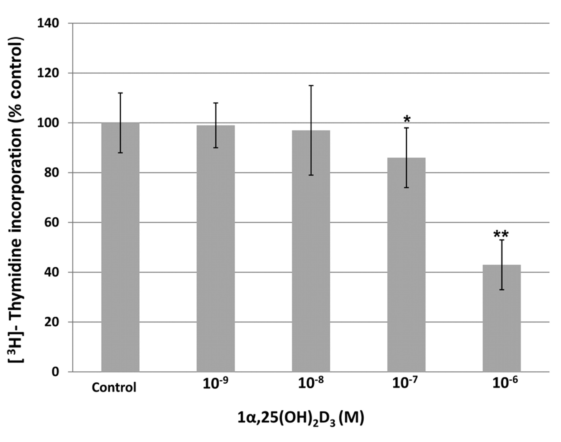
Effect of 1α,25(OH)2 D3 treatment on [3H]-thymidine incorporation into the DNA of HepG2 cells. Results are presented as the % of control. Each value is a mean±SD of six to eight determinations. *P<0.05, **P<0.001 versus control.
Cell proliferation assay. Two assays were performed to compare the antiproliferative activity of 1α,25(OH)2D3 plus fish oil against 1α,25(OH)2D3 or fish oil alone. [3H]-Thymidine incorporation method and cell counting method using a hemocytometer were performed as described previously (16,17). The results were expressed as percent of controls.
[3H]-Thymidine incorporation. [3H]-Thymidine incorporation into DNA was used as an index of cell proliferation. Briefly, when HepG2 cells reached about 50% confluence, media with FBS were replaced with fresh media without FBS and the cells were grown for an additional 24 h. Cells were then treated with 1α,25(OH)2D3 and/or fish oil alone or in combination. The concentrations of 1α,25(OH)2D3 were from 10-6 M to 10-9 M and of fish oil were from 12 μg/mL to 50 μg/mL. Eighteen hours later, the media were replaced with 0.5 mL of fresh basal medium containing methyl-[3H]-thymidine and incubated for 3 hours at 37°C. [3H]-Thymidine incorporation into DNA was stopped by placing the plates on ice. Unincorporated [3H]-thymidine was then removed and the cells were washed three times with ice-cold PBS. DNA labeled with [3H]-thymidine and other macromolecules were precipitated with ice-cold 5% perchloric acid for 20 min and then extracted with 0.5 mL of 5% perchloric acid at 70°C for 20 min. The radioactivity in the extracts was determined by a liquid scintillation counter.
Statistics method. The data of each group were compared by Student's t-test. A P-value <0.05 was considered as indicating a significant difference. The Excel 2007 was used to conduct statistics.
Results
It has been previously shown that 1α,25(OH)2D3 inhibited the proliferation of several hepatoma cell lines, including HepG2 liver cancer cells (18). To determine if fish oil has any effects on 1α,25(OH)2D3-induced antiproliferation, initially the effects of 1α,25(OH)2D3 alone on HepG2 cells was investigated using the [3H]-thymidine incorporation method. As shown in Figure 1, 1α,25(OH)2D3 treatment caused a dose-dependent inhibition of [3H]-thymidine incorporation into the DNA of cultured HepG2 cells. A 14±12% (p<0.05) and 57±10% (p<0.01) inhibition was observed at 10-7 and 10-6 M of 1α,25(OH)2D3, respectively, as compared to the controls treated with ethanol alone. No significant inhibition was detected in the presence of 10-8 and 10-9 M of 1α,25(OH)2D3. The effects of fish oil at 12, 18, 25 and 50 μg/mL without 1α,25(OH)2 D3 on the [3H]-thymidine incorporation into the DNA of HepG2 cells was studied (Table I). No significant inhibition was found at 12 and 18 μg/mL of fish oil, whereas a dose-dependent inhibition of HepG2 cell proliferation was observed at 25 μg/mL (42±10%, p<0.01) and 50 μg/mL (86±6%, p<0.001) of fish oil compared to the controls. In the corn oil group, there was also no significant inhibition observed. Since fish oil at 18 μg/mL concentration did not cause any inhibition in [3H]-thymidine incorporation into DNA, the antiproliferative effect of 1α,25(OH)2D3 in the presence and absence of 18 μg/mL of fish oil were therefore compared (Table II and Figure 2). It is clear that 18 μg/mL of fish oil increased the antiproliferative effect of 1α,25(OH)2D3 at 10-7 M from 14±12% to 31±13% (p<0.05). At 10-6 M 1α,25(OH)2D3, fish oil (18 μg/mL) increased the inhibition slightly from 57±10% to 64±6%, however it did not reach statistical significance. No enhancement of the activity by 18 μg/mL fish oil was observed at 10-8 and 10-9M of 1α,25(OH)2D3. When HepG2 cells were incubated with 12 μg/mL fish oil and increasing concentrations of 1α,25(OH)2D3 (10-9 to 10-6 M), no further stimulation of the antiproliferative effect by fish oil was observed at any 1α,25(OH)2D3 concentration (data not shown). The effect of different concentrations of fish oil (at 12, 18, 25 and 50 μg/mL) on the antiproliferative activity of 1α,25(OH)2D3 at 10-7 M was subsequently investigated. The results in Table III and Figure 3 demonstrate that when HepG2 cells were incubated with a combination of 1α,25(OH)2D3 at 10-7 M and fish oil at either 18 or 25 μg/mL, a significantly greater inhibition was observed as compared to the cells treated with either 1α,25(OH)2D3 at 10-7 M or fish oil at 18 or 25 μg/mL alone.
Effect of increasing fish oil concentrations on [3H]-thymidine incorporation into DNA of HepG2 cells.
Effect of increasing concentrations of 1α,25(OH)2D3 (1,25D), with or without fish oil at 18 μg/ml, on [3H]-thymidine incorporation of HepG2 cells.

Effect of 1α,25(OH)2D3 and fish oil (18 μg/mL), alone and in combination, on [3H]-thymidine incorporation into the DNA of HepG2 cells. Results are presented as the % of control. Each value is a mean±SD of six to eight determinations. *P<0.05, **P<0.001 versus control.
Table IV shows the cell counts of HepG2 cells treated with increasing concentrations of 1α,25(OH)2D3 and 18 μg/mL of fish oil alone and in combination. Fish oil at 18 μg/mL alone had no effect on cell number. When it was combined with 1α,25(OH)2 D3, it enhanced the inhibition induced by 107 M and 10-6 M 1α,25(OH)2D3 from 8±2% to 18±2% and from 26±4 % to 36±3 %, respectively. The results are similar to the [3H]-thymidine incorporation data presented in Figure 2 and Table II.
Discussion
HCC is the most deadly cancer in Asia, especially in China and India, and the incidence of HCC is increasing in the United States (1). The overall survival of patients with HCC is dismal. The only potential curative therapy is hepatectomy but most patients are not suitable for this operation. Currently, no effective secondary prevention or systemic treatments are available. Even after surgical resection, the prognosis of HCC remains poor. Five-year survival of patients undergoing surgical resection for HCC is only 30-40%. A high incidence of recurrence after hepatectomy is the main cause of unsatisfactory results after surgical treatment (7, 8). Chen et al. reported a tumor recurrence rate of up to 59% in the first year and a 5-year cumulative rate of 70 to 100% after hepatectomy (19). Postoperative recurrence is high and remains the main cause of late deaths. Reports on long-term disease-free survival after resection for HCC remain limited (20). Despite many years of sustained efforts, the long-term outcome employing current therapies remains dismal as both recurrences of the lesions within the liver and occurrences of distant metastases are frequent.
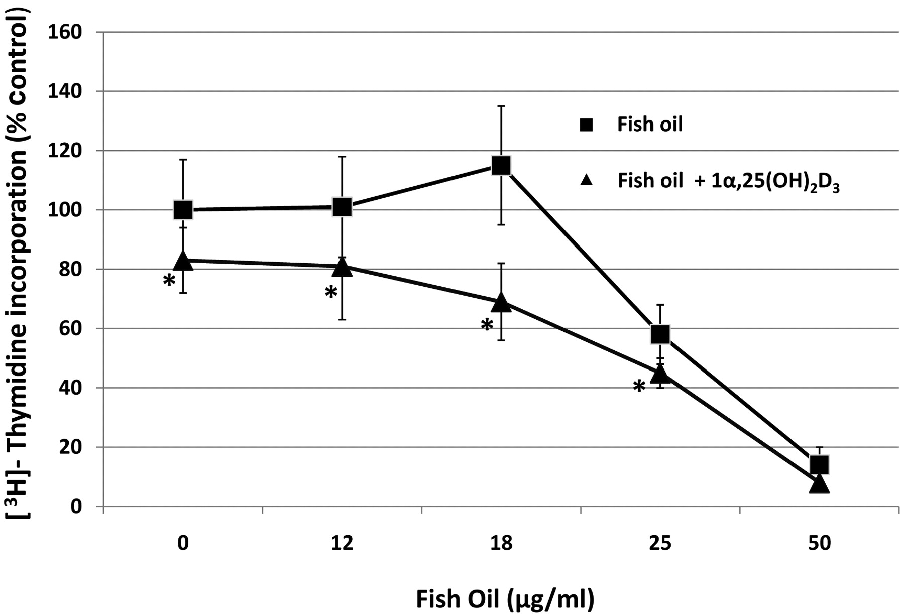
Effect of increasing fish oil concentrations with or without 1,25(OH)2D3 at 10-7 M on [3H]-thymidine incorporation into the DNA of HepG2 cells. Results are presented as the % of control. Each value is a mean±SD of six to eight determinations; *P<0.05.
Effect of increasing fish oil concentrations with or without 1α,25(OH)2D3 (1,25D) at 10-7 M on the 3[3H]-thymidine incorporation into DNA of HepG2 cells.
The antiproliferative effect of 1α,25(OH)2D3 is well documented (21-23). It is also known that 1α,25(OH)2D3 is a potent antitumor agent against HCC. Early work by Morris et al. has shown that 1α,25(OH)2D3 can inhibit liver cancer cells in vitro and in vivo (24). In this study, Morris et al. showed that 1α,25(OH)2D3 (0.02-0.5 μg/kg per day) significantly inhibited SKHEP tumor in nude mice without the development of hypercalcemia. In a phase 1 study using the hepatic arterial administration of 1α,25(OH)2D3 to six HCC patients, Morris et al. demonstrated that the 6 patients remained normocalcemic at doses up to 10 μg/day for a period of 1-4 weeks (25). However, at 15 μg/day two patients developed hypercalcemia after 3-7 days of treatment. To overcome this side-effect, these investigators developed a hepatic intra-arterial injection of 1α,25(OH)2D3 in lipiodol. Eight patients with refractory HCC were given a single intrahepatic arterial dose (50, 75 or 100 μg) of 1α,25(OH)2D3 dissolved in 5 mL of lipiodol and their serum calcium was followed for 4 weeks. Only 3 patients were found to develop a mild hypercalcemia even at these high dose levels, and none of the patients developed grade 3 hypercalcemia. These investigators concluded that locoregional delivery of 1α,25(OH)2D3 in lipidol could allow the administration of suprapharmacological doses of this drug without the development of hypercalcemia. In addition to the locoregional delivery approach, several thousand synthetic vitamin D3 analogues have been developed, with the aim of increasing the antiproliferation effects and decreasing the risk of hypercalcemia.
Effect of increasing concentrations of 1α,25(OH)2D3 (1,25 D) with or without fish oil (18 μg/ml) treatment on cell counts of HepG2 cells.
Fish oil is known to inhibit the growth of certain tumors, such as colon, breast and prostate cancer (26). The antitumor effect is due to the presence of ω3-PUFAs in fish oil. ω3 (n3) and ω6 (n6) PUFAs are essential fatty acids. A high ω6/ω3 ratio is thought to contribute to cardiovascular disease, inflammation and cancer (27). Multiple cellular mechanisms have been proposed to explain the anticancer effects of ω3-PUFAs, including inhibition of arachidonic acid-derived eicosanoid biosynthesis, influences on transcription factors and gene expression, modification of signal transduction pathways, induction of cancer cells differentiation, and enhancement of lipid peroxidation (28, 29). The effect of ω3 PUFAs on cancer in animal and cell culture models has led to the use of ω3 PUFAs in clinical trials for cancer prevention and treatment, and for nutritional support of cancer patients to reduce weight loss and modulate their immune system.
However, very little is known regarding the use of fish oil in liver cancer prevention and treatment. In this study, the anticancer effect of fish oil and 1α,25(OH)2D3 on liver cancer cells are shown (Figures 1, 2, 3, and 4 and Tables I, II, and III). When 1α,25(OH)2D3 and fish oil were added separately into cultured HepG2 cells, they inhibited HepG2 cell proliferation in a dose-dependent manner. An enhanced antiproliferative effect was observed when 1α,25(OH)2D3 and fish oil were used in combination (Tables II, III, and IV and Figures 2, 3).
Although the mechanism underlying the increased effect of 1α,25(OH)2D3 on liver cancer cell growth in the presence of fish oil is not known, it is likely that 1α,25(OH)2D3 may have altered the synthesis and degradation of prostaglandins in HepG2 cells as suggested by Krishnan et al. to occur in prostate cancer cells (30). As the result of enhanced antiproliferative activity in the presence of both 1α,25(OH)2D3 and ω3 PUFAs, an antitumor effect was achieved with a lower dose of 1α,25(OH)2D3 without secondary hypercalcemia. Potentially, a combination of ω3 PUFAs with more potent and less calcemic analogs of 1α,25(OH)2D3, such as MART-10 (31), could be developed for the treatment of HCC.
- Received January 29, 2009.
- Revision received March 21, 2009.
- Accepted May 14, 2009.
- Copyright© 2009 International Institute of Anticancer Research (Dr. John G. Delinassios), All rights reserved