

Abstract
Background: The altered expression of multidrug resistance 1 (MDR1) contributes to various types of carcinogenesis and their disease progression. Hypermethylation of CpG islands located in the promoter regions is involved in gene silencing at the transcriptional level. The methylation status of MDR1 gene promoter was investigated in gastric cancer in relation to various clinicopathological features. Patients and Methods: The methylation of the MDR1 promoter was estimated in both antral non-neoplastic mucosa and cancer lesions in 83 patients with gastric cancer using a methylation-specific PCR method. Results: The methylation ratio (MR) of the cancer lesions (31.3±20.5) was significantly higher than that of non non-neoplastic mucosa (20.9±31.3, p=0.0004). The MR was especially high in Lauren's intestinal cancer (p<0.0001). A higher MR was also observed in more advanced stage (p=0.005) and lymph vessel invasion-positive cases (p=0.002). Conclusion: The promoter methylation of MDR1 seems to have a significant role in carcinogenesis and tumor progression in the stomach.
- MDR1
- methylation
- gastric cancer
Abbreviations: MDR1, multidrug resistance 1; H. pylori, Helicobacter pylori; P-gp, P-glycoprotein.
Gastric cancer is still a significant health problem worldwide. Although the incidence and mortality rates of this malignancy have been decreasing over the last few decades, it still remains second only to lung cancer as the leading cause of cancer death around the world (1, 2).
The multidrug resistance 1 (MDR1) gene encodes for P-glycoprotein (P-gp), a Mr 170,000 transmembrane calcium-dependent efflux pump which is expressed in various tissues (3-5). Aberrant expression of P-gp is associated with various carcinomas. Recent studies have shown that hypermethylation of normally unmethylated dinucleotides (CpG islands) located in the promoter regions is involved in gene silencing at the transcriptional level (6, 7). In some carcinomas, one of the mechanisms underlying down-regulation of MDR1 is thought to be CpG hypermethylation of the MDR1 promoter (8-10). Recent publications have indicated another functional role of P-gp as a marker for disease progression, in addition to its conventional role as a drug resistance marker (11-15). In hepatocellular carcinoma and osteosarcoma, P-gp has been thought to be up-regulated in patients with localized rather than metastatic disease (13, 14). We hypothesized that alteration of MDR1 expression through CpG methylation is responsible for the pathogenesis and progression of gastric cancer. This study was performed to investigate the methylation status of the MDR1 gene promoter in gastric cancer in relation to various clinicopathological features.
Patients and Methods
Tissue samples, DNA extraction and Helicobacter pylori infection status. The studied population comprised 83 patients with gastric cancer being seen by the Endoscopy Center of Fujita Health University Hospital. All the patients underwent an upper endoscopy with a biopsy of both non-neoplastic mucosa in the antrum and the cancer lesions.
The diagnoses of all the gastric carcinomas were performed histologically by the pathology division of our hospital. The gastric cancer was also classified according to Lauren' s classification (16). The biopsy specimens were immediately frozen and stored at -80°C. Patients with systemic severe diseases were excluded. Genomic DNA was isolated from the frozen specimens using proteinase K. H. pylori infection status was assessed by serological or histological analysis, or urea breath test. Patients were diagnosed as infected when at least one of the diagnostic tests was positive. The Ethics Committee of the Fujita Health University School of Medicine approved the protocol, and written informed consent was obtained from all the participating patients.
Bisulfate modification and methylation-specific PCR (MSP). To examine DNA methylation, genomic DNA was treated with sodium bisulfite using a BisulFast DNA Modification Kit for Methylated DNA Detection (Toyobo Co., Osaka, Japan). The MSP was carried out using primers for the promoter region of MDR1 designed to include six CpG dinucleotides that have been linked to regulation of MDR1 expression (10). The primer sequences for amplification of unmethylated MDR1 were: forward 5′-GGGTGTGGGTTGA GTATAGTTGTTTT-3′ and reverse 5′-CCAACTTTACATACCCCTA CCTCACA-3′; and for methylated MDR1: forward 5′-GGGCG TGGGTTGAGTATAGTCGTTTC-3′ and reverse 5′-CGCTCCTT AAAACAACCACCAAAACG-3′.

Fluorescence intensities of methylated (M) and unmethylated (UM) bands of the MDR1 gene promoter measured by digital densitometer.
The annealing temperatures and times were determined using DNA from peripheral blood of a young individual without H. pylori infection and DNA methylated with SssI methylase (New England BioLabs Inc., Beverly, MA, USA). The MSP was carried out in a volume of 20 μl containing 0.1 μg of bislufite-modificated DNA. The DNA was denatured at 95°C for 5 minutes, followed by 32 cycles at 95°C for 30 seconds, 56°C for unmethylated DNA and 60°C for methylated DNA for 1 minute, and 72°C for 1 minute with a final extension at 72 for 7 minutes. The bands of MSP were detected by electrophoresis in 2.5% agarose gels stained with ethdium bromide. The fluorescence intensities of the methylated and unmethylated bands were measured by a digital densitometer. The methylation ratio (MR) was calculated as the ratio of value of the methylated band to the methylated plus unmethylated bands, as in previous studies (17, 18).
Statistical analysis. All the data were expressed as mean±SD. A paired Wilcoxon's rank test was employed for the comparison of MR between the cancer lesions and the non-neoplastic mucosa in each individual. The relationship between the MR in the non-neoplastic mucosa and gender and H. pylori status was analyzed by Student's t-test while ANOVA was used for the relationship between age and MR. The level of significance was set at p<0.05.
Characteristics of subjects.
Methylation level of the MDR1 promoter in cancer lesions and non-neoplastic mucosa.
Results
MR in cancer lesions compared with non-neoplastic mucosa. The methylation status of the MDR1 promoter was determined in all 83 gastric cancer tissues as well as the non-neoplastic mucosa adjacent to the cancer (Figure 1). The patient characteristics are summarized in Table I. Overall, the MR for MDR1 promoter of the cancer lesions (31.3±20.5) was significantly higher than that of the non-neoplastic mucosa (20.9±31.3, p=0.0004; Table II). Concerning the different histological subtypes, MR in the cancer lesions was higher than the non-neoplastic mucosa, especially in Lauren's intestinal cancer (36.2±21.9 vs. 21.9±18.0, p<0.0001). A significant difference in MR was also observed in more advanced stage samples (T2<: 31.1±21.7 vs. 19.1±17.8, p=0.005). Moreover, in the operative cases, a higher MR was observed in the lymph vessel invasion-positive cases (34.1±21.3 vs. 21.9±19.1, p=0.002). On the other hand, MR did not correlate with venous invasion or lymph node metastasis. Moreover, no association was observed between MR and peritoneal dissemination, liver or other distant metastasis (data not shown).

Association between methylation level of the MDR1 promoter in non-neoplastic mucosa and gender. Male vs. female: 23.7±19.9 vs. 12.1±12.0: Student's t-test.
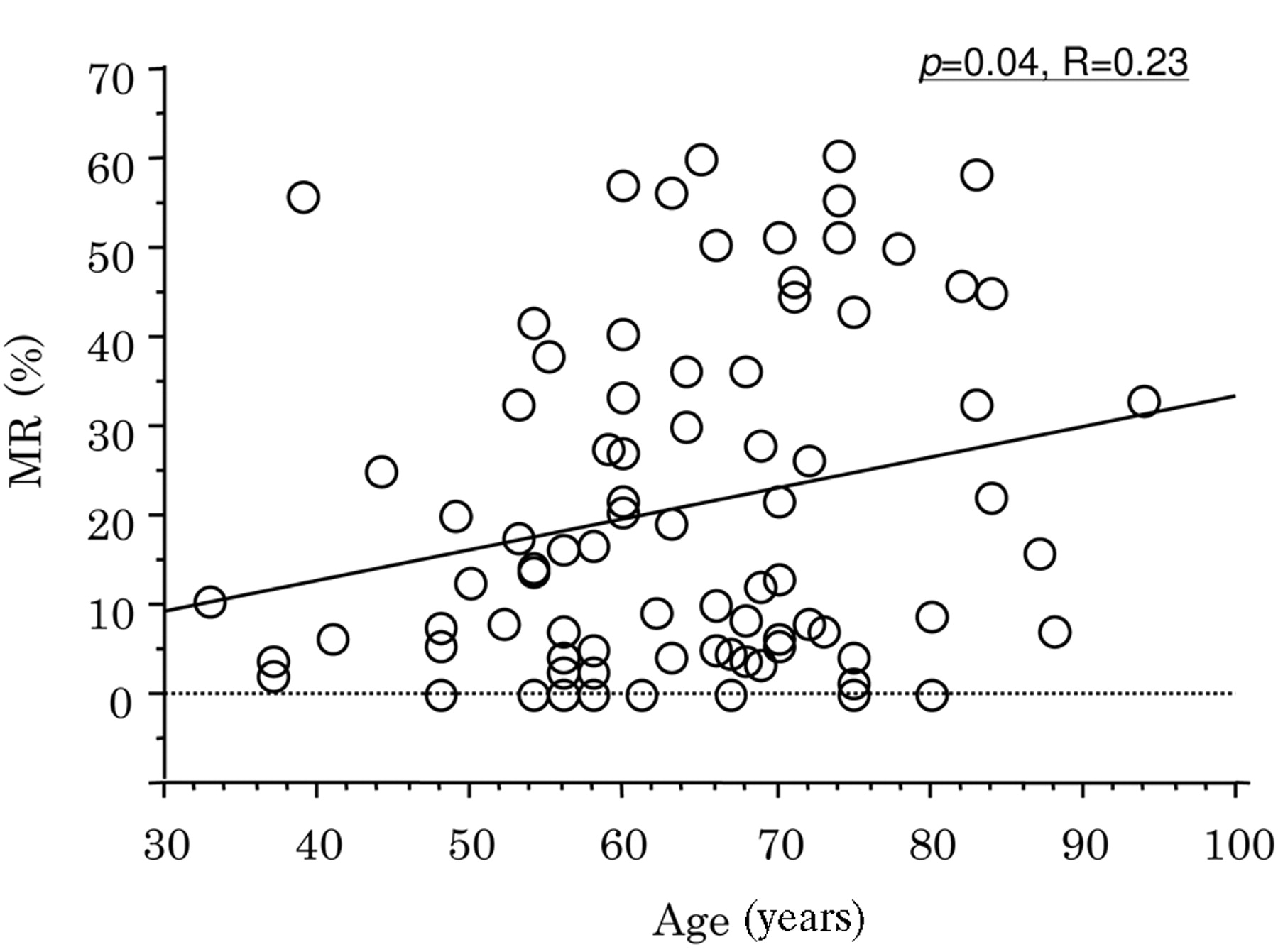
Relationship between MR in non-neoplastic mucosa and age, gender and H. pylori status. The MR was higher in males (male vs. female: 23.7±19.9 vs. 12.1±12.0, p=0.02, Figure 2). In addition, the MR weakly correlated with age (R=0.23, p=0.04; Figure 3). No significant association between MR and H. pylori infection status was found due to the small number of H. pylori infection-negative participants (data not shown).
Disucussion
In order to make a correct assessment of gastric cancer characteristics, the gastric mucosal context must be taken into account. In addition, the analysis by MSP lacked quantitative reliability. Therefore, the MR of the cancer lesions was compared with that of the non-neoplastic mucosa in each individual. The promoter methylation of MDR1 in the gastric cancer lesions was significantly higher than that in the non-neoplastic mucosa. Furthermore, a higher MR was observed in the more advanced phenotypes such as the higher T categories and lymph vessel invasion-positive cases.
Association between methylation level of the MDR1 promoter in non-neoplastic mucosa and age. Statistical analysis was performed by ANOVA.
Several earlier studies have explored whether aberrant expression of the MDR1 gene might be a biomarker for the etiology and progression of various carcinomas (11, 12, 19, 20).
Some investigators reported that overexpression of the MDR1 gene could be an active biomarker for tumor progression in breast and colon carcinomas (19, 20), whereas other investigators reported that loss of the MDR1 gene was associated with an advanced or metastatic stage in hepatocellular carcinoma, osteosarcoma and prostate carcinoma (11, 12). Considering that promoter CpG hypermethylation is a likely mechanism for inactivation of MDR1 gene transcription and down-regulation of P-gp expression in human prostate cancer, the present results suggested that MDR1 methylation might characterize biological aggressiveness of gastric cancer. Regarding the role of MDR1 methylation in gastric carcinogenesis and its progression, the balance between proliferation and apoptosis of gastric cancer cells may play a key role in the net growth of gastric cancer. An inverse correlation between Ki-67 and MDR1 expression has been reported in advanced prostate cancer (14). High expression of Ki-67 and PCNA in mdr1a double-knockout mice during experimental hepatocarcinogenesis (21) and a positive correlation between up-regulation of MDR1 and enhancement in human melanoma metastasis suppressor (KiSS-1) activity (22) have also been documented, suggesting that MDR1 down-regulation may be relevant in cell proliferation. Recent studies have also shown that P-gp is associated with an apoptotic pathway in vitro (23-25). CpG hypermethylation of MDR1 promoter also might be involved in gastric cancer progression via inhibition of apoptosis.
On the other hand, no correlation was found between higher MR and lymph node, peritoneal, liver or distant metastasis of gastric cancer. Thus MDR1 methylation may contribute to the tumor progression itself, but not to metastasis of gastric cancer.
Higher MR was also observed in Lauren's intestinal gastric cancer, which suggested that different histological subtypes of gastric cancer may have different genetic or epigenetic backgrounds. The present data indicated that higher methylation of the MDR1 promoter may be involved in the etiology of intestinal type gastric cancer.
Some genes are methylated with age (26) and chronic inflammation (27) in the colorectal epithelium. In the stomach, aberrant CpG island hypermethylation of chronic gastritis is related to age, gender, intestinal metaplasia and chronic inflammation (28). The stomach is one of the organs that show frequent methylation of CpG islands of genes in non-neoplastic epithelial cells (29, 30). H. pylori infection has been reported to potently induce the methylation of CpG islands to various degrees (31). In this study, MDR1 methylation also occurred in the non-neoplastic mucosa to various degrees and was significantly associated with aging and gender, although no significant association was found between methylation status and H. pylori infection, possibly because of the low number of H. pylori-negative patiens. This suggested that MDR1 methylation may also be an age-related phenomenon, which occurs early in the process of gastric carcinogenesis. Our results show that further longitudinal studies of MDR1 methylation are needed to investigate its potential usefulness as a molecular biomarker and clinical efficacy.
Footnotes
- Received July 14, 2008.
- Revision received October 24, 2008.
- Accepted November 10, 2008.
- Copyright© 2009 International Institute of Anticancer Research (Dr. John G. Delinassios), All rights reserved