
Main menu

  • Home
  • Current Issue
  • Archive
  • Info for
    • Authors
    • Editorial Policies
    • Subscribers
    • Advertisers
    • Editorial Board
  • Other Publications
    • In Vivo
    • Cancer Genomics & Proteomics
    • Cancer Diagnosis & Prognosis
  • More
    • IIAR
    • Conferences
    • 2008 Nobel Laureates
  • About Us
    • General Policy
    • Contact
  • Other Publications
    • Anticancer Research
    • In Vivo
    • Cancer Genomics & Proteomics

User menu

  • Register
  • Subscribe
  • My alerts
  • Log in
  • My Cart

Search

  • Advanced search
Anticancer Research
  • Other Publications
    • Anticancer Research
    • In Vivo
    • Cancer Genomics & Proteomics
  • Register
  • Subscribe
  • My alerts
  • Log in
  • My Cart
Anticancer Research

Advanced Search

  • Home
  • Current Issue
  • Archive
  • Info for
    • Authors
    • Editorial Policies
    • Subscribers
    • Advertisers
    • Editorial Board
  • Other Publications
    • In Vivo
    • Cancer Genomics & Proteomics
    • Cancer Diagnosis & Prognosis
  • More
    • IIAR
    • Conferences
    • 2008 Nobel Laureates
  • About Us
    • General Policy
    • Contact
  • Visit us on Facebook
  • Follow us on Linkedin
Research ArticleExperimental Studies
Open Access

Somatic Variants in DNA Damage Response Genes in Ovarian Cancer Patients Using Whole-exome Sequencing

JOANNA LOPACINSKA-JOERGENSEN, DOUGLAS V.N.P. OLIVEIRA, TIM SVENSTRUP POULSEN, CLAUS K. HOEGDALL and ESTRID V. HOEGDALL
Anticancer Research May 2023, 43 (5) 1891-1900; DOI: https://doi.org/10.21873/anticanres.16348
JOANNA LOPACINSKA-JOERGENSEN
1Department of Pathology, Herlev Hospital, University of Copenhagen, Herlev, Denmark;
  • Find this author on Google Scholar
  • Find this author on PubMed
  • Search for this author on this site
DOUGLAS V.N.P. OLIVEIRA
1Department of Pathology, Herlev Hospital, University of Copenhagen, Herlev, Denmark;
  • Find this author on Google Scholar
  • Find this author on PubMed
  • Search for this author on this site
TIM SVENSTRUP POULSEN
1Department of Pathology, Herlev Hospital, University of Copenhagen, Herlev, Denmark;
  • Find this author on Google Scholar
  • Find this author on PubMed
  • Search for this author on this site
CLAUS K. HOEGDALL
2Department of Gynaecology, Juliane Marie Centre, Rigshospitalet, University of Copenhagen, Copenhagen, Denmark
  • Find this author on Google Scholar
  • Find this author on PubMed
  • Search for this author on this site
ESTRID V. HOEGDALL
1Department of Pathology, Herlev Hospital, University of Copenhagen, Herlev, Denmark;
  • Find this author on Google Scholar
  • Find this author on PubMed
  • Search for this author on this site
  • For correspondence: estrid.hoegdall@regionh.dk
  • Article
  • Figures & Data
  • Info & Metrics
  • PDF
Loading

Abstract

Background/Aim: Several clinical trials have investigated homologous recombination deficiency and BRCA1/2 status to select ovarian cancer patients for treatment with poly(ADP-ribose) polymerase-inhibitors (PARPi), but less attention has been given to other DNA-damage response (DDR) pathways. Therefore, we investigated somatic single/multiple nucleotide variants and small insertions/deletions in exonic and splice-site regions of 356 DDR genes to examine whether genes other than BRCA1/2 are altered. Materials and Methods: Whole-exome sequencing data from eight high-grade serous adenocarcinoma (HGSC) and four clear cell carcinoma (oCCC) patients were analyzed. Results: Forty-two variants (pathogenic, likely pathogenic or variants of uncertain significance) in 28 genes from DDR pathways were identified. Seven out of nine TP53 variants were previously described in The Cancer Genome Atlas Ovarian Cancer; other variants were found in 23 out of 28 unique genes, whereas no variants were reported in FAAP24, GTF2H4, POLE4, RPA3, and XRCC4. Conclusion: As the identified variants were not only limited to well-known TP53, BRCA1/2, and HR-associated genes, our study might contribute to the better understanding of which DDR pathways potentially influence disease progression. Moreover, they may display a potential role as biomarkers to predict platinum-based chemotherapy or PARPi treatment response or disease progression, as differences in disrupted DDR pathways were observed between patients with long and short overall survival in HGSC and oCCC groups.

Key Words:
  • Somatic variants
  • DNA damage response
  • ovarian cancer

Epithelial ovarian cancer (EOC), which represents approximately 90% of ovarian cancer (OC) cases, is diagnosed in late, more lethal, stages of the disease (1, 2). It can be further divided into five main classes: high-grade serous (HGSC, 70%), clear cell (oCCC, 10%), endometrioid (10%), mucinous (3%), and low-grade serous carcinomas (<5%) (3). The 5-year survival rate of late stage EOC patients [International Federation of Gynecology and Obstetrics (FIGO) stage IV] is below 30%, whereas that of patients in early stage (FIGO stage I) is above 90% based on the report from Danish Gynecologic Cancer Database (DGCD) (4). A comprehensive molecular characterization of HGSCs has revealed mutations in several genes: most notably in the TP53 gene, which displayed mutations in almost 96% cases (5, 6). Moreover, it was shown that about 50% of HGSC patients have alterations in the homologous recombination (HR) DNA damage response pathway (7). Germline and somatic BRCA1 and BRCA2 mutations were detected in up to 25% and 3-7% cases of HGSCs, respectively (8). BRCA1 and BRCA2 are essential proteins in the DNA damage response machinery that senses, signals, and repairs DNA lesions (9). Three protein complexes: the MRE11-Rad50-NBS1 (MRN), RPA/ATRIP, and KU70-KU80 are among the first sensors and responders to DNA damage (10). They recruit DNA damage signalling components: ATM, ATR, and DNA-PKs, which triggers repair signalling by phosphorylating key proteins such as BRCA1, CHK1, CHK2, H2AX, p53, and RAD17 (11). Various DNA repair mechanisms are initialized in response to activated signaling-transduction pathways: base excision repair (BER), Fanconi Anemia (FA), HR, mismatch repair (MMR), nucleotide excision repair (NER), and non-homologous end joining (NHEJ) (12, 13). Dysregulation of the DDR may impair genomic instability in cells and promote cancer development, but also may offer targetable weaknesses that can be further exploited therapeutically (14). Defective DDR is an attractive target in EOC, as demonstrated by favorable responses to both platinum chemotherapy and poly (ADP-ribose) polymerase inhibitors (PARPi), which utilize deficiencies in DDR pathways to lead to cancer cell death (7, 12, 15). Although initially the majority of HGSCs are platinum-sensitive, many patients will relapse eventually as platinum-resistant after frequent relapse-response history (12). Resistance to platinum therapy is strongly predictive of resistance to PARPi treatment (16). Olaparib was the first PARPi that received regulatory approval in Europe to treat HGSC patients with a germline (g) or somatic (s) BRCA1/2 mutation (17). Clinical studies have provided evidence of favorable response to PARPi treatment in patients harboring pathogenic/likely pathogenic BRCA1/2 mutations with complete or partial response to platinum-based chemotherapy as compared to patients with no BRCA1/2 mutations (18). Further effort is needed to determine which non-BRCA1/2 patients benefit from such treatment, however, there is yet no consensus reached on how to identify predictors of PARPi sensitivity in OC (19). There is growing evidence that mutations in non-BRCA DDR genes might contribute to better understanding of cancer predisposition and guide the potential treatment of HGSCs by predicting sensitivity to PARPi (20–22). In contrast to HGSCs, oCCCs usually express wild-type p53 protein and have a lower frequency of HR gene mutations (23). The most frequent variants in oCCC are in ARID1A and PIK3CA, both appearing as single or double hit mutations in approximately 50% of patients (23, 24). Moreover, oCCC exhibits higher risk of resistance to platinum-based chemotherapy, which in consequence might lead to poor prognosis (23, 25). Understanding of the DDR mechanisms and their defects may help to target individualized treatment to various subtypes of EOC (26).

We examined the occurrence of somatic single and multiple nucleotide variants (SNV and MNV), as well as small insertions/deletions (INDELs) in exonic and splice site regions of 356 DDR genes in HGSC and oCCC patients using whole exome sequencing. The aim of this study was to investigate whether DDR pathways other than HR may be affected and if differences between HGSC and oCCC groups can be observed.

Materials and Methods

Patient cohort. Fresh-frozen samples were acquired from two Danish projects: the Pelvic Mass study (2004-2014) and the Gynecological Ovarian Vulva Endometrial Cervix cancer (GOVEC) study (2015 – ongoing) through the Bio- and Genome Bank Denmark (27). The study was performed according to the guidelines of the Declaration of Helsinki, including written informed consent from all patients. The study has been approved by the Danish National Committee for Research Ethics, Capital Region (H-17029749/H-15020061). Both patients’ groups: oCCC and HGSC had similar age characteristics: 60.7 (57.2-68.0) and 63.8 (53.8-68.3) years, respectively. Moreover, HGSC patients, were matched on FIGO stage, but their overall survival (OS) differed significantly:“HGSC_ShortOS” with median OS=26.9 months and “HGSC_LongOS”, median OS=131.1 months) (p<0.05, the unpaired two-samples Wilcoxon test) (Table I). Hematoxylin and eosin (H&E)-stained tissue slides neighboring the excised tumor were examined by a trained pathologist to determine percentage of tumor cells.

View this table:
  • View inline
  • View popup
  • Download powerpoint
Table I.

Patients included in this study, chosen from the Danish Pelvic Mass/GOVEC study.

Whole exome sequencing. Genomic DNA was extracted using Maxwell RSC Tissue DNA (AS1610, Promega, Madison, WI, USA). DNA concentration measurements were performed on a Qubit system with the High Sensitivity dsDNA assay kit (Q33120, Thermo Fisher Scientific, Waltham, MA, USA). Exome sequencing libraries were prepared from 100 ng DNA using the Ion AmpliSeq Exome RDY kit (A38262, Thermo Fisher Scientific) according to the manufacturer’s protocol. Equal amounts of amplified DNA libraries were loaded onto an Ion 550 Chip (A34537, Thermo Fisher Scientific) using the Ion Chef System (Thermo Fisher Scientific). Sequencing was performed on an Ion S5XL System (Thermo Fisher Scientific).

Data processing and variant calling. Sequencing data were acquired, pre-processed, aligned, and analyzed by using Ion Suite Software v5.6 (Thermo Fisher Scientific), coupled with AmpliSeq Exome single sample (Somatic) analysis module. Further filtering for true variants was performed using R environment (28), as described below, and presented in Table II. Only mutations occurring in exonic or splice-site (located within the first 3 nucleotides of the 5′ or 3′ end) regions of the 356 DDR genes were selected. As we expected to find TP53 gene variants in more than 90% of HGSC cases based on previous reports (5), we performed first manual TP53 variant check of non-filtered data from the subjects with HGSC to determine cut-off for a coverage filter (value=49).

View this table:
  • View inline
  • View popup
  • Download powerpoint
Table II.

Overview of the workflow applied for variant calling.

The following thresholds define applied exclusion criteria for each variant: Coverage <49; Coverage <10% of median coverage; Phred score <100; Ion Reporter™ p-value >0.01; Allele ratio <25% of average allele ratio per sample; Potential germline (allele ratio on target allele=1); Homopolymer >5; Synonymous variant; Strand bias score >60; UCSC Common SNPs; Minor allele frequency >0.01.

All mutations that passed the above-described criteria were summarized, cross-referenced, and clinically annotated using the ClinVar and Varsome databases (data status check: December 01, 2022). Variants classified as “Benign” or “Likely Benign” by the ClinVar and/or Varsome databases were excluded from further analysis. We excluded potential germline variants based on “1000 Genomes” and “GnomAD/ExAc” databases (29, 30). Moreover, the variants were checked in a Catalogue of Somatic Mutations in Cancer (COSMIC) (31). We manually assessed the remaining variants for sequencing and annotation errors with integrated genomic viewer (IGV) (Broad Institute, Cambridge, MA, USA) to confirm or exclude the variant. Furthermore, we investigated possible variants in two genes: ARID1A and PIK3CA, which are the most frequently mutated genes in oCCC (both occur in approximately 50% of patients) with identical thresholds as described above.

External datasets. We collected exome sequencing data from 436 OC patients data from The Cancer Genome Atlas (TCGA) and for detailed information regarding biospecimen collection, clinical data, and sample processing, we refer to the original publications (5). The data were processed by the R package ”Maftools” (32). Moreover, we downloaded the data from the International Agency for Research on Cancer (IARC TP53) database (version R20) (33) to check whether TP53 mutations found in our study had been previously reported.

Assignment of genes to DDR pathways. We included 356 genes belonging not only to the major repair pathways: BER, FA, HR, MMR, NER, and NHEJ, but also involved in main signalling pathways: ATM, ATR, RB, and TP53 pathways (9, 11-13, 34, 35), as described in Table III. Almost all pathways were collected from The Molecular Signatures Database (MSigDB) v7.4 from three databases: Kyoto Encyclopedia of Genes and Genomes (KEGG) (36), Reactome (37), and Biocarta (38) by use of R environment (28). As the FA pathway from KEGG was not available through MSigDB, we downloaded its content directly from KEGG server. As the same pathways from different resources contained different genes, we compiled them into one, e.g., BER pathway consists of all genes presented in KEGG_BASE_ EXCISION _REPAIR and REACTOME_BASE_EXCISION_REPAIR. Each gene has been assigned to one or more pathways, as different pathways share their components.

View this table:
  • View inline
  • View popup
  • Download powerpoint
Table III.

The full list of the pathways with their accession numbers.

Results

We analyzed 4 oCCC and 8 HGSC samples by exome sequencing with Ion AmpliSeq Exome RDY kit from Thermo Fisher Scientific. Patients’ age characteristics were similar in oCCC and HGSC groups. Moreover, individuals in HGSC group were matched on FIGO stage, but divided into two groups with short and long OS, respectively (Table I). All patients underwent standard first-line combination platinum and paclitaxel chemotherapy. None of the patients received PARP-inhibitor treatment. Treatment-free interval (time from end of first-line to start of second-line therapy or to the date of last follow-up (no later than August 22, 2022) is presented for each patient.

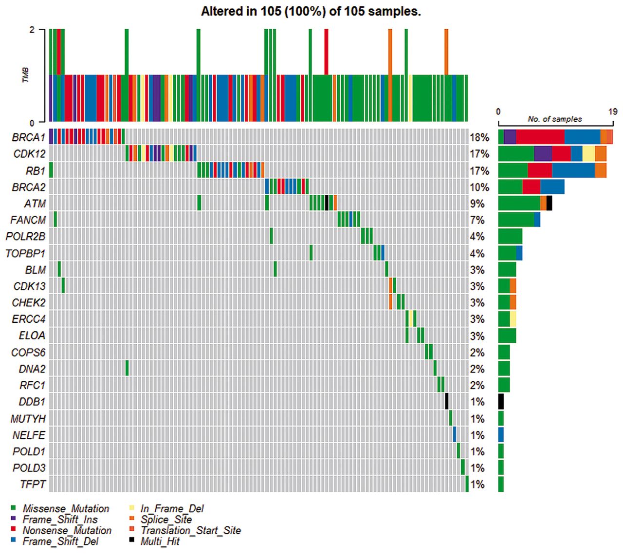
Sequencing data analyzed (n=778,623 variants) were subjected to SNV, MNV, and INDELs variant calling in exonic or splice-site regions of 356 DDR genes, which resulted in n=4,723 variants (Table II). These variants were subjected to further filtering and inspected with IGV to approve or reject them from the final list. Nine different TP53 variants were found in all serous patients and in one of the oCCC samples. All of these variants were previously reported in the IARC database, whereas seven of them were found in a TCGA dataset (c.97-1G>A, c.413C>T, c.646G>A, c.659A>C, c.701A>G, c.731G>A, c.772G>A).

In addition to TP53, we identified 33 variants in a total of 27 DDR genes, which were classified as pathogenic/likely pathogenic (P/LP) (Table IV) or as variants of uncertain significance (VUS) (Table V). All the 33 variants were unique, but some overlap between genes carrying these variants was observed (Figure 1). None of these variants were found in the TCGA cohort, however, other variants were found in 22 genes (ATM, BLM, BRCA1, BRCA2, CDK12, CDK13, CHEK2, COPS6, DDB1, DNA2, ELOA, ERCC4, FANCM, MUTYH, NELFE, POLD1, POLD3, POLR2B, RB1, RFC1, TFPT, TOPBP1) (Figure 2). There were no reported variants in FAAP24, GTF2H4, POLE4, RPA3, and XRCC4.

View this table:
  • View inline
  • View popup
  • Download powerpoint
Table IV.

Summary of pathogenic (P) or likely-pathogenic (LP) somatic variants found in exonic and splice-site regions of DNA-damage response genes by exome sequencing.

View this table:
  • View inline
  • View popup
  • Download powerpoint
Table V.

Summary of unknown significance variants found in exonic and splice-site regions of DNA-damage response genes by exome sequencing.

Figure 1.
  • Download figure
  • Open in new tab
  • Download powerpoint
Figure 1.

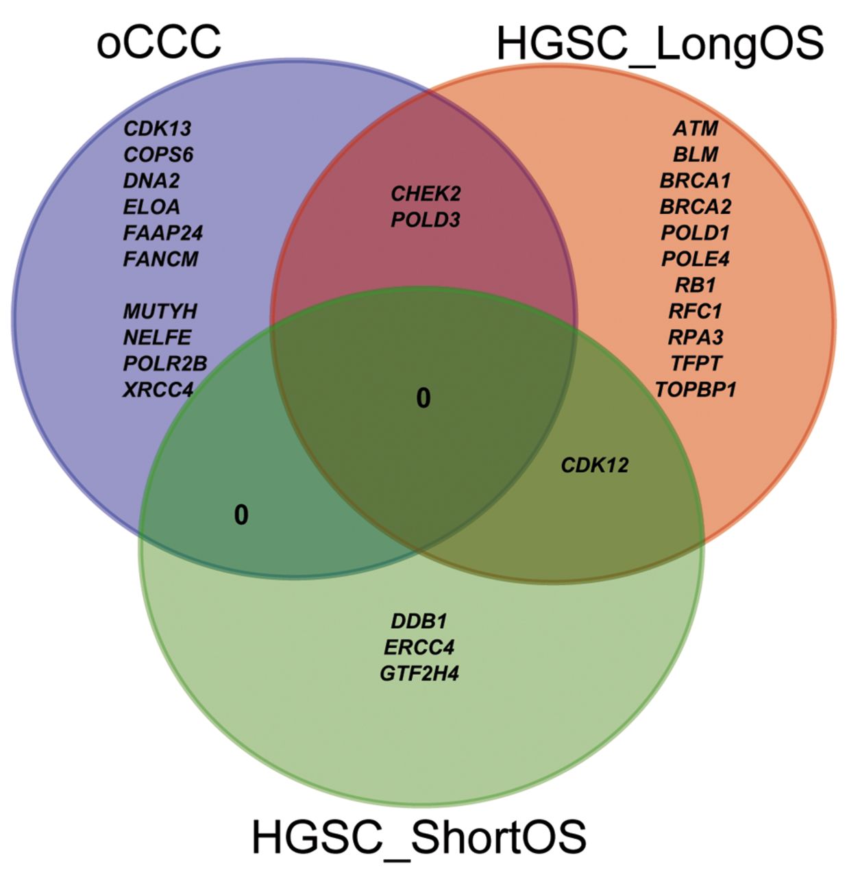
Venn diagram of overlapping genes carrying a variant among the three groups of patients: HGSC_LongOS: High-grade serous carcinoma patient with long overall survival; HGSC_ShortOS: high-grade serous carcinoma patient with short overall survival; oCCC: ovarian clear cell carcinoma.

Moreover, when investigating possible mutations in ARID1A and PIK3CA genes, pathogenic, likely-pathogenic or VUS variants were found exclusively in oCCC patients: PIK3CA: c.3140A>G (P) in oCCC_01, two VUS variants in oCCC_02: ARID1A: c.1780delC and PIK3CA: c.344_346delGAG, and one VUS variant in oCCC_03: ARID1A:c.3428_3429delGG, c.3429delG.

Discussion

Many clinical trials investigated BRCA1/2 variants and/or homologous recombination deficiency (HRD) status, as crucial criteria to select EOC patients with complete or partial response to platinum-based chemotherapy for PARPi treatment (7, 39). Less attention has been given to other DDR pathways in predicting response to treatment (26). In this study, we examined mutations in the exonic and splice-site regions of a total of 356 DDR genes in the same workflow to determine whether DDR pathways other than HR are affected in HGSC or oCCC.

BRCA1/2 were found only in HGSC_LongOS patients, which is in agreement with previous studies that demonstrated prolonged OS with BRCAness phenotype (40, 41). Twelve out of 16 variants (P+VUS) (75%) found in HGSC_LongOS were located in genes that have implications in the HR-pathway. Moreover, each patient in the HGSC_LongOS group carried a variant in a HR-related gene in contrast to HGSC_ShortOS patients (only one patient with HR-deficiency) and oCCC (3 out of 4 patients with HR-deficiency with 3 HR-related variants: 3 out of 13, 23%). It seems that genes that were defective in the HGSC_LongOS group are involved in different DDR pathways (Table IV and Table V, column “SUM”), whereas in HGSC_ShortOS and oCCC they are usually members of only one of the DDR pathways.

None of the 33 variants identified in this study were found in the TCGA cohort that contains only HGSC patients; however, other variants were found in 22 out of 27 unique genes: ATM, BLM, BRCA1, BRCA2, CDK12, CDK13, CHEK2, COPS6, DDB1, DNA2, ELOA, ERCC4, FANCM, MUTYH, NELFE, POLD1, POLD3, POLR2B, RB1, RFC1, TFPT, and TOPBP1 (Figure 2). There were no reported variants in the TCGA cohort for FAAP24, GTF2H4, POLE4, RPA3, and XRCC4 genes. Variants in two of these five non-TCGA genes FAAP24 and XRCC4, were observed only in the oCCC group, but not in the HGSC cohort from TCGA. Further exploration may reveal whether these genes are of special importance for the oCCC group and might be helpful in guiding treatment of oCCC patients. The FANCM/FAAP24 complex detects interstrand crosslinks and recruits other components of the FA core complex (42). Cells with FA deficiency display hypersensitivity to DNA cross-linking agents (43) and it might explain the prolonged OS of the oCCC_03 patient with FAAP24:c.635C>T variant. This patient is also a carrier of DNA2: c.68C>T. Loss of XRCC4 might activate to a back-up repair pathway called alternative end-joining repair (a-EJ) in human somatic cells (44). The a-EJ pathway is considered as a promising therapeutic target in HR or NHEJ deficient cancers, as its inhibition causes death of cells dependent on the a-EJ pathway to repair their DNA, but not normal cells (45). GTF2H4, POLE4, and RPA3 have also been previously reported in cancer and OC studies. Other variants in GTF2H4 have previously been shown to confer susceptibility to lung cancer (46, 47). Interestingly, loss of POLE4 was shown to sensitize HeLa cells to PARPi treatment (48) and hypersensitivity to ATR inhibition (49). Inhibition of RPA:RAD52 protein-protein interactions was reported to cause selective death of HRD cancer cell lines (50). Moreover, RPA exhaustion was reported to determine cisplatin response in HGSC cells (51).

Figure 2.
  • Download figure
  • Open in new tab
  • Download powerpoint
Figure 2.

DNA-damage response genes, in which we found 33 somatic variants. were submitted as a query to The Cancer Genome Atlas Ovarian Cancer dataset to find if any single/multiple nucleotide variants and small insertions/deletions variants for those genes have been previously reported. Missense_Mutation: The point mutation alters the protein structure by one amino acid; Nonsense_Mutation: a premature stop codon is created by the variant; Frame_Shift_Ins: insertion that moves the coding sequence out of frame; Frame_Shift_Del: deletion that moves the coding sequence out of frame; In_Frame_Del: deletion that keeps the sequence in frame; Splice_Site: the variant is within a configurable number of bases of a splice site; Multi_Hit: more than one variant type.

TP53 SNVs were found in all patients with HGSC, whereas variants in ARID1A and PIK3CA genes were only identified in oCCC patients, which is in line with previous reports (5, 23, 52). We also found one TP53 variant in an oCCC patient, which is unusual for oCCC that normally exhibits TP53 wild-type (25). Our finding may be explained by the fact that the tumor sample from this specific patient displayed a mixed clear-cell and endometroid histology although with dominant oCCC histology. In the tissue sample analyzed, both histology types may be present, thereby explaining the observation of the TP53 variants, although uncommon in clear cell, being reported in endometroid OC (25). Furthermore, all nine TP53 variants were previously reported in the IARC database and seven of them were present in the TCGA dataset (c.97-1G>A, c.413C>T, c.646G>A, c.659A>C, c.701A>G, c.731G>A, c.772G>A). In addition to TP53, we identified 33 variants, of which 5 were classified as “P” and 28 as “VUS”. In our opinion, it is relevant to report not only pathogenic variants with a documented role in the disease, but also VUSs, at least those that some pathogenic potential can be indicated, e.g., based on pathogenicity prediction bioinformatic tools. There is still a large number of variants revealed by NGS with undefined clinical significance, which accounts for about 40% of total variants (53). As variant classification may alter over time, periodical checks of the VUS outcomes might be necessary.

Our study has a few limitations. We decided to employ tumor-only testing based on our current settings to identify somatic variants, which is based on filtering variants against available databases (30). Therefore, our findings might be further validated by matched tumor and blood/adjacent normal tissue parallel testing, where somatic tumor-only variants are identified by subtraction of blood/normal tissue variants from the total variants found in the tumor tissue (30).

In conclusion, by using a discovery cohort of relatively few patients but investigating a total of 356 DDR genes using exome sequencing with high coverage, we have identified 42 somatic mutations that were not only limited to TP53, BRCA1/2, and HR-associated genes. They may display a potential role as biomarkers to predict platinum-based chemotherapy or PARPi treatment response, disease severity (measured on OS) or progression, as differences in disrupted DDR pathways between HGSC_LongOS, HGSC_ShortOS, and oCCC groups were observed.

Acknowledgements

The Authors would like to thank Mikael Kronborg Christophersen for performing whole exome sequencing experiments. The Authors thank the Bio- and Genome Bank Denmark for providing samples used in this study. The Authors received financial support from: The Mermaid Foundation, available at: http://www.mermaidprojektet.dk/ (JLJ, DNPO, CKH and EVH), Danish Cancer Research Foundation, available at: http://www.dansk-kraeftforsknings-fond.dk/ (EVH), and Herlev Hospital Research Council, available at: https://www.herlevhospital.dk/forskning/ (EVH).

Footnotes

  • Authors’ Contributions

    All Authors participated in data analysis, discussed the results, and contributed to the writing of the final manuscript.

  • Conflicts of Interest

    The Authors declare that there are no conflicts of interest in relation to this study.

  • Received February 17, 2023.
  • Revision received March 3, 2023.
  • Accepted March 6, 2023.
  • Copyright © 2023 International Institute of Anticancer Research (Dr. George J. Delinasios), All rights reserved.

This article is an open access article distributed under the terms and conditions of the Creative Commons Attribution (CC BY-NC-ND) 4.0 international license (https://creativecommons.org/licenses/by-nc-nd/4.0).

References

  1. ↵
    1. Alshamrani AA
    : Roles of microRNAs in ovarian cancer tumorigenesis: Two decades later, what have we learned? Front Oncol 10: 1084, 2020. PMID: 32850313. DOI: 10.3389/fonc.2020.01084
    OpenUrlCrossRefPubMed
  2. ↵
    1. Berek JS,
    2. Kehoe ST,
    3. Kumar L and
    4. Friedlander M
    : Cancer of the ovary, fallopian tube, and peritoneum. Int J Gynaecol Obstet 143(Suppl 2): 59-78, 2018. PMID: 30306591. DOI: 10.1002/ijgo.12614
    OpenUrlCrossRefPubMed
  3. ↵
    1. Prat J
    : Ovarian carcinomas: five distinct diseases with different origins, genetic alterations, and clinicopathological features. Virchows Arch 460(3): 237-249, 2012. PMID: 22322322. DOI: 10.1007/s00428-012-1203-5
    OpenUrlCrossRefPubMed
  4. ↵
    1. Danish Gynaecologic Cancer Database (DGCD)
    : Annual Report 2017-2019, 2019. Available at: http://www.dgcg.dk/images/rsrapport_DGCD_2017-19.pdf [Last accessed on March 11, 2022]
  5. ↵
    1. Cancer Genome Atlas Research Network
    : Integrated genomic analyses of ovarian carcinoma. Nature 474(7353): 609-615, 2011. PMID: 21720365. DOI: 10.1038/nature10166
    OpenUrlCrossRefPubMed
  6. ↵
    1. Mandilaras V,
    2. Garg S,
    3. Cabanero M,
    4. Tan Q,
    5. Pastrello C,
    6. Burnier J,
    7. Karakasis K,
    8. Wang L,
    9. Dhani NC,
    10. Butler MO,
    11. Bedard PL,
    12. Siu LL,
    13. Clarke B,
    14. Shaw PA,
    15. Stockley T,
    16. Jurisica I,
    17. Oza AM and
    18. Lheureux S
    : TP53 mutations in high grade serous ovarian cancer and impact on clinical outcomes: a comparison of next generation sequencing and bioinformatics analyses. Int J Gynecol Cancer 29(2): 346-352, 2019. PMID: 30659026. DOI: 10.1136/ijgc-2018-000087
    OpenUrlAbstract/FREE Full Text
  7. ↵
    1. Konstantinopoulos PA,
    2. Ceccaldi R,
    3. Shapiro GI and
    4. D’Andrea AD
    : Homologous recombination deficiency: Exploiting the fundamental vulnerability of ovarian cancer. Cancer Discov 5(11): 1137-1154, 2015. PMID: 26463832. DOI: 10.1158/2159-8290.CD-15-0714
    OpenUrlAbstract/FREE Full Text
  8. ↵
    1. Peixoto A,
    2. Pinto P,
    3. Guerra J,
    4. Pinheiro M,
    5. Santos C,
    6. Pinto C,
    7. Santos R,
    8. Escudeiro C,
    9. Bartosch C,
    10. Canário R,
    11. Barbosa A,
    12. Gouveia A,
    13. Petiz A,
    14. Abreu MH,
    15. Sousa S,
    16. Pereira D,
    17. Silva J and
    18. Teixeira MR
    : Tumor testing for somatic and germline BRCA1/BRCA2 variants in ovarian cancer patients in the context of strong founder effects. Front Oncol 10: 1318, 2020. PMID: 32850417. DOI: 10.3389/fonc.2020.01318
    OpenUrlCrossRefPubMed
  9. ↵
    1. Ronco C,
    2. Martin AR,
    3. Demange L and
    4. Benhida R
    : ATM, ATR, CHK1, CHK2 and WEE1 inhibitors in cancer and cancer stem cells. Medchemcomm 8(2): 295-319, 2016. PMID: 30108746. DOI: 10.1039/c6md00439c
    OpenUrlCrossRefPubMed
  10. ↵
    1. Paull TT and
    2. Lee JH
    : The Mre11/Rad50/Nbs1 complex and its role as a DNA double-strand break sensor for ATM. Cell Cycle 4(6): 737-740, 2005. PMID: 15908798. DOI: 10.4161/cc.4.6.1715
    OpenUrlCrossRefPubMed
  11. ↵
    1. Blackford AN and
    2. Jackson SP
    : ATM, ATR, and DNA-PK: The trinity at the heart of the DNA damage response. Mol Cell 66(6): 801-817, 2017. PMID: 28622525. DOI: 10.1016/j.molcel.2017.05.015
    OpenUrlCrossRefPubMed
  12. ↵
    1. Damia G and
    2. Broggini M
    : Platinum resistance in ovarian cancer: Role of DNA repair. Cancers (Basel) 11(1): 119, 2019. PMID: 30669514. DOI: 10.3390/cancers11010119
    OpenUrlCrossRefPubMed
  13. ↵
    1. Curtin NJ
    : DNA repair dysregulation from cancer driver to therapeutic target. Nat Rev Cancer 12(12): 801-817, 2012. PMID: 23175119. DOI: 10.1038/nrc3399
    OpenUrlCrossRefPubMed
  14. ↵
    1. Pearl LH,
    2. Schierz AC,
    3. Ward SE,
    4. Al-Lazikani B and
    5. Pearl FM
    : Therapeutic opportunities within the DNA damage response. Nat Rev Cancer 15(3): 166-180, 2015. PMID: 25709118. DOI: 10.1038/nrc3891
    OpenUrlCrossRefPubMed
  15. ↵
    1. Lord CJ and
    2. Ashworth A
    : PARP inhibitors: Synthetic lethality in the clinic. Science 355(6330): 1152-1158, 2017. PMID: 28302823. DOI: 10.1126/science.aam7344
    OpenUrlAbstract/FREE Full Text
  16. ↵
    1. Jiang X,
    2. Li X,
    3. Li W,
    4. Bai H and
    5. Zhang Z
    : PARP inhibitors in ovarian cancer: Sensitivity prediction and resistance mechanisms. J Cell Mol Med 23(4): 2303-2313, 2019. PMID: 30672100. DOI: 10.1111/jcmm.14133
    OpenUrlCrossRefPubMed
  17. ↵
    1. European Medicines Agency
    : Lynparza recommended for approval in ovarian cancer. Available at: https://www.ema.europa.eu/en/news/lynparza-recommended-approval-ovarian-cancer [Last accessed on March 11, 2022]
  18. ↵
    1. Gadducci A and
    2. Cosio S
    : Randomized clinical trials and real world prospective observational studies on bevacizumab, PARP inhibitors, and immune checkpoint inhibitors in the first-line treatment of advanced ovarian carcinoma: a critical review. Anticancer Res 41(10): 4673-4685, 2021. PMID: 34593415. DOI: 10.21873/anticanres.15281
    OpenUrlAbstract/FREE Full Text
  19. ↵
    1. Vergote I,
    2. González-Martín A,
    3. Ray-Coquard I,
    4. Harter P,
    5. Colombo N,
    6. Pujol P,
    7. Lorusso D,
    8. Mirza MR,
    9. Brasiuniene B,
    10. Madry R,
    11. Brenton JD,
    12. Ausems MGEM,
    13. Büttner R,
    14. Lambrechts D and European experts’ consensus group
    : European experts consensus: BRCA/homologous recombination deficiency testing in first-line ovarian cancer. Ann Oncol 33(3): 276-287, 2022. PMID: 34861371. DOI: 10.1016/j.annonc.2021.11.013
    OpenUrlCrossRefPubMed
  20. ↵
    1. Turchiano A,
    2. Loconte DC,
    3. De Nola R,
    4. Arezzo F,
    5. Chiarello G,
    6. Pantaleo A,
    7. Iacoviello M,
    8. Bagnulo R,
    9. De Luisi A,
    10. Perrelli S,
    11. Martino S,
    12. Ranieri C,
    13. Garganese A,
    14. Stella A,
    15. Forleo C,
    16. Loizzi V,
    17. Marinaccio M,
    18. Cicinelli E,
    19. Cormio G and
    20. Resta N
    : Beyond BRCA1/2: Homologous recombination repair genetic profile in a large cohort of apulian ovarian cancers. Cancers (Basel) 14(2): 365, 2022. PMID: 35053526. DOI: 10.3390/cancers14020365
    OpenUrlCrossRefPubMed
    1. Toh M and
    2. Ngeow J
    : Homologous recombination deficiency: Cancer predispositions and treatment implications. Oncologist 26(9): e1526-e1537, 2021. PMID: 34021944. DOI: 10.1002/onco.13829
    OpenUrlCrossRefPubMed
  21. ↵
    1. Suszynska M,
    2. Ratajska M and
    3. Kozlowski P
    : BRIP1, RAD51C, and RAD51D mutations are associated with high susceptibility to ovarian cancer: mutation prevalence and precise risk estimates based on a pooled analysis of ~30,000 cases. J Ovarian Res 13(1): 50, 2020. PMID: 32359370. DOI: 10.1186/s13048-020-00654-3
    OpenUrlCrossRefPubMed
  22. ↵
    1. Iida Y,
    2. Okamoto A,
    3. Hollis RL,
    4. Gourley C and
    5. Herrington CS
    : Clear cell carcinoma of the ovary: a clinical and molecular perspective. Int J Gynecol Cancer 31(4): 605-616, 2021. PMID: 32948640. DOI: 10.1136/ijgc-2020-001656
    OpenUrlAbstract/FREE Full Text
  23. ↵
    1. Oliveira DVNP,
    2. Schnack TH,
    3. Poulsen TS,
    4. Christiansen AP,
    5. Høgdall CK and
    6. Høgdall EV
    : Genomic sub-classification of ovarian clear cell carcinoma revealed by distinct mutational signatures. Cancers (Basel) 13(20): 5242, 2021. PMID: 34680390. DOI: 10.3390/cancers13205242
    OpenUrlCrossRefPubMed
  24. ↵
    1. Wong OGW,
    2. Li J and
    3. Cheung ANY
    : Targeting DNA damage response pathway in ovarian clear cell carcinoma. Front Oncol 11: 666815, 2021. PMID: 34737943. DOI: 10.3389/fonc.2021.666815
    OpenUrlCrossRefPubMed
  25. ↵
    1. Gee ME,
    2. Faraahi Z,
    3. McCormick A and
    4. Edmondson RJ
    : DNA damage repair in ovarian cancer: unlocking the heterogeneity. J Ovarian Res 11(1): 50, 2018. PMID: 29925418. DOI: 10.1186/s13048-018-0424-x
    OpenUrlCrossRefPubMed
  26. ↵
    1. Danish Gynecologic Cancer Group
    : Annual report of the Danish Gynecologic Cancer Database 2016-17. Danish Gynecol Cancer Database, 2017. Available at: http://www.dgcg.dk/images/Grupper/Databasegruppen/rsrapport_DGCD_2016-17_endelig_anonymiseret.pdf [Last accessed on March 4, 2023]
  27. ↵
    1. RCT
    : R: A language and environment for statistical computing. R Foundation for Statistical Computing, Vienna, Austria. Available at: https://www.r-project.org/ [Last accessed on March 4, 2023]
  28. ↵
    1. Vestergaard LK,
    2. Oliveira DNP,
    3. Poulsen TS,
    4. Høgdall CK and
    5. Høgdall EV
    : Oncomine™ Comprehensive Assay v3 vs. Oncomine™ Comprehensive Assay Plus. Cancers (Basel) 13(20): 5230, 2021. PMID: 34680378. DOI: 10.3390/cancers13205230
    OpenUrlCrossRefPubMed
  29. ↵
    1. Sukhai MA,
    2. Misyura M,
    3. Thomas M,
    4. Garg S,
    5. Zhang T,
    6. Stickle N,
    7. Virtanen C,
    8. Bedard PL,
    9. Siu LL,
    10. Smets T,
    11. Thijs G,
    12. Van Vooren S,
    13. Kamel-Reid S and
    14. Stockley TL
    : Somatic tumor variant filtration strategies to optimize tumor-only molecular profiling using targeted next-generation sequencing panels. J Mol Diagn 21(2): 261-273, 2019. PMID: 30576869. DOI: 10.1016/j.jmoldx.2018.09.008
    OpenUrlCrossRefPubMed
  30. ↵
    1. Tate JG,
    2. Bamford S,
    3. Jubb HC,
    4. Sondka Z,
    5. Beare DM,
    6. Bindal N,
    7. Boutselakis H,
    8. Cole CG,
    9. Creatore C,
    10. Dawson E,
    11. Fish P,
    12. Harsha B,
    13. Hathaway C,
    14. Jupe SC,
    15. Kok CY,
    16. Noble K,
    17. Ponting L,
    18. Ramshaw CC,
    19. Rye CE,
    20. Speedy HE,
    21. Stefancsik R,
    22. Thompson SL,
    23. Wang S,
    24. Ward S,
    25. Campbell PJ and
    26. Forbes SA
    : COSMIC: the catalogue of somatic mutations in cancer. Nucleic Acids Res 47(D1): D941-D947, 2019. PMID: 30371878. DOI: 10.1093/nar/gky1015
    OpenUrlCrossRefPubMed
  31. ↵
    1. Mayakonda A,
    2. Lin DC,
    3. Assenov Y,
    4. Plass C and
    5. Koeffler HP
    : Maftools: efficient and comprehensive analysis of somatic variants in cancer. Genome Res 28(11): 1747-1756, 2018. PMID: 30341162. DOI: 10.1101/gr.239244.118
    OpenUrlAbstract/FREE Full Text
  32. ↵
    1. Bouaoun L,
    2. Sonkin D,
    3. Ardin M,
    4. Hollstein M,
    5. Byrnes G,
    6. Zavadil J and
    7. Olivier M
    : TP53 variations in human cancers: new lessons from the IARC TP53 database and genomics data. Hum Mutat 37(9): 865-876, 2016. PMID: 27328919. DOI: 10.1002/humu.23035
    OpenUrlCrossRefPubMed
  33. ↵
    1. Sherr CJ and
    2. McCormick F
    : The RB and p53 pathways in cancer. Cancer Cell 2(2): 103-112, 2002. PMID: 12204530. DOI: 10.1016/s1535-6108(02)00102-2
    OpenUrlCrossRefPubMed
  34. ↵
    1. Vélez-Cruz R,
    2. Manickavinayaham S,
    3. Biswas AK,
    4. Clary RW,
    5. Premkumar T,
    6. Cole F and
    7. Johnson DG
    : RB localizes to DNA double-strand breaks and promotes DNA end resection and homologous recombination through the recruitment of BRG1. Genes Dev 30(22): 2500-2512, 2016. PMID: 27940962. DOI: 10.1101/gad.288282.116
    OpenUrlAbstract/FREE Full Text
  35. ↵
    1. Kanehisa M,
    2. Sato Y,
    3. Kawashima M,
    4. Furumichi M and
    5. Tanabe M
    : KEGG as a reference resource for gene and protein annotation. Nucleic Acids Res 44(D1): D457-D462, 2016. PMID: 26476454. DOI: 10.1093/nar/gkv1070
    OpenUrlCrossRefPubMed
  36. ↵
    1. Gillespie M,
    2. Jassal B,
    3. Stephan R,
    4. Milacic M,
    5. Rothfels K,
    6. Senff-Ribeiro A,
    7. Griss J,
    8. Sevilla C,
    9. Matthews L,
    10. Gong C,
    11. Deng C,
    12. Varusai T,
    13. Ragueneau E,
    14. Haider Y,
    15. May B,
    16. Shamovsky V,
    17. Weiser J,
    18. Brunson T,
    19. Sanati N,
    20. Beckman L,
    21. Shao X,
    22. Fabregat A,
    23. Sidiropoulos K,
    24. Murillo J,
    25. Viteri G,
    26. Cook J,
    27. Shorser S,
    28. Bader G,
    29. Demir E,
    30. Sander C,
    31. Haw R,
    32. Wu G,
    33. Stein L,
    34. Hermjakob H and
    35. D’Eustachio P
    : The reactome pathway knowledgebase 2022. Nucleic Acids Res 50(D1): D687-D692, 2022. PMID: 34788843. DOI: 10.1093/nar/gkab1028
    OpenUrlCrossRefPubMed
  37. ↵
    1. Nishimura D
    : BioCarta. Biotech Software & Internet Report 2(3): 117-120, 2013. DOI: 10.1089/152791601750294344
    OpenUrlCrossRef
  38. ↵
    1. Banerjee S,
    2. Gonzalez-Martin A,
    3. Harter P,
    4. Lorusso D,
    5. Moore KN,
    6. Oaknin A and
    7. Ray-Coquard I
    : First-line PARP inhibitors in ovarian cancer: summary of an ESMO Open - Cancer Horizons round-table discussion. ESMO Open 5(6): e001110, 2020. PMID: 33310779. DOI: 10.1136/esmoopen-2020-001110
    OpenUrlAbstract/FREE Full Text
  39. ↵
    1. Hjortkjær M,
    2. Malik Aagaard Jørgensen M,
    3. Waldstrøm M,
    4. Ørnskov D,
    5. Søgaard-Andersen E,
    6. Jakobsen A and
    7. Dahl-Steffensen K
    : The clinical importance of BRCAness in a population-based cohort of Danish epithelial ovarian cancer. Int J Gynecol Cancer 29(1): 166-173, 2019. PMID: 30640700. DOI: 10.1136/ijgc-2018-000017
    OpenUrlAbstract/FREE Full Text
  40. ↵
    1. Pennington KP,
    2. Walsh T,
    3. Harrell MI,
    4. Lee MK,
    5. Pennil CC,
    6. Rendi MH,
    7. Thornton A,
    8. Norquist BM,
    9. Casadei S,
    10. Nord AS,
    11. Agnew KJ,
    12. Pritchard CC,
    13. Scroggins S,
    14. Garcia RL,
    15. King MC and
    16. Swisher EM
    : Germline and somatic mutations in homologous recombination genes predict platinum response and survival in ovarian, fallopian tube, and peritoneal carcinomas. Clin Cancer Res 20(3): 764-775, 2014. PMID: 24240112. DOI: 10.1158/1078-0432.CCR-13-2287
    OpenUrlAbstract/FREE Full Text
  41. ↵
    1. Wang Y,
    2. Leung JW,
    3. Jiang Y,
    4. Lowery MG,
    5. Do H,
    6. Vasquez KM,
    7. Chen J,
    8. Wang W and
    9. Li L
    : FANCM and FAAP24 maintain genome stability via cooperative as well as unique functions. Mol Cell 49(5): 997-1009, 2013. PMID: 23333308. DOI: 10.1016/j.molcel.2012.12.010
    OpenUrlCrossRefPubMed
  42. ↵
    1. Liu W,
    2. Palovcak A,
    3. Li F,
    4. Zafar A,
    5. Yuan F and
    6. Zhang Y
    : Fanconi anemia pathway as a prospective target for cancer intervention. Cell Biosci 10: 39, 2020. PMID: 32190289. DOI: 10.1186/s13578-020-00401-7
    OpenUrlCrossRefPubMed
  43. ↵
    1. Ruis B,
    2. Molan A,
    3. Takasugi T and
    4. Hendrickson EA
    : Absence of XRCC4 and its paralogs in human cells reveal differences in outcomes for DNA repair and V(D)J recombination. DNA Repair (Amst) 85: 102738, 2020. PMID: 31731258. DOI: 10.1016/j.dnarep.2019.102738
    OpenUrlCrossRefPubMed
  44. ↵
    1. Sallmyr A and
    2. Tomkinson AE
    : Repair of DNA double-strand breaks by mammalian alternative end-joining pathways. J Biol Chem 293(27): 10536-10546, 2018. PMID: 29530982. DOI: 10.1074/jbc.TM117.000375
    OpenUrlAbstract/FREE Full Text
  45. ↵
    1. Wang M,
    2. Liu H,
    3. Liu Z,
    4. Yi X,
    5. Bickeboller H,
    6. Hung RJ,
    7. Brennan P,
    8. Landi MT,
    9. Caporaso N,
    10. Christiani DC,
    11. Doherty JA, TRICL Research Team,
    12. Amos CI and
    13. Wei Q
    : Genetic variant in DNA repair gene GTF2H4 is associated with lung cancer risk: a large-scale analysis of six published GWAS datasets in the TRICL consortium. Carcinogenesis 37(9): 888-896, 2016. PMID: 27288692. DOI: 10.1093/carcin/bgw070
    OpenUrlCrossRefPubMed
  46. ↵
    1. Song X,
    2. Wang S,
    3. Hong X,
    4. Li X,
    5. Zhao X,
    6. Huai C,
    7. Chen H,
    8. Gao Z,
    9. Qian J,
    10. Wang J,
    11. Han B,
    12. Bai C,
    13. Li Q,
    14. Wu J and
    15. Lu D
    : Single nucleotide polymorphisms of nucleotide excision repair pathway are significantly associated with outcomes of platinum-based chemotherapy in lung cancer. Sci Rep 7(1): 11785, 2017. PMID: 28924235. DOI: 10.1038/s41598-017-08257-7
    OpenUrlCrossRefPubMed
  47. ↵
    1. Clements KE,
    2. Schleicher EM,
    3. Thakar T,
    4. Hale A,
    5. Dhoonmoon A,
    6. Tolman NJ,
    7. Sharma A,
    8. Liang X,
    9. Imamura Kawasawa Y,
    10. Nicolae CM,
    11. Wang HG,
    12. De S and
    13. Moldovan GL
    : Identification of regulators of poly-ADP-ribose polymerase inhibitor response through complementary CRISPR knockout and activation screens. Nat Commun 11(1): 6118, 2020. PMID: 33257658. DOI: 10.1038/s41467-020-19961-w
    OpenUrlCrossRefPubMed
  48. ↵
    1. Hustedt N,
    2. Álvarez-Quilón A,
    3. McEwan A,
    4. Yuan JY,
    5. Cho T,
    6. Koob L,
    7. Hart T and
    8. Durocher D
    : A consensus set of genetic vulnerabilities to ATR inhibition. Open Biol 9(9): 190156, 2019. PMID: 31506018. DOI: 10.1098/rsob.190156
    OpenUrlCrossRefPubMed
  49. ↵
    1. Al-Mugotir M,
    2. Lovelace JJ,
    3. George J,
    4. Bessho M,
    5. Pal D,
    6. Struble L,
    7. Kolar C,
    8. Rana S,
    9. Natarajan A,
    10. Bessho T and
    11. Borgstahl GEO
    : Selective killing of homologous recombination-deficient cancer cell lines by inhibitors of the RPA:RAD52 protein-protein interaction. PLoS One 16(3): e0248941, 2021. PMID: 33784323. DOI: 10.1371/journal.pone.0248941
    OpenUrlCrossRefPubMed
  50. ↵
    1. Bélanger F,
    2. Fortier E,
    3. Dubé M,
    4. Lemay JF,
    5. Buisson R,
    6. Masson JY,
    7. Elsherbiny A,
    8. Costantino S,
    9. Carmona E,
    10. Mes-Masson AM,
    11. Wurtele H and
    12. Drobetsky E
    : Replication protein A availability during DNA replication stress is a major determinant of cisplatin resistance in ovarian cancer cells. Cancer Res 78(19): 5561-5573, 2018. PMID: 30072396. DOI: 10.1158/0008-5472.CAN-18-0618
    OpenUrlAbstract/FREE Full Text
  51. ↵
    1. Zhang Y,
    2. Cao L,
    3. Nguyen D and
    4. Lu H
    : TP53 mutations in epithelial ovarian cancer. Transl Cancer Res 5(6): 650-663, 2016. PMID: 30613473. DOI: 10.21037/tcr.2016.08.40
    OpenUrlCrossRefPubMed
  52. ↵
    1. Federici G and
    2. Soddu S
    : Variants of uncertain significance in the era of high-throughput genome sequencing: a lesson from breast and ovary cancers. J Exp Clin Cancer Res 39(1): 46, 2020. PMID: 32127026. DOI: 10.1186/s13046-020-01554-6
    OpenUrlCrossRefPubMed
PreviousNext
Back to top

In this issue

Anticancer Research: 43 (5)
Anticancer Research
Vol. 43, Issue 5
May 2023
  • Table of Contents
  • Table of Contents (PDF)
  • About the Cover
  • Index by author
  • Back Matter (PDF)
  • Ed Board (PDF)
  • Front Matter (PDF)
Print
Download PDF
Article Alerts
Sign In to Email Alerts with your Email Address
Email Article

Thank you for your interest in spreading the word on Anticancer Research.

NOTE: We only request your email address so that the person you are recommending the page to knows that you wanted them to see it, and that it is not junk mail. We do not capture any email address.

Enter multiple addresses on separate lines or separate them with commas.
Somatic Variants in DNA Damage Response Genes in Ovarian Cancer Patients Using Whole-exome Sequencing
(Your Name) has sent you a message from Anticancer Research
(Your Name) thought you would like to see the Anticancer Research web site.
CAPTCHA
This question is for testing whether or not you are a human visitor and to prevent automated spam submissions.
1 + 1 =
Solve this simple math problem and enter the result. E.g. for 1+3, enter 4.
Citation Tools
Somatic Variants in DNA Damage Response Genes in Ovarian Cancer Patients Using Whole-exome Sequencing
JOANNA LOPACINSKA-JOERGENSEN, DOUGLAS V.N.P. OLIVEIRA, TIM SVENSTRUP POULSEN, CLAUS K. HOEGDALL, ESTRID V. HOEGDALL
Anticancer Research May 2023, 43 (5) 1891-1900; DOI: 10.21873/anticanres.16348

Citation Manager Formats

  • BibTeX
  • Bookends
  • EasyBib
  • EndNote (tagged)
  • EndNote 8 (xml)
  • Medlars
  • Mendeley
  • Papers
  • RefWorks Tagged
  • Ref Manager
  • RIS
  • Zotero
Reprints and Permissions
Share
Somatic Variants in DNA Damage Response Genes in Ovarian Cancer Patients Using Whole-exome Sequencing
JOANNA LOPACINSKA-JOERGENSEN, DOUGLAS V.N.P. OLIVEIRA, TIM SVENSTRUP POULSEN, CLAUS K. HOEGDALL, ESTRID V. HOEGDALL
Anticancer Research May 2023, 43 (5) 1891-1900; DOI: 10.21873/anticanres.16348
Reddit logo Twitter logo Facebook logo Mendeley logo
  • Tweet Widget
  • Facebook Like
  • Google Plus One

Jump to section

  • Article
    • Abstract
    • Materials and Methods
    • Results
    • Discussion
    • Acknowledgements
    • Footnotes
    • References
  • Figures & Data
  • Info & Metrics
  • PDF

Related Articles

  • No related articles found.
  • PubMed
  • Google Scholar

Cited By...

  • No citing articles found.
  • Google Scholar

More in this TOC Section

  • TIG1 Inhibits the mTOR Signaling Pathway in Malignant Melanoma Through the VAC14 Protein
  • Novel α-Trifluoromethyl Chalcone Exerts Antitumor Effects Against Prostate Cancer Cells
  • Differential Effects of Anti-PD-1/PD-L1 Checkpoint Inhibitors on Adhesion Molecules and Cytokine Secretion by THP-1 Monocytes
Show more Experimental Studies

Similar Articles

Keywords

  • Somatic variants
  • DNA damage response
  • ovarian cancer
Anticancer Research

© 2023 Anticancer Research

Powered by HighWire