
Main menu

  • Home
  • Current Issue
  • Archive
  • Info for
    • Authors
    • Editorial Policies
    • Subscribers
    • Advertisers
    • Editorial Board
    • Special Issues 2025
  • Journal Metrics
  • Other Publications
    • In Vivo
    • Cancer Genomics & Proteomics
    • Cancer Diagnosis & Prognosis
  • More
    • IIAR
    • Conferences
    • 2008 Nobel Laureates
  • About Us
    • General Policy
    • Contact
  • Other Publications
    • Anticancer Research
    • In Vivo
    • Cancer Genomics & Proteomics

User menu

  • Register
  • Subscribe
  • My alerts
  • Log in
  • My Cart

Search

  • Advanced search
Anticancer Research
  • Other Publications
    • Anticancer Research
    • In Vivo
    • Cancer Genomics & Proteomics
  • Register
  • Subscribe
  • My alerts
  • Log in
  • My Cart
Anticancer Research

Advanced Search

  • Home
  • Current Issue
  • Archive
  • Info for
    • Authors
    • Editorial Policies
    • Subscribers
    • Advertisers
    • Editorial Board
    • Special Issues 2025
  • Journal Metrics
  • Other Publications
    • In Vivo
    • Cancer Genomics & Proteomics
    • Cancer Diagnosis & Prognosis
  • More
    • IIAR
    • Conferences
    • 2008 Nobel Laureates
  • About Us
    • General Policy
    • Contact
  • Visit us on Facebook
  • Follow us on Linkedin
Review ArticleReview
Open Access

Evolution of Models of Prostate Cancer: Their Contribution to Current Therapies

ANA I. FAUSTINO-ROCHA, CATARINA JOTA-BAPTISTA, ELISABETE NASCIMENTO-GONÇALVES and PAULA A. OLIVEIRA
Anticancer Research January 2023, 43 (1) 323-333; DOI: https://doi.org/10.21873/anticanres.16167
ANA I. FAUSTINO-ROCHA
1Center for the Research and Technology of Agro-Environmental and Biological Sciences (CITAB), Inov4Agro, Vila Real, Portugal;
2Comprehensive Health Research Centre (CHRC), Évora, Portugal;
3Department of Zootechnics, School of Sciences and Technology (ECT), University of Évora, Évora, Portugal;
  • Find this author on Google Scholar
  • Find this author on PubMed
  • Search for this author on this site
  • For correspondence: anafaustino.faustino@sapo.pt
CATARINA JOTA-BAPTISTA
1Center for the Research and Technology of Agro-Environmental and Biological Sciences (CITAB), Inov4Agro, Vila Real, Portugal;
4Department of Veterinary Medicine, Surgery and Anatomy, Institute of Biomedicine (IBIOMED), University of León, Léon, Spain;
  • Find this author on Google Scholar
  • Find this author on PubMed
  • Search for this author on this site
ELISABETE NASCIMENTO-GONÇALVES
1Center for the Research and Technology of Agro-Environmental and Biological Sciences (CITAB), Inov4Agro, Vila Real, Portugal;
5LAQV-REQUIMTE, Department of Chemistry, University of Aveiro, Aveiro, Portugal;
  • Find this author on Google Scholar
  • Find this author on PubMed
  • Search for this author on this site
PAULA A. OLIVEIRA
1Center for the Research and Technology of Agro-Environmental and Biological Sciences (CITAB), Inov4Agro, Vila Real, Portugal;
6Department of Veterinary Sciences, University of Trás-os-Montes and Alto Douro, Vila Real, Portugal
  • Find this author on Google Scholar
  • Find this author on PubMed
  • Search for this author on this site
  • Article
  • Figures & Data
  • Info & Metrics
  • PDF
Loading

Abstract

Prostate cancer (PCa) is among the most frequent cancers worldwide. Nowadays, several therapeutic strategies are available for PCa treatment, namely chemotherapy, immunotherapy, radiotherapy, and hormonal therapy. Despite existing therapeutic approaches, in vitro and in vivo models are essential to better understand cancer development and search for more effective therapies, with a positive impact in cancer patient survival and quality of life. Among several models available, the rat model is the one most frequently used, since it shares anatomical, physiological, pathological, and behavioral features with humans. Animal models can be classified as: spontaneous, chemically-induced; hormonally-induced; implantation of cancer cell lines obtained from humans or from the same species, in the place of disease development or in a different place; and genetically-modified models. The chemically-induced models are among the most frequently used for PCa research. This manuscript provides a comprehensive overview of PCa models, presenting their application, advantages, and disadvantages, and their importance for the development of current therapies for prostate cancer.

Key Words:
  • Animal models
  • mice
  • prostate carcinogenesis
  • rats
  • rodents
  • review

Prostate cancer (PCa) is among the most common cancers in men. In 2020, PCa was the third most diagnosed neoplasia, with 1,414,259 new cases (7.3% of all cancer cases) and 375,304 deaths (1).

During the last decades, PCa therapy has seen a remarkable evolution in many sectors, namely chemotherapy, immunotherapy, radiotherapy, and hormonal therapy. New precision medicine strategies and treatments are also suffering massive developments nowadays, also for PCa management, namely new proteomic and genomic technologies, non-coding RNA therapeutics or gene editing technologies (2).

Over the decades, different animal and non-animal models have been used to describe PCa morphologically, evaluate its development, set up prognostics, and develop more successful and globally used treatments.

This work aimed to review the different models used in PCa research among the decades and highlight their significant contribution to the discovery of various therapies regularly used nowadays to treat men with PCa and significantly improve their lives.

Prostate Cancer: An Overview

PCa is a neoplasia that results from the uncontrolled growth of the prostatic gland cells. Almost all PCas are adenocarcinomas. However, histologically, two types can be considered: acinar adenocarcinoma and non-acinar carcinoma. PCa acinar adenocarcinomas include atrophic, colloid, pseudohyperplastic, signet ring, and oncocytic carcinomas, among others. Non-acinar carcinoma only includes 5-10% of PCas, as the ductal adenocarcinoma, urothelial carcinoma, sarcomatoid carcinoma, squamous, basal cell carcinoma, or the small-cell carcinoma (3).

PCa is usually asymptomatic at the early stages with an average age of diagnosis at 66 years old. The most reported symptoms include dysuria, pollakiuria, and nocturia, which also occur in patients with prostatic hypertrophy. At an advanced stage, urinary retention is common, as well as back pain, since the axis skeleton is the most usual site of metastasis (4). Detection of elevated levels of prostate-specific antigen (PSA >4 ng/ml) is the first suspicion for the presence of PCa. However, tissue biopsy is necessary to confirm the diagnosis, since PSA levels may be found elevated in men with normal prostate (4). The growing incidence of PCa can be attributed to the increased awareness of PCa and routine use of the PSA analysis (5). It was found that a single PSA measurement between 44 and 50 years-old could predict PCa up to 25 years later (6).

Risk factors of PCa have been widely studied and discussed over the last decades. The factors most frequently mentioned are age, ethnicity, genetic, inflammation and infection, lifestyle, and androgen-related (5). The incidence of PCa is variable between countries and ethnic groups: in Asia (India, China, and Japan), the annual incidence is 1.9 new cases per 100,000 people. In the US, it is 161 per 100,000. However, if only the African American ethnicity is considered, this number rises to 272 new cases per 100,000 people. Hereditary PCa represents 43% of all men that had PCa before 55 years-old (7). PCa is also related to prostatitis (odds ratio=1.6) and the presence of sexually transmitted diseases (relative risk=1.4), like gonorrhea, syphilis, and human papillomavirus (5, 8). Even though others did not confirm this hypothesis, and one study demonstrated that high serum testosterone levels in men increased the risk of PCa (5).

What Constitutes a Disease Model?

Models of disease display some of the pathological processes observed in both humans and animals. They are essential for understanding how the disease develops, evaluate new diagnostic tools and testing potential treatments (9).

Animal models have been used in biomedical research with a perspective of comparative medicine, since the foundation of anatomy and physiology, in ancient Greece, more than 2,000 years ago (10). Animal species share anatomical, physiological, pathological, and behavioral features with humans. Mice and rats are the most commonly used species, representing approximately 95% of all animals used in laboratory research (11). Animal models of disease can be classified according to the mode of action: the spontaneous models (those that spontaneously develop the disease to be studied), genetically-engineered models (basically a mutant, i.e., a natural organism with a genetic alteration), the induced model (when a condition is experimentally induced through exposition to biological, physical or chemical agents); the implantation of cell lines (xenograft when cells are derived from a different species, syngeneic when the cells are derived from an animal genetically similar, orthotopic in which the cells are implanted in the place of origin of the neoplasia, or heterotopic when the cells are implanted in a place different from origin, like subcutaneous, intraperitoneal or intramuscular); the negative model (when studied condition/agent is innocuous to the model); and the orphan model (when the situation has not yet been described in humans) (12–14). Moreover, models can also be categorized regarding their genetics (inbred or outbred), as well as their microecology (gnotobiotic, germ free, specific germ free, or conventional) (15, 16).

Other disease models are becoming popular in clinical research, providing an alternative to animal models in scientific projects. The 3 R’s principle (Reduction, Replacement and Refinement) launched in the early 1960s by Russel and Burch have encouraged the substitution of mammals by simpler organisms (as invertebrates) or even non-living organisms. These alternatives include cell cultures and ex vivo models, such as computer modelling (17, 18).

A variety of in vitro cell lines, tissues or even organ cultures (3D models, organ-on-a-chip models) are available to study different conditions. This involves growth of cells outside the body in a controlled environment. Cells and tissues (from skin, liver, brain etc.) that are obtained from a donor and can be kept for a few days to a few years. In general, such research techniques are easy to perform and inexpensive. They are widely used in preliminary drug or cosmetic screening to test for potential toxicity and efficacy as, for instance, in eye irritancy tests (18). When compared with 2D models, the 3D cell culture model allows a better cell-cell and cell-extracellular matrix interaction, mimicking the complexity of tumor architecture and physiology. These 3D models are promising tools for PCa diagnosis, and drug discovery (19). Organ-on-chip model arose as a great technological advance, reforming biomedical research and drug discovery, by mimicking the structure and functionality of organs in vitro. Regarding oncology research, organ-on-a-chip is able to replicate key aspects of the tumor microenvironment, like biochemical gradients and niche factors, cell–cell and cell–matrix interactions, and complex tissue structures composed of tumor and stromal cells (20).

Computer modelling is widely used in certain medical areas, such as epidemiology. In this case, they help predict and characterize an infectious agent’s behavior in a population, becoming crucial to decide possible mitigation strategies or vaccination policies (21). Moreover, such models can help understand biology responses based on recognized principles and patterns. For instance, they can help predict possible biological or toxic effects of a drug candidate as primary screening, before in vivo experimentation, helping reduce and refine animal experimentation (18).

Which Models Are Available for Studying Prostate Cancer?

According to the World Health Organization (WHO), cancer is a leading cause of death worldwide, accounting for nearly one in six deaths, with a total of approximately 10 million deaths in 2020 (22). Considering this, and the great diversity of cancers, in terms of their location, histological type, progression, and response to therapy, researchers and funding agencies are continuously investing in projects for cancer research, looking for strategies towards an early diagnosis and better treatment, with a positive impact in the quality of life and survival of cancer patients.

In vitro and in vivo models. Several in vitro assays using cell lines have been formulated to study this PCa. Although this model allows understanding of several biological features of the disease, it is not able to mimic the complex cell–to–cell interactions occurring in the tumor microenvironment. To overtake this limitation, scientists were forced to develop animal models of PCa. Moore and Melchionna were pioneers in this field, with the induction of prostate carcinoma in the anterior lobe of prostate of rats, in 1937, through injection of 1:2 benzpyrene into prostate (23). This model was not able to develop metastasis, which constituted a limitation, and lead researchers to the development of other in vivo models of PCa. In 1945, Dunning et al. (24) induced the development of PCa able to metastasize in Fisher and Irish AxC 9935 rats, trough implantation of methylcholanthrene crystal into the prostate. Since the development of the first models of PCa, and with the discovery of chemical carcinogens with tropism for prostate, other models were arising. Some examples of these carcinogens are the N-nitrosobis (2-oxopropyl) amine (BOP) discovered by Pour in 1981 (25), the N-methyl-N-nitrosourea (MNU) discovered by Pollard in 1986 (26), the 3,2′-dimethyl-4-aminobiphenyl (DMAB) discovered by Katayama in 1986 (27) and the 2-amino-1-methyl-6-phe-nylimidazol[4,5-b]pyridine (PhiP) discovered by Shirai in 1997 (28). Beyond these, other chemical or natural agents may be used for the chemical induction of PCa development, namely: dehydroepiandrosterone (DHEA), cadmium, celecoxib (29), cholesterol, fluasterone, flutamide, lycopene, pioglitazone, pomegranate, quercetin, selenium, vitamin E, and zinc (30). These induced models are easy to obtain and allow to assess the effects of natural biological and synthetic agents.

Although the rat model of PCa may mimic human male prostate tumors, and they are able to metastasize to bones, it is worth noting that there are several differences in prostate anatomy between these species that should not be neglected. The human male prostate is a non-lobulated gland, appearing as a compact solitary structure below the bladder in front of rectum, like dogs’ prostate. Differently, the rat prostate consists of four distinct lobes, named according to their relative position to urinary bladder as: ventral, lateral, dorsal, and anterior lobes (26, 31). The lobes are morphologically different, and some authors consider only dorsal and lateral lobes homologous to human prostate (32). Despite these anatomical disparities, humans and rats present many similarities in the molecular mechanisms underlying PCa development, validating the rat as a model for PCa study. A proper rat model of PCa should be able to develop androgen-dependent adenocarcinomas within a short latency period, mimic the physiology and characteristics of man tumors, and the tumors must metastasize, preferably to bones (33).

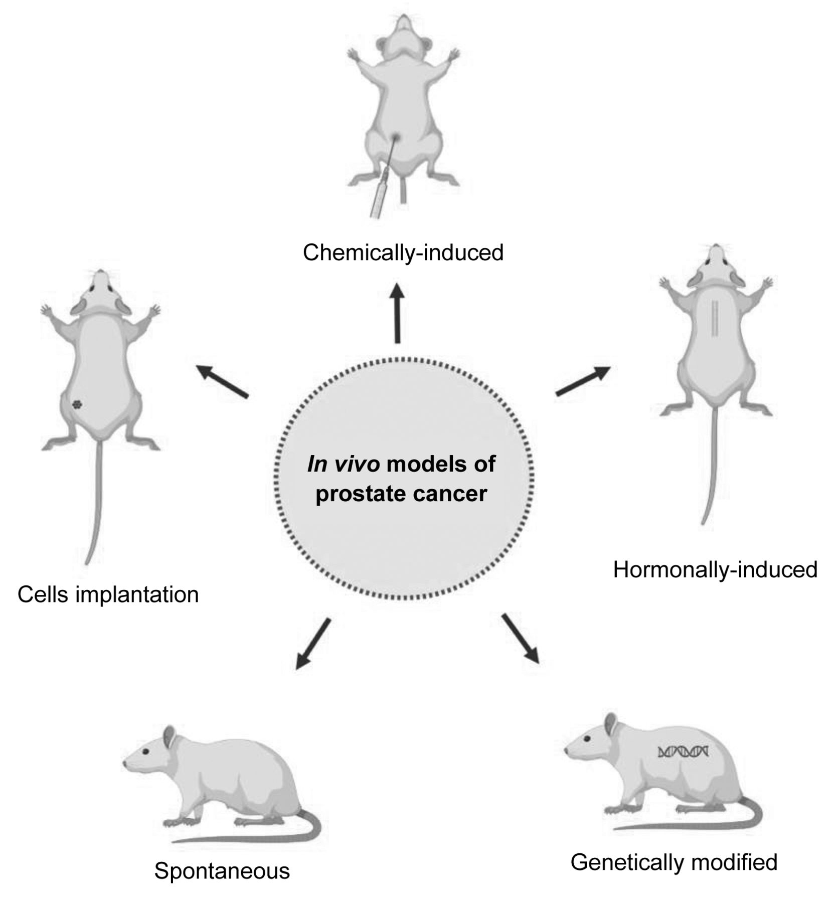
Nowadays, several animal models are available for PCa study, namely spontaneous models, chemically-induced, hormonally-induced, implanted, and genetically-modified (34, 35) (Figure 1).

Figure 1.
  • Download figure
  • Open in new tab
  • Download powerpoint
Figure 1.

In vivo models available for prostate cancer. Spontaneous models develop prostate cancer spontaneously; chemically-induced models are obtained through administration of chemical carcinogens, like N-methyl-N-nitrosourea, 3,2′-dimethyl-4-aminobiphenyl, 2-amino-1-methyl-6-phenylimidazol[4,5-b]pyridine and N-nitrosobis (2-oxopropyl) amine; hormonally-induced models are obtained through administration of hormones, like testosterone; implanted models can be obtained through implantation of cancer cell lines obtained from humans (xenograft model) or from the same species (syngeneic), in the prostate (orthotopic) or in a different place (heterotopic); and genetically-modified models results from genetic alterations.

The spontaneous models of PCa present a long period of latency (nearly 2-3 years) and a low incidence, being not advantageous for the study of this condition. The first spontaneous PCa tumor was reported by Dunning in 1963 in a 22 months-old Copenhagen rat (36–38). Ten years later, in 1973, Pollard described a spontaneous prostatic carcinoma in 26 months-old Wistar rats (37). In 1957 were described spontaneous tumors of ventral prostate in 34-37 months-old AxC rats (38). Moreover to these rat strains, aged ACI/Seg rats have a high susceptibility to develop spontaneous tumors in ventral prostate lobe (39).

The development of PCa may be induced though administration of chemical carcinogens, being the MNU, 3,2′-dimethyl-4-aminobiphenyl (DMBA), PhiP and BOP the most used. MNU is a direct alkylating agent, frequently used to induce the development of cancer in several organs, including prostate and the mammary gland. MNU may be used in combination with hormones to induce PCa development. Our research team has successfully used a rat model of PCa that was chemically and hormonally-induced in 12 weeks-old Wistar rats as follows: the anti-androgenic drug flutamide (50 mg/kg) was administered subcutaneously for 21 consecutive days; twenty-four hours later the testosterone propionate (100 mg/kg) was dissolved in corn oil and administered subcutaneously; two days later the MNU (30 mg/kg) was intraperitoneally injected; and two weeks later the implants filled with crystalline testosterone were implanted in the interscapular region of animals under anesthesia (30, 40–42). DMBA is an indirect carcinogenic agent that needs to be activated in the liver. Its metabolites interact with DNA inducing irreversible changes in DNA (43). DMBA is able to induce the development of carcinomas in ventral prostate lobe through a subcutaneous injection (27). PhiP is a heterocyclic amine whose active metabolites form DNA adducts, inducing development of cancer in prostate, mammary gland, and intestine (44–46). It can be administered in the diet or administered by gavage (47, 48). BOP is a nitrosamine with tropism to several organs, like pancreas, liver, and prostate (49). After its oral administration, BOP is converted into carbon dioxide that acts as a DNA methylating agent. The BOP has only the ability to induce the development of squamous cell carcinomas in ventral prostate lobes, that have no correspondence with human prostate, constituting a limitation of this model (33).

PCa can be also induced through administration of hormones, due to the sensitivity of prostate tissue to androgens. The implants filled with testosterone and estrogen may be subcutaneously implanted for a long period of time in rats. Simultaneously to the development of PCa, the animals may develop pituitary tumors leading to their early death (33).

The implanted models of PCa may be obtained through implantation of PCa cells obtained from humans (xenograft model) or from rat PCa (syngeneic model). The cell lines may be implanted in the rat prostate (orthotopic) or in a different place (heterotopic) (50–53). The xenograft models are immunodeficient, which did not allow the evaluation of the interplay between immune system and cancer. Genetically-engineered models are less time-consuming and labor-intensive when compared with chemically and hormonally-induced models. An incidence of 100% in guaranteed and tumors are androgen-sensitive (54).

At the current stage of knowledge, the advances in the study of PCa using 3D and organ-on-chip models have been small and need further research and increased funding.

Current Therapies for Prostate Cancer

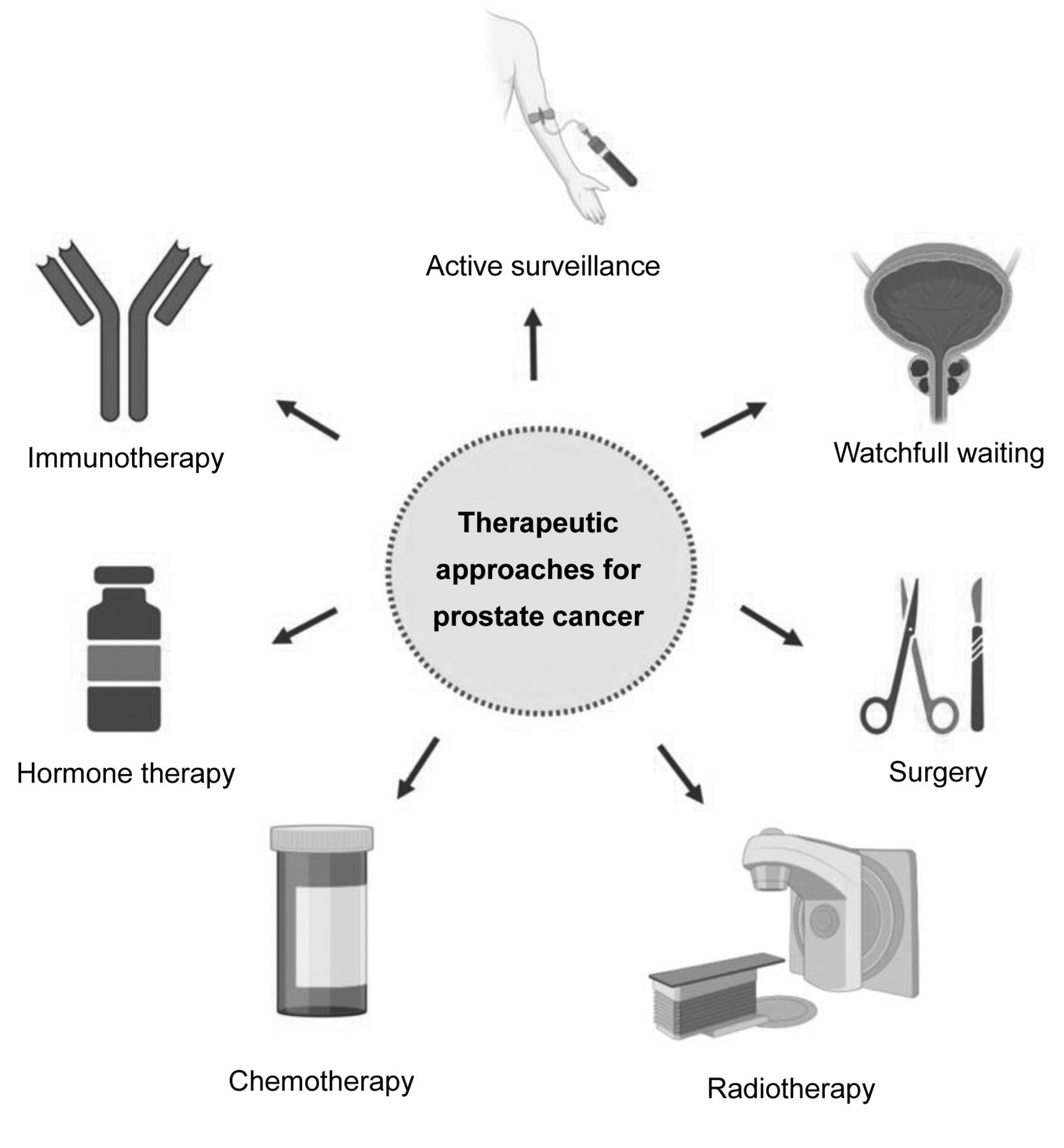
First, the stage of the disease and the patient’s profile must be clearly understood to choose the best treatment for each patient (55, 56). For this, it is important to have a good early diagnosis and assess the risk of the disease. After this, there exist a variety of approaches to be considered, which include active surveillance, watchful waiting, surgery, radiation, chemotherapy, hormone therapy and immunotherapy (55, 57) (Figure 2). All these approaches can be used alone or in combination.

Figure 2.
  • Download figure
  • Open in new tab
  • Download powerpoint
Figure 2.

Therapeutic approaches for prostate cancer.

Active surveillance consists of cancer monitoring through blood tests, PSA blood test and digital rectal exam. Moreover, prostate biopsies and imaging screening can be performed. This strategy is usually applied to patients with localized PCa and low risk of cancer growing or spreading outside of prostate. Watchful waiting is used in patients with low-risk and slow-growing disease, patients with other diseases and those with a limited life expectancy. Routine tests are not usually done on these patients (55, 57).

There are different surgical approaches, depending on the stage of the disease and patient status. Radical prostatectomy, by laparoscopic or robotic-assisted, removes entire prostate gland and seminal vesicles (58). Transurethral resection of the prostate is performed to remove the inner part of the prostate that surrounds the urethra relieving trouble urinating (59). Minimally invasive techniques, with minimal incisions or using tiny instruments into the urethra are increasingly used to treat PCa or relieve symptoms.

Radiation therapy in PCa include, for example, external beam radiation, brachytherapy or radiopharmaceuticals use. Radiopharmaceuticals is an emergent cancer therapy that deliver radiation therapy directly to cancer cells (60). Specifically in set of PCa, at the end of March 2022 the Food and Drug Administration (FDA) approved 177Lu-PSMA-617 to treat patients with metastatic PCa who have been treated with androgen receptor (AR) pathway inhibitors and taxane-based chemotherapy (61). This drug combines a targeting compound with a therapeutic radioisotope. After its administration binds to PCa cells that express prostate-specific membrane antigen (PSMA), a transmembrane protein and, once bound, emissions from the radioisotope damage tumor cells disrupting their capacity to replicate and triggering cell death (62–64).

Hormonotherapy is one of the most advanced areas where new drugs have been developed, such as apalutamide and darolutamide. Apalutamide, an androgen signaling inhibitor, has been studied and was approved by FDA in 2018 and by European Medicine Agency (EMA) in 2019 (65–67). It is used to treat non-metastatic castration-resistant prostate cancer (CRPCa) and metastatic hormone-sensitive PCa in combination with androgen deprivation therapy (65). Darolutamide is an androgen-receptor inhibitor used in the treatment of non-metastatic CRPCa in combination with surgery or medical castration approved in USA in 2019 and in the European Union in 2020 (68–71).

Another emerging therapy for PCa is called focal irreversible electroporation. This technology uses low voltage direct electric current that permanently damages the cell membrane and leads to cell death, preserving the surrounding prostate structures (72–74). Tissue selectivity, reduced toxicity on vital anatomical structures surrounding the prostate lesions, and preservation of urinary and sexual functions are the main advantages of this technique (75). Moreover, this technique is minimally invasive in contrast with other therapies, allow precision in targeting cancerous cells and the recovery time is short (74, 76). However, this technique is still at an early stage of development and there are still not many scientific studies comparing it with more traditional approaches, but the results seem promising (74, 77–79).

Immunotherapy is a promising PCa therapy that uses patients’ own immune system to recognize and kill cancer cells. Sipuleucel-T, a cell-based vaccine, is the only FDA approved immunotherapy for asymptomatic or minimally symptomatic metastatic CRPCa (80, 81). However, it is not yet available in Europe (82, 83). The treatment involves the use of patient’s immune cells stimulated to target the antigen prostatic acid phosphatase (80). Other vaccine therapies are being tested in clinical trials, such as Prostvac (84). Immune checkpoint inhibitors are another type of immunotherapy that block checkpoint proteins from binding with their partner proteins, allowing the immune system, namely T cells, to attack and kill the cancer cells. Pembrolizumab is a drug that targets PD-1/PD-L1 pathway and is the only FDA approved for advanced PCa that has high microsatellite instability, DNA mismatch repair deficiency or high tumor mutational burden (85–87). There are more ongoing studies and clinical trials with these immune checkpoint inhibitors drugs but whose results still do not allow their approval by the competent authorities and their commercialization (88–91).

Animal models have been vital for discovery, development, and approval of novel drugs for cancer treatment, by allowing to evaluate their safety, toxicity, pharmacodynamics, pharmacokinetic and efficacy. Determining efficacy and safety is critical during the initial phases of drugs discovery and development, and ultimately for approval and commercialization. Some of the drugs approved by the FDA or EMA, and listed above, were tested in rodent models (92–102).

Conclusion

Models for any given assay should be carefully chosen by researchers whilst considering the aim of the study, the associated costs, and the advantages and disadvantages of each model. Although several models are available for PCa studies, rodent models remain the ones most used. Despite their disadvantages, rat models have provided a great deal of information concerning PCa biopathology, allowing better understanding of the disease as well as leading to the development of more effective therapies. The PCa hormone and chemically-induced model is very complex, time and labor-consuming, but it successfully induces development of PCa tumors hormone-dependent and histologically similar to those developed by men, allowing data translation. In the future, animal models will certainly continue to be a fundamental tool in the study of PCa and development of new therapeutic approaches, since only laboratory animals allow for the study of several variables simultaneously.

Acknowledgements

This work was supported by National Funds by FCT - Portuguese Foundation for Science and Technology, under the projects UIDB/04033/2020 and LA/P/0126/2020), and PhD grants SFRH/BD/136747/2018 (E. Nascimento-Gonçalves) and 2021.04520.BD (C. Jota-Baptista).

Footnotes

  • Authors’ Contributions

    All Authors were responsible for manuscript writing and revision and approved its submission.

  • Conflicts of Interest

    The Authors have no conflicts of interest to declare.

  • Received November 14, 2022.
  • Revision received November 26, 2022.
  • Accepted December 9, 2022.
  • Copyright © 2023 International Institute of Anticancer Research (Dr. George J. Delinasios), All rights reserved.

This article is an open access article distributed under the terms and conditions of the Creative Commons Attribution (CC BY-NC-ND) 4.0 international license (https://creativecommons.org/licenses/by-nc-nd/4.0).

References

  1. ↵
    1. Sung H,
    2. Ferlay J,
    3. Siegel RL,
    4. Laversanne M,
    5. Soerjomataram I,
    6. Jemal A and
    7. Bray F
    : Global cancer statistics 2020: GLOBOCAN estimates of incidence and mortality worldwide for 36 cancers in 185 countries. CA Cancer J Clin 71(3): 209-249, 2021. PMID: 33538338. DOI: 10.3322/caac.21660
    OpenUrlCrossRefPubMed
  2. ↵
    1. Malik A,
    2. Srinivasan S and
    3. Batra J
    : A new era of prostate cancer precision medicine. Front Oncol 9: 1263, 2019. PMID: 31850193. DOI: 10.3389/fonc.2019.01263
    OpenUrlCrossRefPubMed
  3. ↵
    1. Humphrey PA
    : Histological variants of prostatic carcinoma and their significance. Histopathology 60(1): 59-74, 2012. PMID: 22212078. DOI: 10.1111/j.1365-2559.2011.04039.x
    OpenUrlCrossRefPubMed
  4. ↵
    1. Rawla P
    : Epidemiology of prostate cancer. World J Oncol 10(2): 63-89, 2019. PMID: 31068988. DOI: 10.14740/wjon1191
    OpenUrlCrossRefPubMed
  5. ↵
    1. Patel AR and
    2. Klein EA
    : Risk factors for prostate cancer. Nat Clin Pract Urol 6(2): 87-95, 2009. PMID: 19198622. DOI: 10.1038/ncpuro1290
    OpenUrlCrossRefPubMed
  6. ↵
    1. Legler JM,
    2. Feuer EJ,
    3. Potosky AL,
    4. Merrill RM and
    5. Kramer BS
    : The role of prostate-specific antigen (PSA) testing patterns in the recent prostate cancer incidence decline in the United States. Cancer Causes Control 9(5): 519-527, 1998. PMID: 9934717. DOI: 10.1023/a:1008805718310
    OpenUrlCrossRefPubMed
  7. ↵
    1. Carter BS,
    2. Bova GS,
    3. Beaty TH,
    4. Steinberg GD,
    5. Childs B,
    6. Isaacs WB and
    7. Walsh PC
    : Hereditary prostate cancer: epidemiologic and clinical features. J Urol 150(3): 797-802, 1993. PMID: 8345587. DOI: 10.1016/s0022-5347(17)35617-3
    OpenUrlCrossRefPubMed
  8. ↵
    1. Klein EA and
    2. Silverman R
    : Inflammation, infection, and prostate cancer. Curr Opin Urol 18(3): 315-319, 2008. PMID: 18382242. DOI: 10.1097/MOU.0b013e3282f9b3b7
    OpenUrlCrossRefPubMed
  9. ↵
    1. Khaja FA,
    2. Koo OM and
    3. Önyüksel H
    : Nanomedicines for inflammatory diseases. Methods Enzymol 508: 355-375, 2012. PMID: 22449935. DOI: 10.1016/B978-0-12-391860-4.00018-5
    OpenUrlCrossRefPubMed
  10. ↵
    1. Ericsson AC,
    2. Crim MJ and
    3. Franklin CL
    : A brief history of animal modeling. Mo Med 110(3): 201-205, 2013. PMID: 23829102.
    OpenUrlPubMed
  11. ↵
    1. Hickman D,
    2. Johnson J,
    3. Vemulapalli T,
    4. Crisler J and
    5. Shepherd R
    : Commonly Used Animal Models. Principles of Animal Research: 117-175, 2018. DOI: 10.1016/B978-0-12-802151-4.00007-4
    OpenUrlCrossRef
  12. ↵
    1. Dietrich MR,
    2. Ankeny RA,
    3. Crowe N,
    4. Green S and
    5. Leonelli S
    : How to choose your research organism. Stud Hist Philos Biol Biomed Sci 80: 101227, 2020. PMID: 31883711. DOI: 10.1016/j.shpsc.2019.101227
    OpenUrlCrossRefPubMed
    1. Ighodaro OM,
    2. Adeosun AM and
    3. Akinloye OA
    : Alloxan-induced diabetes, a common model for evaluating the glycemic-control potential of therapeutic compounds and plants extracts in experimental studies. Medicina (Kaunas) 53(6): 365-374, 2017. PMID: 29548636. DOI: 10.1016/j.medici.2018.02.001
    OpenUrlCrossRefPubMed
  13. ↵
    1. Barré-Sinoussi F and
    2. Montagutelli X
    : Animal models are essential to biological research: issues and perspectives. Future Sci OA 1(4): FSO63, 2015. PMID: 28031915. DOI: 10.4155/fso.15.63
    OpenUrlCrossRefPubMed
  14. ↵
    1. Al-Asmakh M and
    2. Zadjali F
    : Use of germ-free animal models in microbiota-related research. J Microbiol Biotechnol 25(10): 1583-1588, 2015. PMID: 26032361. DOI: 10.4014/jmb.1501.01039
    OpenUrlCrossRefPubMed
  15. ↵
    1. Swearengen JR
    : Choosing the right animal model for infectious disease research. Animal Model Exp Med 1(2): 100-108, 2018. PMID: 30891554. DOI: 10.1002/ame2.12020
    OpenUrlCrossRefPubMed
  16. ↵
    1. Freires IA,
    2. Sardi JC,
    3. de Castro RD and
    4. Rosalen PL
    : Alternative animal and non-animal models for drug discovery and development: Bonus or burden? Pharm Res 34(4): 681-686, 2017. PMID: 27858217. DOI: 10.1007/s11095-016-2069-z
    OpenUrlCrossRefPubMed
  17. ↵
    1. Doke SK and
    2. Dhawale SC
    : Alternatives to animal testing: A review. Saudi Pharm J 23(3): 223-229, 2015. PMID: 26106269. DOI: 10.1016/j.jsps.2013.11.002
    OpenUrlCrossRefPubMed
  18. ↵
    1. Atat OE,
    2. Farzaneh Z,
    3. Pourhamzeh M,
    4. Taki F,
    5. Abi-Habib R,
    6. Vosough M and
    7. El-Sibai M
    : 3D modeling in cancer studies. Hum Cell 35(1): 23-36, 2022. PMID: 34761350. DOI: 10.1007/s13577-021-00642-9
    OpenUrlCrossRefPubMed
  19. ↵
    1. Imparato G,
    2. Urciuolo F and
    3. Netti PA
    : Organ on chip technology to model cancer growth and metastasis. Bioengineering (Basel) 9(1): 28, 2022. PMID: 35049737. DOI: 10.3390/bioengineering9010028
    OpenUrlCrossRefPubMed
  20. ↵
    1. Castagno P,
    2. Pernice S,
    3. Ghetti G,
    4. Povero M,
    5. Pradelli L,
    6. Paolotti D,
    7. Balbo G,
    8. Sereno M and
    9. Beccuti M
    : A computational framework for modeling and studying pertussis epidemiology and vaccination. BMC Bioinformatics 21(Suppl 8): 344, 2020. PMID: 32938370. DOI: 10.1186/s12859-020-03648-6
    OpenUrlCrossRefPubMed
  21. ↵
    Cancer. Available at: https://www.who.int/news-room/fact-sheets/detail/cancer [Last accessed on November 13, 2022]
  22. ↵
    1. Moore RA and
    2. Melchionna RH
    : Production of tumors of the prostate of the white rat with 1:2-benzpyrene. Am J Cancer 30: 731-741, 1937.
    OpenUrlAbstract/FREE Full Text
  23. ↵
    1. Dunning WF,
    2. Curtis MR and
    3. Segaloff A
    : Methylcholanthrene squamous cell carcinoma of the rat prostate with skeletal metastases, and failure of the rat liver to respond to the carcinogen. Cancer Res 6: 256-262, 1946. PMID: 21024837.
    OpenUrlFREE Full Text
  24. ↵
    1. Pour PM
    : A new prostatic cancer model: systemic induction of prostatic cancer in rats by a nitrosamine. Cancer Lett 13(4): 303-308, 1981. PMID: 6171336. DOI: 10.1016/0304-3835(81)90058-6
    OpenUrlCrossRefPubMed
  25. ↵
    1. Bosland MC
    : Chemical and hormonal induction of prostate cancer in animal models. Urol Oncol 2(4): 103-110, 1996. PMID: 21224148. DOI: 10.1016/s1078-1439(97)82840-2
    OpenUrlCrossRefPubMed
  26. ↵
    1. Katayama S,
    2. Fiala E,
    3. Reddy BS,
    4. Rivenson A,
    5. Silverman J,
    6. Williams GM and
    7. Weisburger JH
    : Prostate adenocarcinoma in rats: induction by 3,2′-dimethyl-4-aminobiphenyl. J Natl Cancer Inst 68(5): 867-873, 1982. PMID: 6951094.
    OpenUrlCrossRefPubMed
  27. ↵
    1. Shirai T,
    2. Sano M,
    3. Tamano S,
    4. Takahashi S,
    5. Hirose M,
    6. Futakuchi M,
    7. Hasegawa R,
    8. Imaida K,
    9. Matsumoto K,
    10. Wakabayashi K,
    11. Sugimura T and
    12. Ito N
    : The prostate: a target for carcinogenicity of 2-amino-1-methyl-6-phenylimidazo[4,5-b]pyridine (PhIP) derived from cooked foods. Cancer Res 57(2): 195-198, 1997. PMID: 9000552.
    OpenUrlAbstract/FREE Full Text
  28. ↵
    1. Samy RP,
    2. Rajendran P,
    3. Li F,
    4. Anandi NM,
    5. Stiles BG,
    6. Ignacimuthu S,
    7. Sethi G and
    8. Chow VT
    : Identification of a novel Calotropis procera protein that can suppress tumor growth in breast cancer through the suppression of NF-ĸB pathway. PLoS One 7(12): e48514, 2012. PMID: 23284617. DOI: 10.1371/journal.pone.0048514
    OpenUrlCrossRefPubMed
  29. ↵
    1. Nascimento-Gonçalves E,
    2. Faustino-Rocha AI,
    3. Seixas F,
    4. Ginja M,
    5. Colaço B,
    6. Ferreira R,
    7. Fardilha M and
    8. Oliveira PA
    : Modelling human prostate cancer: Rat models. Life Sci 203: 210-224, 2018. PMID: 29684445. DOI: 10.1016/j.lfs.2018.04.014
    OpenUrlCrossRefPubMed
  30. ↵
    1. Shirai T,
    2. Takahashi S,
    3. Cui L,
    4. Futakuchi M,
    5. Kato K,
    6. Tamano S and
    7. Imaida K
    : Experimental prostate carcinogenesis - rodent models. Mutat Res 462(2-3): 219-226, 2000. PMID: 10767633. DOI: 10.1016/s1383-5742(00)00039-9
    OpenUrlCrossRefPubMed
  31. ↵
    1. Shappell SB,
    2. Thomas GV,
    3. Roberts RL,
    4. Herbert R,
    5. Ittmann MM,
    6. Rubin MA,
    7. Humphrey PA,
    8. Sundberg JP,
    9. Rozengurt N,
    10. Barrios R,
    11. Ward JM and
    12. Cardiff RD
    : Prostate pathology of genetically engineered mice: definitions and classification. The consensus report from the Bar Harbor meeting of the Mouse Models of Human Cancer Consortium Prostate Pathology Committee. Cancer Res 64(6): 2270-2305, 2004. PMID: 15026373. DOI: 10.1158/0008-5472.can-03-0946
    OpenUrlAbstract/FREE Full Text
  32. ↵
    1. Bosland MC
    : Animal models for the study of prostate carcinogenesis. J Cell Biochem Suppl 16H: 89-98, 1992. PMID: 1289679. DOI: 10.1002/jcb.240501221
    OpenUrlCrossRefPubMed
  33. ↵
    1. Nascimento-Gonçalves E,
    2. Seixas F,
    3. Ferreira R,
    4. Colaço B,
    5. Parada B and
    6. Oliveira PA
    : An overview of the latest in state-of-the-art murine models for prostate cancer. Expert Opin Drug Discov 16(11): 1349-1364, 2021. PMID: 34224283. DOI: 10.1080/17460441.2021.1943354
    OpenUrlCrossRefPubMed
  34. ↵
    1. Nascimento-Gonçalves E,
    2. Ferreira R,
    3. Oliveira PA and
    4. Colaço BJA
    : An overview of current alternative models for use in the context of prostate cancer research. Altern Lab Anim 48(2): 58-69, 2020. PMID: 32614643. DOI: 10.1177/0261192920929701
    OpenUrlCrossRefPubMed
  35. ↵
    1. Tennant TR,
    2. Kim H,
    3. Sokoloff M and
    4. Rinker-Schaeffer CW
    : The Dunning model. Prostate 43(4): 295-302, 2000. PMID: 10861749. DOI: 10.1002/1097-0045(20000601)43:4<295::aid-pros9>3.0.co;2-w
    OpenUrlCrossRefPubMed
  36. ↵
    1. Pollard M
    : Spontaneous prostate adenocarcinomas in aged germfree Wistar rats. J Natl Cancer Inst 51(4): 1235-1241, 1973. PMID: 4745857. DOI: 10.1093/jnci/51.4.1235
    OpenUrlCrossRefPubMed
  37. ↵
    1. Shain SA,
    2. McCullough B and
    3. Segaloff A
    : Spontaneous adenocarcinomas of the ventral prostate of aged A X C rats. J Natl Cancer Inst 55(1): 177-180, 1975. PMID: 1159811. DOI: 10.1093/jnci/55.1.177
    OpenUrlCrossRefPubMed
  38. ↵
    1. Isaacs JT
    : The aging ACI/Seg versus Copenhagen male rat as a model system for the study of prostatic carcinogenesis. Cancer Res 44(12 Pt 1): 5785-5796, 1984. PMID: 6498839.
    OpenUrlAbstract/FREE Full Text
  39. ↵
    1. Bosland MC
    : Chemical and hormonal induction of prostate cancer in animal models. Urol Oncol 2(4): 103-110, 1996. PMID: 21224148. DOI: 10.1016/s1078-1439(97)82840-2
    OpenUrlCrossRefPubMed
    1. Rao KV,
    2. Johnson WD,
    3. Bosland MC,
    4. Lubet RA,
    5. Steele VE,
    6. Kelloff GJ and
    7. McCormick DL
    : Chemoprevention of rat prostate carcinogenesis by early and delayed administration of dehydroepiandrosterone. Cancer Res 59(13): 3084-3089, 1999. PMID: 10397249.
    OpenUrlAbstract/FREE Full Text
  40. ↵
    1. Bosland MC
    : Chemical and hormonal induction of prostate cancer in animal models. Urol Oncol 2(4): 103-110, 1996. PMID: 21224148. DOI: 10.1016/s1078-1439(97)82840-2
    OpenUrlCrossRefPubMed
  41. ↵
    1. Shirai T,
    2. Tada M,
    3. Kojima M,
    4. Hasegawa R,
    5. Masui T and
    6. Ito N
    : DNA adducts in target and nontarget tissues of 3,2′-dimethyl-4-aminobiphenyl in rats. Environ Health Perspect 102 Suppl 6(Suppl 6): 167-172, 1994. PMID: 7889841. DOI: 10.1289/ehp.94102s6167
    OpenUrlCrossRefPubMed
  42. ↵
    1. Takayama K,
    2. Yamashita K,
    3. Wakabayashi K,
    4. Sugimura T and
    5. Nagao M
    : DNA modification by 2-amino-1-methyl-6-phenylimidazo[4,5-b]pyridine in rats. Jpn J Cancer Res 80(12): 1145-1148, 1989. PMID: 2516840. DOI: 10.1111/j.1349-7006.1989.tb01644.x
    OpenUrlCrossRefPubMed
    1. Snyderwine EG,
    2. Venugopal M and
    3. Yu M
    : Mammary gland carcinogenesis by food-derived heterocyclic amines and studies on the mechanisms of carcinogenesis of 2-amino-1-methyl-6-phenylimidazo[4,5-b]pyridine (PhIP). Mutat Res 506-507: 145-152, 2002. PMID: 12351154. DOI: 10.1016/s0027-5107(02)00161-6
    OpenUrlCrossRefPubMed
  43. ↵
    1. Tang MX,
    2. Ogawa K,
    3. Asamoto M,
    4. Chewonarin T,
    5. Suzuki S,
    6. Tanaka T and
    7. Shirai T
    : Effects of nobiletin on PhIP-induced prostate and colon carcinogenesis in F344 rats. Nutr Cancer 63(2): 227-233, 2011. PMID: 21298591. DOI: 10.1080/01635581.2011.523506
    OpenUrlCrossRefPubMed
  44. ↵
    1. Naiki-Ito A,
    2. Chewonarin T,
    3. Tang M,
    4. Pitchakarn P,
    5. Kuno T,
    6. Ogawa K,
    7. Asamoto M,
    8. Shirai T and
    9. Takahashi S
    : Ellagic acid, a component of pomegranate fruit juice, suppresses androgen-dependent prostate carcinogenesis via induction of apoptosis. Prostate 75(2): 151-160, 2015. PMID: 25284475. DOI: 10.1002/pros.22900
    OpenUrlCrossRefPubMed
  45. ↵
    1. Canene-Adams K,
    2. Sfanos KS,
    3. Liang CT,
    4. Yegnasubramanian S,
    5. Nelson WG,
    6. Brayton C and
    7. De Marzo AM
    : Dietary chemoprevention of PhIP induced carcinogenesis in male Fischer 344 rats with tomato and broccoli. PLoS One 8(11): e79842, 2013. PMID: 24312188. DOI: 10.1371/journal.pone.0079842
    OpenUrlCrossRefPubMed
  46. ↵
    1. Lawson TA,
    2. Gingell R,
    3. Nagel D,
    4. Hines LA and
    5. Ross A
    : Methylation of hamster DNA by the carcinogen N-nitroso-bis (2-oxopropyl)amine. Cancer Lett 11(3): 251-255, 1981. PMID: 7248930. DOI: 10.1016/0304-3835(81)90116-6
    OpenUrlCrossRefPubMed
  47. ↵
    1. Johannsen M,
    2. Thiesen B,
    3. Jordan A,
    4. Taymoorian K,
    5. Gneveckow U,
    6. Waldöfner N,
    7. Scholz R,
    8. Koch M,
    9. Lein M,
    10. Jung K and
    11. Loening SA
    : Magnetic fluid hyperthermia (MFH)reduces prostate cancer growth in the orthotopic Dunning R3327 rat model. Prostate 64(3): 283-292, 2005. PMID: 15726645. DOI: 10.1002/pros.20213
    OpenUrlCrossRefPubMed
    1. McCullough DJ,
    2. Stabley JN,
    3. Siemann DW and
    4. Behnke BJ
    : Modulation of blood flow, hypoxia, and vascular function in orthotopic prostate tumors during exercise. J Natl Cancer Inst 106(4): dju036, 2014. PMID: 24627275. DOI: 10.1093/jnci/dju036
    OpenUrlCrossRefPubMed
    1. McCullough DJ,
    2. Nguyen LM,
    3. Siemann DW and
    4. Behnke BJ
    : Effects of exercise training on tumor hypoxia and vascular function in the rodent preclinical orthotopic prostate cancer model. J Appl Physiol (1985) 115(12): 1846-1854, 2013. PMID: 24177690. DOI: 10.1152/japplphysiol.00949.2013
    OpenUrlCrossRefPubMed
  48. ↵
    1. Neubauer BL,
    2. Bemis KG,
    3. Best KL,
    4. Goode RL,
    5. Merriman RL,
    6. Smith GF,
    7. Tanzer LR and
    8. Hoover DM
    : Metastatic spread of the PAIII prostatic adenocarcinoma after implantation in the tail of the rat. Prostate 8(3): 265-276, 1986. PMID: 3703746. DOI: 10.1002/pros.2990080307
    OpenUrlCrossRefPubMed
  49. ↵
    1. Johnson LE,
    2. Becker JT,
    3. Dubovsky JA,
    4. Olson BM and
    5. McNeel DG
    : Prostate carcinoma in transgenic Lewis rats - a tumor model for evaluation of immunological treatments. Chin Clin Oncol 2(1): doi:10.3978/j.issn.2304-3865.2012.11.06, 2013. PMID: 24324949. DOI: 10.3978/j.issn.2304-3865.2012.11.06
    OpenUrlCrossRefPubMed
  50. ↵
    1. Mottet N,
    2. van den Bergh RCN,
    3. Briers E,
    4. Van den Broeck T,
    5. Cumberbatch MG,
    6. De Santis M,
    7. Fanti S,
    8. Fossati N,
    9. Gandaglia G,
    10. Gillessen S,
    11. Grivas N,
    12. Grummet J,
    13. Henry AM,
    14. van der Kwast TH,
    15. Lam TB,
    16. Lardas M,
    17. Liew M,
    18. Mason MD,
    19. Moris L,
    20. Oprea-Lager DE,
    21. van der Poel HG,
    22. Rouvière O,
    23. Schoots IG,
    24. Tilki D,
    25. Wiegel T,
    26. Willemse PM and
    27. Cornford P
    : EAU-EANM-ESTRO-ESUR-SIOG guidelines on prostate cancer-2020 update. Part 1: Screening, diagnosis, and local treatment with curative intent. Eur Urol 79(2): 243-262, 2021. PMID: 33172724. DOI: 10.1016/j.eururo.2020.09.042
    OpenUrlCrossRefPubMed
  51. ↵
    1. Cornford P,
    2. van den Bergh RCN,
    3. Briers E,
    4. Van den Broeck T,
    5. Cumberbatch MG,
    6. De Santis M,
    7. Fanti S,
    8. Fossati N,
    9. Gandaglia G,
    10. Gillessen S,
    11. Grivas N,
    12. Grummet J,
    13. Henry AM,
    14. der Kwast THV,
    15. Lam TB,
    16. Lardas M,
    17. Liew M,
    18. Mason MD,
    19. Moris L,
    20. Oprea-Lager DE,
    21. der Poel HGV,
    22. Rouvière O,
    23. Schoots IG,
    24. Tilki D,
    25. Wiegel T,
    26. Willemse PM and
    27. Mottet N
    : EAU-EANM-ESTRO-ESUR-SIOG guidelines on prostate cancer. Part II-2020 update: Treatment of relapsing and metastatic prostate cancer. Eur Urol 79(2): 263-282, 2021. PMID: 33039206. DOI: 10.1016/j.eururo.2020.09.046
    OpenUrlCrossRefPubMed
  52. ↵
    1. Parker C,
    2. Castro E,
    3. Fizazi K,
    4. Heidenreich A,
    5. Ost P,
    6. Procopio G,
    7. Tombal B,
    8. Gillessen S and ESMO Guidelines Committee
    : Prostate cancer: ESMO Clinical Practice Guidelines for diagnosis, treatment and follow-up. Ann Oncol 31(9): 1119-1134, 2020. PMID: 32593798. DOI: 10.1016/j.annonc.2020.06.011
    OpenUrlCrossRefPubMed
  53. ↵
    1. Basiri A,
    2. de la Rosette JJ,
    3. Tabatabaei S,
    4. Woo HH,
    5. Laguna MP and
    6. Shemshaki H
    : Comparison of retropubic, laparoscopic and robotic radical prostatectomy: who is the winner? World J Urol 36(4): 609-621, 2018. PMID: 29362896. DOI: 10.1007/s00345-018-2174-1
    OpenUrlCrossRefPubMed
  54. ↵
    1. Wang JW and
    2. Man LB
    : Transurethral resection of the prostate stricture management. Asian J Androl 22(2): 140-144, 2020. PMID: 31898584. DOI: 10.4103/aja.aja_126_19
    OpenUrlCrossRefPubMed
  55. ↵
    1. Jaymand M,
    2. Davatgaran taghipour Y,
    3. Rezaei A,
    4. Derakhshankhah H,
    5. Foad abazari M,
    6. Samadian H and
    7. Hamblin M
    : Radiolabeled carbon-based nanostructures: New radiopharmaceuticals for cancer therapy? Coordination Chemistry Reviews 440: 213974, 2022. DOI: 10.1016/j.ccr.2021.213974
    OpenUrlCrossRef
  56. ↵
    FDA approves pluvicto for metastatic castration-resistant prostate cancer | FDA. Available at: https://www.fda.gov/drugs/resources-information-approved-drugs/fda-approves-pluvicto-metastatic-castration-resistant-prostate-cancer [Last accessed on November 13, 2022]
  57. ↵
    1. Kratochwil C,
    2. Giesel FL,
    3. Stefanova M,
    4. Benešová M,
    5. Bronzel M,
    6. Afshar-Oromieh A,
    7. Mier W,
    8. Eder M,
    9. Kopka K and
    10. Haberkorn U
    : PSMA-targeted radionuclide therapy of metastatic castration-resistant prostate cancer with 177Lu-labeled PSMA-617. J Nucl Med 57(8): 1170-1176, 2016. PMID: 26985056. DOI: 10.2967/jnumed.115.171397
    OpenUrlAbstract/FREE Full Text
    1. Violet J,
    2. Jackson P,
    3. Ferdinandus J,
    4. Sandhu S,
    5. Akhurst T,
    6. Iravani A,
    7. Kong G,
    8. Kumar AR,
    9. Thang SP,
    10. Eu P,
    11. Scalzo M,
    12. Murphy D,
    13. Williams S,
    14. Hicks RJ and
    15. Hofman MS
    : Dosimetry of (177)Lu-PSMA-617 in metastatic castration-resistant prostate cancer: correlations between pretherapeutic imaging and whole-body tumor dosimetry with treatment outcomes. J Nucl Med 60(4): 517-523, 2019. PMID: 30291192. DOI: 10.2967/jnumed.118.219352
    OpenUrlAbstract/FREE Full Text
  58. ↵
    1. Sartor O,
    2. de Bono J,
    3. Chi KN,
    4. Fizazi K,
    5. Herrmann K,
    6. Rahbar K,
    7. Tagawa ST,
    8. Nordquist LT,
    9. Vaishampayan N,
    10. El-Haddad G,
    11. Park CH,
    12. Beer TM,
    13. Armour A,
    14. Pérez-Contreras WJ,
    15. DeSilvio M,
    16. Kpamegan E,
    17. Gericke G,
    18. Messmann RA,
    19. Morris MJ,
    20. Krause BJ and VISION Investigators
    : Lutetium-177-PSMA-617 for metastatic castration-resistant prostate cancer. N Engl J Med 385(12): 1091-1103, 2021. PMID: 34161051. DOI: 10.1056/NEJMoa2107322
    OpenUrlCrossRefPubMed
  59. ↵
    1. Smith MR,
    2. Saad F,
    3. Chowdhury S,
    4. Oudard S,
    5. Hadaschik BA,
    6. Graff JN,
    7. Olmos D,
    8. Mainwaring PN,
    9. Lee JY,
    10. Uemura H,
    11. Lopez-Gitlitz A,
    12. Trudel GC,
    13. Espina BM,
    14. Shu Y,
    15. Park YC,
    16. Rackoff WR,
    17. Yu MK,
    18. Small EJ and SPARTAN Investigators
    : Apalutamide treatment and metastasis-free survival in prostate cancer. N Engl J Med 378(15): 1408-1418, 2018. PMID: 29420164. DOI: 10.1056/NEJMoa1715546
    OpenUrlCrossRefPubMed
  60. Novel Drug Approvals for 2018 | FDA. Available at: https://www.fda.gov/drugs/new-drugs-fda-cders-new-molecular-entities-and-new-therapeutic-biological-products/novel-drug-approvals-2018 [Last accessed on November 13, 2022]
  61. ↵
    Erleada | European Medicines Agency. Available at: https://www.ema.europa.eu/en/medicines/human/EPAR/erleada [Last accessed on November 13, 2022]
  62. ↵
    Nubeqa | European Medicines Agency. Available at: https://www.ema.europa.eu/en/medicines/human/EPAR/nubeqa [Last accessed on November 13, 2022]
  63. FDA approves darolutamide for non-metastatic castration-resistant prostate cancer | FDA. Available at: https://www.fda.gov/drugs/resources-information-approved-drugs/fda-approves-darolutamide-non-metastatic-castration-resistant-prostate-cancer [Last accessed on November 13, 2022]
    1. Smith MR,
    2. Hussain M,
    3. Saad F,
    4. Fizazi K,
    5. Sternberg CN,
    6. Crawford ED,
    7. Kopyltsov E,
    8. Park CH,
    9. Alekseev B,
    10. Montesa-Pino Á,
    11. Ye D,
    12. Parnis F,
    13. Cruz F,
    14. Tammela TLJ,
    15. Suzuki H,
    16. Utriainen T,
    17. Fu C,
    18. Uemura M,
    19. Méndez-Vidal MJ,
    20. Maughan BL,
    21. Joensuu H,
    22. Thiele S,
    23. Li R,
    24. Kuss I,
    25. Tombal B and ARASENS Trial Investigators
    : Darolutamide and survival in metastatic, hormone-sensitive prostate cancer. N Engl J Med 386(12): 1132-1142, 2022. PMID: 35179323. DOI: 10.1056/NEJMoa2119115
    OpenUrlCrossRefPubMed
  64. ↵
    1. Fizazi K,
    2. Shore N,
    3. Tammela TL,
    4. Ulys A,
    5. Vjaters E,
    6. Polyakov S,
    7. Jievaltas M,
    8. Luz M,
    9. Alekseev B,
    10. Kuss I,
    11. Le Berre MA,
    12. Petrenciuc O,
    13. Snapir A,
    14. Sarapohja T,
    15. Smith MR and ARAMIS Investigators
    : Nonmetastatic, castration-resistant prostate cancer and survival with darolutamide. N Engl J Med 383(11): 1040-1049, 2020. PMID: 32905676. DOI: 10.1056/NEJMoa2001342
    OpenUrlCrossRefPubMed
  65. ↵
    1. Valerio M,
    2. Dickinson L,
    3. Ali A,
    4. Ramachandran N,
    5. Donaldson I,
    6. Freeman A,
    7. Ahmed HU and
    8. Emberton M
    : A prospective development study investigating focal irreversible electroporation in men with localised prostate cancer: Nanoknife Electroporation Ablation Trial (NEAT). Contemp Clin Trials 39(1): 57-65, 2014. PMID: 25072507. DOI: 10.1016/j.cct.2014.07.006
    OpenUrlCrossRefPubMed
    1. Al-Sakere B,
    2. André F,
    3. Bernat C,
    4. Connault E,
    5. Opolon P,
    6. Davalos RV,
    7. Rubinsky B and
    8. Mir LM
    : Tumor ablation with irreversible electroporation. PLoS One 2(11): e1135, 2007. PMID: 17989772. DOI: 10.1371/journal.pone.0001135
    OpenUrlCrossRefPubMed
  66. ↵
    1. Ong S,
    2. Leonardo M,
    3. Chengodu T,
    4. Bagguley D and
    5. Lawrentschuk N
    : Irreversible electroporation for prostate cancer. Life (Basel) 11(6): 490, 2021. PMID: 34071934. DOI: 10.3390/life11060490
    OpenUrlCrossRefPubMed
  67. ↵
    1. Rubinsky B,
    2. Onik G and
    3. Mikus P
    : Irreversible electroporation: a new ablation modality – clinical implications. Technol Cancer Res Treat 6(1): 37-48, 2007. PMID: 17241099. DOI: 10.1177/153303460700600106
    OpenUrlCrossRefPubMed
  68. ↵
    1. Valerio M,
    2. Stricker PD,
    3. Ahmed HU,
    4. Dickinson L,
    5. Ponsky L,
    6. Shnier R,
    7. Allen C and
    8. Emberton M
    : Initial assessment of safety and clinical feasibility of irreversible electroporation in the focal treatment of prostate cancer. Prostate Cancer Prostatic Dis 17(4): 343-347, 2014. PMID: 25179590. DOI: 10.1038/pcan.2014.33
    OpenUrlCrossRefPubMed
  69. ↵
    1. Guenther E,
    2. Klein N,
    3. Zapf S,
    4. Weil S,
    5. Schlosser C,
    6. Rubinsky B and
    7. Stehling MK
    : Prostate cancer treatment with Irreversible Electroporation (IRE): Safety, efficacy and clinical experience in 471 treatments. PLoS One 14(4): e0215093, 2019. PMID: 30986263. DOI: 10.1371/journal.pone.0215093
    OpenUrlCrossRefPubMed
    1. Collettini F,
    2. Enders J,
    3. Stephan C,
    4. Fischer T,
    5. Baur ADJ,
    6. Penzkofer T,
    7. Busch J,
    8. Hamm B and
    9. Gebauer B
    : Image-guided irreversible electroporation of localized prostate cancer: Functional and oncologic outcomes. Radiology 292(1): 250-257, 2019. PMID: 31161973. DOI: 10.1148/radiol.2019181987
    OpenUrlCrossRefPubMed
  70. ↵
    1. Dong S,
    2. Wang H,
    3. Zhao Y,
    4. Sun Y and
    5. Yao C
    : First human trial of high-frequency irreversible electroporation therapy for prostate cancer. Technol Cancer Res Treat 17: 1533033818789692, 2018. PMID: 30045668. DOI: 10.1177/1533033818789692
    OpenUrlCrossRefPubMed
  71. ↵
    1. Kantoff PW,
    2. Higano CS,
    3. Shore ND,
    4. Berger ER,
    5. Small EJ,
    6. Penson DF,
    7. Redfern CH,
    8. Ferrari AC,
    9. Dreicer R,
    10. Sims RB,
    11. Xu Y,
    12. Frohlich MW,
    13. Schellhammer PF and IMPACT Study Investigators
    : Sipuleucel-T immunotherapy for castration-resistant prostate cancer. N Engl J Med 363(5): 411-422, 2010. PMID: 20818862. DOI: 10.1056/NEJMoa1001294
    OpenUrlCrossRefPubMed
  72. ↵
    1. Kim TJ and
    2. Koo KC
    : Current status and future perspectives of checkpoint inhibitor immunotherapy for prostate cancer: a comprehensive review. Int J Mol Sci 21(15): 5484, 2020. PMID: 32751945. DOI: 10.3390/ijms21155484
    OpenUrlCrossRefPubMed
  73. ↵
    Provenge | European Medicines Agency. Available at: https://www.ema.europa.eu/en/medicines/human/EPAR/provenge [Last accessed on November 13, 2022]
  74. ↵
    1. Cornford P,
    2. Bellmunt J,
    3. Bolla M,
    4. Briers E,
    5. De Santis M,
    6. Gross T,
    7. Henry AM,
    8. Joniau S,
    9. Lam TB,
    10. Mason MD,
    11. van der Poel HG,
    12. van der Kwast TH,
    13. Rouvière O,
    14. Wiegel T and
    15. Mottet N
    : EAU-ESTRO-SIOG guidelines on prostate cancer. Part II: Treatment of relapsing, metastatic, and castration-resistant prostate cancer. Eur Urol 71(4): 630-642, 2017. PMID: 27591931. DOI: 10.1016/j.eururo.2016.08.002
    OpenUrlCrossRefPubMed
  75. ↵
    1. Gulley JL,
    2. Borre M,
    3. Vogelzang NJ,
    4. Ng S,
    5. Agarwal N,
    6. Parker CC,
    7. Pook DW,
    8. Rathenborg P,
    9. Flaig TW,
    10. Carles J,
    11. Saad F,
    12. Shore ND,
    13. Chen L,
    14. Heery CR,
    15. Gerritsen WR,
    16. Priou F,
    17. Langkilde NC,
    18. Novikov A and
    19. Kantoff PW
    : Phase III trial of PROSTVAC in asymptomatic or minimally symptomatic metastatic castration-resistant prostate cancer. J Clin Oncol 37(13): 1051-1061, 2019. PMID: 30817251. DOI: 10.1200/JCO.18.02031
    OpenUrlCrossRefPubMed
  76. ↵
    1. Janiczek M,
    2. Szylberg Ł,
    3. Kasperska A,
    4. Kowalewski A,
    5. Parol M,
    6. Antosik P,
    7. Radecka B and
    8. Marszałek A
    : Immunotherapy as a promising treatment for prostate cancer: a systematic review. J Immunol Res 2017: 4861570, 2017. PMID: 29109964. DOI: 10.1155/2017/4861570
    OpenUrlCrossRefPubMed
    1. Abida W,
    2. Cheng ML,
    3. Armenia J,
    4. Middha S,
    5. Autio KA,
    6. Vargas HA,
    7. Rathkopf D,
    8. Morris MJ,
    9. Danila DC,
    10. Slovin SF,
    11. Carbone E,
    12. Barnett ES,
    13. Hullings M,
    14. Hechtman JF,
    15. Zehir A,
    16. Shia J,
    17. Jonsson P,
    18. Stadler ZK,
    19. Srinivasan P,
    20. Laudone VP,
    21. Reuter V,
    22. Wolchok JD,
    23. Socci ND,
    24. Taylor BS,
    25. Berger MF,
    26. Kantoff PW,
    27. Sawyers CL,
    28. Schultz N,
    29. Solit DB,
    30. Gopalan A and
    31. Scher HI
    : Analysis of the prevalence of microsatellite instability in prostate cancer and response to immune checkpoint blockade. JAMA Oncol 5(4): 471-478, 2019. PMID: 30589920. DOI: 10.1001/jamaoncol.2018.5801
    OpenUrlCrossRefPubMed
  77. ↵
    1. Petrylak DP,
    2. Ratta R,
    3. Gafanov R,
    4. Facchini G,
    5. Piulats JM,
    6. Kramer G,
    7. Flaig TW,
    8. Chandana SR,
    9. Li B,
    10. Burgents J and
    11. Fizazi K
    : KEYNOTE-921: Phase III study of pembrolizumab plus docetaxel for metastatic castration-resistant prostate cancer. Future Oncol 17(25): 3291-3299, 2021. PMID: 34098744. DOI: 10.2217/fon-2020-1133
    OpenUrlCrossRefPubMed
  78. ↵
    1. Gao J,
    2. Ward JF,
    3. Pettaway CA,
    4. Shi LZ,
    5. Subudhi SK,
    6. Vence LM,
    7. Zhao H,
    8. Chen J,
    9. Chen H,
    10. Efstathiou E,
    11. Troncoso P,
    12. Allison JP,
    13. Logothetis CJ,
    14. Wistuba II,
    15. Sepulveda MA,
    16. Sun J,
    17. Wargo J,
    18. Blando J and
    19. Sharma P
    : VISTA is an inhibitory immune checkpoint that is increased after ipilimumab therapy in patients with prostate cancer. Nat Med 23(5): 551-555, 2017. PMID: 28346412. DOI: 10.1038/nm.4308
    OpenUrlCrossRefPubMed
    1. Kwon ED,
    2. Drake CG,
    3. Scher HI,
    4. Fizazi K,
    5. Bossi A,
    6. van den Eertwegh AJ,
    7. Krainer M,
    8. Houede N,
    9. Santos R,
    10. Mahammedi H,
    11. Ng S,
    12. Maio M,
    13. Franke FA,
    14. Sundar S,
    15. Agarwal N,
    16. Bergman AM,
    17. Ciuleanu TE,
    18. Korbenfeld E,
    19. Sengeløv L,
    20. Hansen S,
    21. Logothetis C,
    22. Beer TM,
    23. McHenry MB,
    24. Gagnier P,
    25. Liu D,
    26. Gerritsen WR and CA184-043 Investigators
    : Ipilimumab versus placebo after radiotherapy in patients with metastatic castration-resistant prostate cancer that had progressed after docetaxel chemotherapy (CA184-043): a multicentre, randomised, double-blind, phase 3 trial. Lancet Oncol 15(7): 700-712, 2014. PMID: 24831977. DOI: 10.1016/S1470-2045(14)70189-5
    OpenUrlCrossRefPubMed
    1. Taghizadeh H,
    2. Marhold M,
    3. Tomasich E,
    4. Udovica S,
    5. Merchant A and
    6. Krainer M
    : Immune checkpoint inhibitors in mCRPC - rationales, challenges and perspectives. Oncoimmunology 8(11): e1644109, 2019. PMID: 31646092. DOI: 10.1080/2162402X.2019.1644109
    OpenUrlCrossRefPubMed
  79. ↵
    1. Marhold M,
    2. Kramer G,
    3. Krainer M and
    4. Le Magnen C
    : The prostate cancer landscape in Europe: Current challenges, future opportunities. Cancer Lett 526: 304-310, 2022. PMID: 34863887. DOI: 10.1016/j.canlet.2021.11.033
    OpenUrlCrossRefPubMed
  80. ↵
    1. Lee H
    : Relative efficacy of (225)Ac-PSMA-617 and (177)Lu-PSMA-617 in prostate cancer based on subcellular dosimetry. Mol Imaging Radionucl Ther 31(1): 1-6, 2022. PMID: 35114745. DOI: 10.4274/mirt.galenos.2021.63308
    OpenUrlCrossRefPubMed
    1. Ruigrok EAM,
    2. van Vliet N,
    3. Dalm SU,
    4. de Blois E,
    5. van Gent DC,
    6. Haeck J,
    7. de Ridder C,
    8. Stuurman D,
    9. Konijnenberg MW,
    10. van Weerden WM,
    11. de Jong M and
    12. Nonnekens J
    : Extensive preclinical evaluation of lutetium-177-labeled PSMA-specific tracers for prostate cancer radionuclide therapy. Eur J Nucl Med Mol Imaging 48(5): 1339-1350, 2021. PMID: 33094433. DOI: 10.1007/s00259-020-05057-6
    OpenUrlCrossRefPubMed
    1. Zurth C,
    2. Koskinen M,
    3. Fricke R,
    4. Prien O,
    5. Korjamo T,
    6. Graudenz K,
    7. Denner K,
    8. Bairlein M,
    9. von Bühler CJ,
    10. Wilkinson G and
    11. Gieschen H
    : Drug-drug interaction potential of darolutamide: in vitro and clinical studies. Eur J Drug Metab Pharmacokinet 44(6): 747-759, 2019. PMID: 31571146. DOI: 10.1007/s13318-019-00577-5
    OpenUrlCrossRefPubMed
    1. Banerjee SR,
    2. Kumar V,
    3. Lisok A,
    4. Chen J,
    5. Minn I,
    6. Brummet M,
    7. Boinapally S,
    8. Cole M,
    9. Ngen E,
    10. Wharram B,
    11. Brayton C,
    12. Hobbs RF and
    13. Pomper MG
    : (177)Lu-labeled low-molecular-weight agents for PSMA-targeted radiopharmaceutical therapy. Eur J Nucl Med Mol Imaging 46(12): 2545-2557, 2019. PMID: 31399803. DOI: 10.1007/s00259-019-04434-0
    OpenUrlCrossRefPubMed
    1. Khreish F,
    2. Ghazal Z,
    3. Marlowe RJ,
    4. Rosar F,
    5. Sabet A,
    6. Maus S,
    7. Linxweiler J,
    8. Bartholomä M and
    9. Ezziddin S
    : 177 Lu-PSMA-617 radioligand therapy of metastatic castration-resistant prostate cancer: Initial 254-patient results from a prospective registry (REALITY Study). Eur J Nucl Med Mol Imaging 49(3): 1075-1085, 2022. PMID: 34494131. DOI: 10.1007/s00259-021-05525-7
    OpenUrlCrossRefPubMed
    1. Tschan VJ,
    2. Borgna F,
    3. Busslinger SD,
    4. Stirn M,
    5. Rodriguez JMM,
    6. Bernhardt P,
    7. Schibli R and
    8. Müller C
    : Preclinical investigations using [(177)Lu]Lu-Ibu-DAB-PSMA toward its clinical translation for radioligand therapy of prostate cancer. Eur J Nucl Med Mol Imaging 49(11): 3639-3650, 2022. PMID: 35635566. DOI: 10.1007/s00259-022-05837-2
    OpenUrlCrossRefPubMed
    1. Fendler WP,
    2. Stuparu AD,
    3. Evans-Axelsson S,
    4. Lückerath K,
    5. Wei L,
    6. Kim W,
    7. Poddar S,
    8. Said J,
    9. Radu CG,
    10. Eiber M,
    11. Czernin J,
    12. Slavik R and
    13. Herrmann K
    : Establishing (177)Lu-PSMA-617 radioligand therapy in a syngeneic model of murine prostate cancer. J Nucl Med 58(11): 1786-1792, 2017. PMID: 28546332. DOI: 10.2967/jnumed.117.193359
    OpenUrlAbstract/FREE Full Text
    1. Rathkopf DE and
    2. Scher HI
    : Apalutamide for the treatment of prostate cancer. Expert Rev Anticancer Ther 18(9): 823-836, 2018. PMID: 30101644. DOI: 10.1080/14737140.2018.1503954
    OpenUrlCrossRefPubMed
    1. Eberli D,
    2. Kranzbühler B,
    3. Prause L,
    4. Baumgartner V,
    5. Preda S,
    6. Sousa R,
    7. Lehner F and
    8. Salemi S
    : Apalutamide and autophagy inhibition in a xenograft mouse model of human prostate cancer. J Cancer Res Clin Oncol 148(12): 3351-3360, 2022. PMID: 35751683. DOI: 10.1007/s00432-022-04059-1
    OpenUrlCrossRefPubMed
    1. Saini NK,
    2. Gabani BB,
    3. Todmal U,
    4. Sulochana SP,
    5. Kiran V,
    6. Zainuddin M,
    7. Balaji N,
    8. Polina SB,
    9. Srinivas NR and
    10. Mullangi R
    : Pharmacokinetics of darolutamide in mouse – assessment of the disposition of the diastereomers, key active metabolite and interconversion phenomenon: implications to cancer patients. Drug Metab Lett 14(1): 54-65, 2021. PMID: 32436836. DOI: 10.2174/1872312814666200521091236
    OpenUrlCrossRefPubMed
  81. ↵
    1. Nykänen P,
    2. Korjamo T,
    3. Gieschen H,
    4. Zurth C and
    5. Koskinen M
    : Pharmacokinetics of darolutamide, its diastereomers and active metabolite in the mouse: Response to Saini NK et al. (2020). Drug Metab Lett 14(1): 9-16, 2021. PMID: 33183216. DOI: 10.2174/1872312814666201112121129
    OpenUrlCrossRefPubMed
PreviousNext
Back to top

In this issue

Anticancer Research: 43 (1)
Anticancer Research
Vol. 43, Issue 1
January 2023
  • Table of Contents
  • Table of Contents (PDF)
  • About the Cover
  • Index by author
  • Back Matter (PDF)
  • Ed Board (PDF)
  • Front Matter (PDF)
Print
Download PDF
Article Alerts
Sign In to Email Alerts with your Email Address
Email Article

Thank you for your interest in spreading the word on Anticancer Research.

NOTE: We only request your email address so that the person you are recommending the page to knows that you wanted them to see it, and that it is not junk mail. We do not capture any email address.

Enter multiple addresses on separate lines or separate them with commas.
Evolution of Models of Prostate Cancer: Their Contribution to Current Therapies
(Your Name) has sent you a message from Anticancer Research
(Your Name) thought you would like to see the Anticancer Research web site.
CAPTCHA
This question is for testing whether or not you are a human visitor and to prevent automated spam submissions.
13 + 5 =
Solve this simple math problem and enter the result. E.g. for 1+3, enter 4.
Citation Tools
Evolution of Models of Prostate Cancer: Their Contribution to Current Therapies
ANA I. FAUSTINO-ROCHA, CATARINA JOTA-BAPTISTA, ELISABETE NASCIMENTO-GONÇALVES, PAULA A. OLIVEIRA
Anticancer Research Jan 2023, 43 (1) 323-333; DOI: 10.21873/anticanres.16167

Citation Manager Formats

  • BibTeX
  • Bookends
  • EasyBib
  • EndNote (tagged)
  • EndNote 8 (xml)
  • Medlars
  • Mendeley
  • Papers
  • RefWorks Tagged
  • Ref Manager
  • RIS
  • Zotero
Reprints and Permissions
Share
Evolution of Models of Prostate Cancer: Their Contribution to Current Therapies
ANA I. FAUSTINO-ROCHA, CATARINA JOTA-BAPTISTA, ELISABETE NASCIMENTO-GONÇALVES, PAULA A. OLIVEIRA
Anticancer Research Jan 2023, 43 (1) 323-333; DOI: 10.21873/anticanres.16167
Twitter logo Facebook logo Mendeley logo
  • Tweet Widget
  • Facebook Like
  • Google Plus One

Jump to section

  • Article
    • Abstract
    • Prostate Cancer: An Overview
    • What Constitutes a Disease Model?
    • Which Models Are Available for Studying Prostate Cancer?
    • Current Therapies for Prostate Cancer
    • Conclusion
    • Acknowledgements
    • Footnotes
    • References
  • Figures & Data
  • Info & Metrics
  • PDF

Related Articles

  • No related articles found.
  • PubMed
  • Google Scholar

Cited By...

  • How clinically relevant are prostate cancer cell lines? A comprehensive characterisation and multiomics comparison
  • Google Scholar

More in this TOC Section

  • Mobile and Wireless Technologies for Gastrointestinal Cancer Treatment
  • De Novo Testicular Cancers Arising After Renal Transplantation: A Narrative Review
  • Adjuvant Treatment for Resectable Pancreatic Cancer
Show more Review

Similar Articles

Keywords

  • Animal models
  • mice
  • prostate carcinogenesis
  • rats
  • rodents
  • review
Anticancer Research

© 2025 Anticancer Research

Powered by HighWire