
Main menu

  • Home
  • Current Issue
  • Archive
  • Info for
    • Authors
    • Editorial Policies
    • Subscribers
    • Advertisers
    • Editorial Board
    • Special Issues
  • Journal Metrics
  • Other Publications
    • In Vivo
    • Cancer Genomics & Proteomics
    • Cancer Diagnosis & Prognosis
  • More
    • IIAR
    • Conferences
    • 2008 Nobel Laureates
  • About Us
    • General Policy
    • Contact
  • Other Publications
    • Anticancer Research
    • In Vivo
    • Cancer Genomics & Proteomics

User menu

  • Register
  • Subscribe
  • My alerts
  • Log in
  • My Cart

Search

  • Advanced search
Anticancer Research
  • Other Publications
    • Anticancer Research
    • In Vivo
    • Cancer Genomics & Proteomics
  • Register
  • Subscribe
  • My alerts
  • Log in
  • My Cart
Anticancer Research

Advanced Search

  • Home
  • Current Issue
  • Archive
  • Info for
    • Authors
    • Editorial Policies
    • Subscribers
    • Advertisers
    • Editorial Board
    • Special Issues
  • Journal Metrics
  • Other Publications
    • In Vivo
    • Cancer Genomics & Proteomics
    • Cancer Diagnosis & Prognosis
  • More
    • IIAR
    • Conferences
    • 2008 Nobel Laureates
  • About Us
    • General Policy
    • Contact
  • Visit us on Facebook
  • Follow us on Linkedin
Review ArticleReview
Open Access

Intradural Extramedullary Spinal Metastases from Non-neurogenic Primary Tumors: A Systematic Review

PAOLO PALMISCIANO, ANDREW L. CHEN, MAYUR SHARMA, OTHMAN BIN-ALAMER, GIANLUCA FERINI, GIUSEPPE E. UMANA, SALAH G. AOUN and ALI S. HAIDER
Anticancer Research July 2022, 42 (7) 3251-3259; DOI: https://doi.org/10.21873/anticanres.15814
PAOLO PALMISCIANO
1Department of Neurosurgery, University of Cincinnati College of Medicine, Cincinnati, OH, U.S.A.;
  • Find this author on Google Scholar
  • Find this author on PubMed
  • Search for this author on this site
  • For correspondence: paolo.palmisciano94{at}gmail.com
ANDREW L. CHEN
2University of Texas Southwestern Medical Center, Dallas, TX, USA.;
  • Find this author on Google Scholar
  • Find this author on PubMed
  • Search for this author on this site
MAYUR SHARMA
3Department of Neurosurgery, University of Louisville, Louisville, KY, U.S.A.;
  • Find this author on Google Scholar
  • Find this author on PubMed
  • Search for this author on this site
OTHMAN BIN-ALAMER
4Department of Neurosurgery, University of Pittsburgh Medical Center, Pittsburgh, PA, U.S.A.;
  • Find this author on Google Scholar
  • Find this author on PubMed
  • Search for this author on this site
GIANLUCA FERINI
5Department of Radiation Oncology, REM Radioterapia srl, Viagrande, Italy;
  • Find this author on Google Scholar
  • Find this author on PubMed
  • Search for this author on this site
GIUSEPPE E. UMANA
6Department of Neurosurgery, Trauma Center, Gamma Knife Center, Cannizzaro Hospital, Catania, Italy;
  • Find this author on Google Scholar
  • Find this author on PubMed
  • Search for this author on this site
SALAH G. AOUN
7Department of Neurological Surgery, University of Texas Southwestern Medical Center, Dallas, TX, U.S.A.;
  • Find this author on Google Scholar
  • Find this author on PubMed
  • Search for this author on this site
ALI S. HAIDER
8Department of Neurosurgery, The University of Texas MD Anderson Cancer Center, Houston, TX, U.S.A.
  • Find this author on Google Scholar
  • Find this author on PubMed
  • Search for this author on this site
  • Article
  • Figures & Data
  • Info & Metrics
  • PDF
Loading

Abstract

Background/Aim: Intradural extramedullary spinal metastases (IESMs) may severely affect quality-of-life of oncological patients. Several treatments are available but their impact on prognosis is unclear. We systematically reviewed the literature on IESMs of non-neurogenic origin. Materials and Methods: PubMed, Ovid EMBASE, Scopus, and Web-of-Science were screened to include articles reporting patients with IESMs from non-neurogenic primary tumors. Clinico-radiological presentation, treatments, and outcomes were analyzed. Results: We included 51 articles encompassing 130 patients of a median age of 62 years (range=32-91 years). The most common primary neoplasms were pulmonary (26.2%), renal (20%), and breast (13.8%) carcinomas. Median time interval from primary tumor to IESMs was 18 months (range=0-240 months). The most common symptoms were sensory (58.3%) and motor (54.2%) deficits. Acute cauda equina syndrome was reported in 29 patients (37.7%). Lesions were diagnosed at magnetic resonance imaging (93.3%), myelography (25%), or computed tomography (16.7%). All patients underwent decompressive laminectomy with tumor resection, partial (54.6%) more frequently than complete (43.1%). Adjuvant radiation (67.5%) and/or systemic (13.3%) therapies were administered. After treatment, most patients had symptom improvement (70.8%) and optimal radiological response (64.2%). Four patients experienced IESMs recurrences (3.1%) with median local tumor control of 14.5 months (range=0.1-36 months). Deaths occurred in 50% of patients, with median overall survival of 6.7 months (range=0.1-108 months). Conclusion: Patients with IESMs have significant tumor burden with poor prognoses. Resection and locoregional radiation may offer favorable clinico-radiological responses but are limited in achieving optimal local control and survival.

Key Words:
  • Extramedullary spine metastases
  • intradural metastases
  • neuro-oncology
  • spine oncology
  • spine metastases
  • systematic review

Intradural extramedullary spinal metastases (IESMs) are uncommon entities, with an estimated prevalence of ≤5% among all spinal metastases (1-3). Despite the rarity, their incidence is recently increasing due to the improved management and increased survival of oncological patients, coupled with major advances in diagnostic imaging tools (4, 5). Presenting symptoms are non-specific and stem from tumor location, commonly characterized by axial and radicular pain, sensorimotor deficits, sphincter, and sexual dysfunction (6-8). Radiological diagnosis may be difficult as imaging characteristics may show some similarities with benign primary intradural spinal cord tumors (9, 10). Lesion biopsy and histopathological diagnosis are mandatory, particularly in patients with no cancer history, to guide metastatic diagnostic work-up and treatment planning. Of note, contrary to leptomeningeal metastases (LM), which present diffuse brain and/or spinal disease, IESMs are confined lesions amenable to focal regional treatment (11).

The management of IESMs is complex and comprises multiple therapeutic options, all of which somewhat effective but not risk-free (1, 5, 12). Surgical resection plays both a diagnostic and therapeutic role, preserving or improving functional status in selected patients and also showing some benefits in terms of survival (5). Several intraoperative adjuncts, including neuromonitoring, imaging guidance, and neuronavigation, have been introduced in spine oncology surgery, aimed at enhancing surgical performance and reducing risks (13-15). The confined space and the neighboring critical neurovascular structures pose serious challenges for surgical tumor resection. Locoregional radiotherapy and systemic chemotherapy represent additional options that should be discussed in multidisciplinary teams to weight feasible benefits, related to primary tumor responsiveness, to potential treatment-related adverse events (16, 17). Overall, IESMs are associated with poor prognosis, and the lack of optimal management strategies constitutes a major burden for affected patients.

Due to the rarity of IESMs, most conclusions are drawn from a limited number of case reports and case series, heterogeneous in clinical presentations and treatments (18, 19). In this systematic review, we comprehensively summarized clinical characteristics, management, and outcomes in patients with IESMs from non-neurogenic tumors.

Materials and Methods

Literature search. A systematic review was performed in accordance with the Preferred Reporting Items for Systematic Reviews and Meta-Analyses (PRISMA) guidelines (20). PubMed, Ovid EMBASE, Scopus, and Web of Science were searched from database inception to May 27, 2022, using the search query: [(intradural extramedullary) AND (spinal OR spine OR cauda equina) AND (metastases OR metastasis)]. Articles were exported to Mendeley, and duplicates were deleted.

Study selection. Inclusion and exclusion criteria were set a priori. Studies were included if they: 1) involved ≥1 patients aged ≥18 years with histologically confirmed IESMs from non-neurogenic primary tumors, 2) reported data on clinico-radiological features, treatment strategies, and post-treatment outcomes, 3) were written in English. Studies were excluded if they were: 1) literature reviews, animal studies, or autopsy reports, 2) studies involving patients only with diffuse leptomeningeal spinal metastases, 3) studies involving patients with spinal metastases from neurogenic primary tumors, 4) studies with unclear distinction between patients with IESMs and patients with intramedullary or extradural spinal metastases, 5) studies with insufficient clinical data, lacking ≥2 of clinico-radiological features, management strategies, or post-treatment outcomes.

Titles and abstracts of all collected articles were independently screened by two reviewers (P.P. and A.L.C.), who then assessed full texts of studies meeting the inclusion criteria. Any disagreements were settled by a third reviewer (A.S.H.). Eligible articles were included upon the pre-specified criteria and references were searched to retrieve additional relevant studies.

Data extraction. Data were extracted by one reviewer (A.L.C.) and then confirmed independently by two additional reviewers (P.P. and O.B.A.). Missing data were not reported by the authors. Extracted data included: authors, year, sample size, age, sex, primary tumor, concurrent systemic metastases, symptoms, tumor location, surgical technique, extent of resection, adjuvant therapy protocols, symptom improvement, radiological responses, local tumor control (LC), and overall survival (OS). Extent of resection was defined as “complete resection” for 90%-100% tumor resection and “partial resection” for <90% resection. Symptom improvement and radiological responses were appraised at last available follow-up. Post-treatment radiological responses were defined as: “complete response” (CR), for absence of IESM, “partial response” (PR), reduced IESM volume, “stable disease” (PD), for no change in IESM volume, “progression” (PD), for increase in IESM volume.

Data synthesis and quality assessment. Primary outcomes of interest were clinical presentation, management, and post-treatment outcomes in non-neurogenic IESMs. For each article, level of evidence was determined upon the 2011 Oxford Centre For Evidence-Based Medicine guidelines (21), and risk of bias was appraised by two independent reviewers (P.P. and O.B.A.) upon the JBI checklists (22). A study-level meta-analysis was not performed because all included articles had levels IV and V of evidence and hazard ratios could not be deducted. Patient-level data were collected for individual patient data meta-analysis.

Statistical analysis. SPSS V.25 (IBM Corp, Armonk, NY, USA) was utilized for all statistical analyses. Continuous variables are summarized as medians with ranges, and categorical variables as frequencies with percentages. The time intervals between tumor resection and IESMs recurrence (LC curve) or death (OS curve) were appraised with the Kaplan-Meier method.

Results

Study selection and overview. The initial search returned 1,575 citations (PubMed: 199, Ovid EMBASE: 990, Scopus: 208, Web of Science: 178) (Supplementary File 1). Nine case series and 42 case reports were included, evaluated as level IV and V of evidence (Supplementary File 2) (1, 4-8, 11, 23-67). For all included studies, quality assessment returned low risk of bias (Supplementary File 3), predisposing this review to a low risk of bias overall.

Demographics and characteristics of primary tumors. In total,130 patients diagnosed with non-neurogenic IESMs were collected (Table I). Patients were mostly male (55.4%), with a median age of 62 years (range=32-91 years). The most common primary neoplasms comprised lung cancer (26.2%), renal cell carcinoma (20%), breast cancer (13.8%), and prostate cancer (8.5%). Two patients reported by Gazzeri et al. (5) had unknown primary tumors. All primary neoplasms were treated with surgery (100%), and often with adjuvant radiotherapy (67.7%) and/or chemotherapy (20.8%). Systemic metastases were reported in 58.5% patients, frequently involving the brain (67.1%), the lungs (13.2%), and/or the vertebrae (13.2%). Castillo et al. (66) reported one case of IESM with concurrent LM.

View this table:
  • View inline
  • View popup
  • Download powerpoint
Table I.

Summary of patients’ demographics and primary tumors’ characteristics.

Clinical and diagnostic features of IESMs. Median time interval from the diagnosis of primary tumor to IESM occurrence was 18.0 months (range=0-240.0 months), with synchronous IESMs detected in 16 cases (12.3%). The most common presenting symptoms were sensory deficits (58.3%), motor deficits (54.2%), including paraparesis with difficult walking (15%), lower back pain (LBP, 40.8%), and radicular pain (59.3%) (Table II). Acute cauda equina syndrome (CES) was reported in 29/77 cases (37.7%). Strong et al. (45) and Saway et al. (60) documented two asymptomatic patients with renal cell and pancreatic carcinoma, respectively, diagnosed with IESM at oncological imaging follow-up. The diagnosis of IESMs was mainly obtained at T1-contrast magnetic resonance imaging (MRI) scans (93.3%), showing contrastenhancing lesions within the spinal dural sac and not involving the spinal cord, which encased or displaced the spinal nerve roots. Myelography was performed in 30 (25%) patients, showing partial or complete blockage of the contrast dye. Most lesions occurred in the lumbar (40%) and thoracic (32.3%) spine, with only 14 (10.8%) involving the cervical spine. Stein et al. (54) reported one patient with multiple IESMs involving the cervical and lumbar-sacral spine.

View this table:
  • View inline
  • View popup
  • Download powerpoint
Table II.

Summary of clinical and radiological features of intradural extramedullary spinal metastases.

Management strategies. All patients underwent decompressive laminectomy with tumor removal (100%), most frequently partial (54.6%) than complete (43.1%) (Table III). Biopsy alone was performed in 3 cases (2.3%). Locoregional radiation therapy was delivered in 81 patients (67.5%) at a median dose of 30 Gy (range=10-40 Gy). Most patients received external beam radiotherapy (EBRT) targeting the involved spine region. Pagano et al. (64) utilized palliative stereotactic radiosurgery (SRS) in one patient with lung IESM. Systemic chemotherapy was administered in 16 patients (13.3%). Castillo et al. (66) used intrathecal chemotherapy in one patient with a lung IESM and concurrent LM. No treatment-related complications were reported.

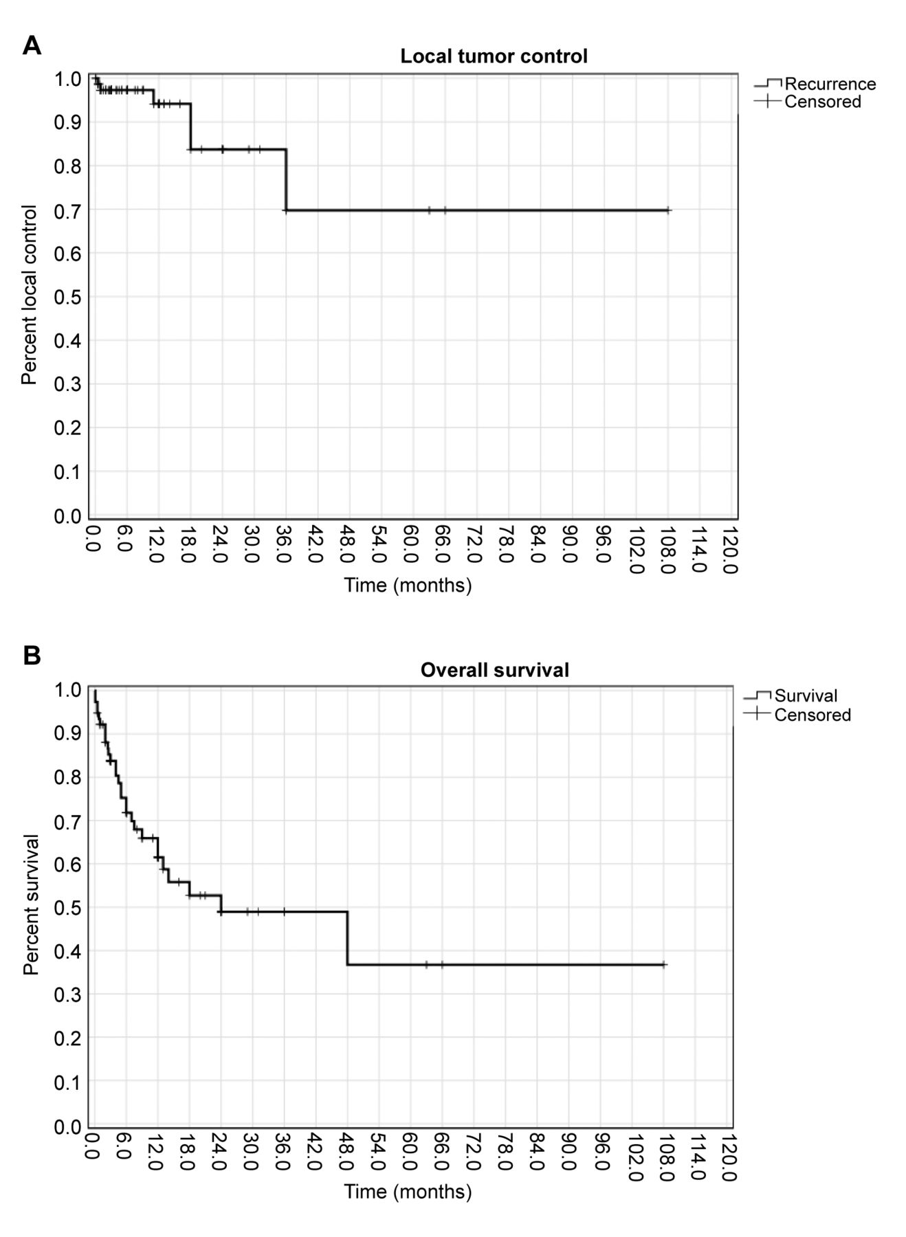
Post-treatment clinico-radiological outcomes and survival. Patients were followed-up for a median of 6.7 months (range=0.1-108 months) (Table III). Most patients (70.8%) achieved post-treatment symptom improvement. Recovery of the ambulatory status occurred in 13 of the 18 patients with pre-treatment paraparesis (72.2%). Documented radiological responses were: CR in 18 patients (15%), PR in 59 (49.2%), SD in 22 (18.3%), and PD in 21 (17.5%). IESM recurrences occurred in 4 patients (8.5%), with median LC of 14.5 months (range=0.1-36.0 months) (Figure 1). A 50% of patients died at last follow-up, with a median OS of 6.7 months (range=0.1-108 months).

View this table:
  • View inline
  • View popup
  • Download powerpoint
Table III.

Summary of treatment strategies and outcomes.

Figure 1.
  • Download figure
  • Open in new tab
  • Download powerpoint
Figure 1.

Kaplan-Meier curves of (A) local tumor control and (B) overall survival of the pooled cohort.

Discussion

Non-neurogenic IESMs may occur at later stages of advanced systemic tumors, often causing debilitating alteration of patients’ functional status. Advances in imaging and treatments may offer timely management, but clinical outcomes and survival have remained dismal. We found that IESMs frequently manifest with severe sensory and motor deficits, causing acute CES when involving the lumbar-sacral spine. Current therapies are mostly palliative, offering short-term clinico-radiological improvement. This review aimed to provide useful insights for managing IESMs within the context of other extradural and intramedullary spinal metastases.

IESMs comprise only 1-2% of all metastatic spine lesions, occurring less frequently than both vertebral (68) and intramedullary (69) spine metastases. In this review of non-neurogenic IESMs, the most common primary neoplasms comprised lung, renal, and breast carcinomas, reflecting their overall prevalence among oncological patients and/or patients with spine metastases (70, 71). Several mechanisms of IESM’s metastatic spread have been suggested, including: hematogenous spread via the arteriovenous system, the Batson’s plexus, or the perineural lymphatic system, cerebrospinal fluid (CSF) dissemination with leptomeningeal spread and drop subarachnoid metastases, and direct invasion from adjacent structures with dural seeding (7, 25). We assumed that the most common origin of non-neurogenic IESMs derives from hematogenous tumor spread, owing to the prevalence (58.5%) of concomitant systemic metastases noted in our pooled patients. Leptomeningeal spread is expected to be more common for the occurrence of neurogenic IESMs from primary central nervous system tumors. However, the high rates of concurrent brain metastases (67.1%) found in this cohort suggest that CSF dissemination may be a common route also for IESMs. Direct tumor invasion with dural seeding has been documented less frequently, especially in patients with melanomas or urologic neoplasm with concurrent vertebral metastases (31, 59). The most common metastatic routes may account for the late onset of IESMs from the diagnoses of primary tumors, as reported across all pooled studies (median 18 months). In some cases, aggressive carcinomas may show different metastatic mechanisms accountable for synchronous IESMs, which characterized the initial clinical event that led to the diagnosis of primary tumors in 16 of our pooled patients (12.3%) diagnosed with late-stage melanoma (53, 54), lung (36, 42), and renal (45, 46) cancers.

Similar to intramedullary spinal cord metastases, most IESMs are symptomatic but non-specific in terms of clinical presentation, causing delay in diagnosis with consequent worsening of patients’ functional status (5, 69). Accurate evaluation of sensorimotor deficits and regional axial pain may assist in localizing affected spine levels, but may be underestimated at early stages in case of lesions with moderate growth and non-severe spinal cord compression (7, 44). As found in this pooled cohort, acute CES has been frequently reported in lesions involving the lumbar-sacral spine, which may facilitate a differential diagnosis with other intramedullary tumors (17, 72). Although CES-related symptoms, such as lumbar or radicular leg pain, and mild motor deficits with preservation of sphincter functions, may simulate lumbar disc diseases, their quick progression may serve as an important red flag mandating further, prompt diagnostic assessments (73). Spine myelography and CT scans were the most commonly used diagnostic tools in early series. Contrarily to intramedullary lesions, spine myelography allowed for the detection of contrast dye block within the spinal canal, suggesting the location and suspicion of IESMs, especially in patients with a known history of cancer (6, 10, 17, 65). The introduction of T1-contrast MRI scans improved the pre-operative diagnostic accuracy, representing the current diagnostic standard. IESMs are frequently identified as single or multiple contrast-enhancing masses located outside the spinal cord and often attached to the dura, causing compression or infiltration of spinal nerve roots particularly when occurring within the conus medullaris (5, 10, 43). MRI and CT may yield low specificity to distinguish primary from metastatic intradural extramedullary lesions, particularly in cases with no prior cancer history (10). However, the importance of early spine imaging relies in clearly distinguishing intramedullary versus extramedullary lesions in patients with non-specific sensorimotor deficits, guiding the design of timely surgical management to avoid irreversible neurological deficits. Furthermore, imaging of the whole spine is also useful to detect concomitant LMs (66) or vertebral metastases (11), offering additional valuable information to plan accurate patient-tailored treatments.

In patients with intradural and extradural spine metastases, the primary treatment goals focus on providing symptom relief with improvement in functional status and/or walking rehabilitation (74). Compared to other spine tumors, such as vertebral metastases, the main difficulties in IESM’s management mostly stem from their complex surgical anatomy, owing to their close relationship with the surrounding spinal cord and spinal nerve roots (5, 6, 11). When feasible, decompressive laminectomy with tumor resection should be performed to confirm the diagnosis and to provide symptom relief (57, 64). However, most IESMs entangle the spinal nerve roots and/or are strictly attached to them, lacking a distinct cleavage plane and posing some risks to cause post-surgical complications when attempting complete tumor resection (1). Similar to a series of patients with intramedullary spinal tumors (69), partial tumor resection was preferred in patients with IESMs, achieving optimal symptom improvement with fewer risks of post-operative neurological complications (57). In some studies, total en-bloc IESMs resection was performed by transecting the infiltrated nerve roots, with no post-operative deficits noted (33, 36, 44, 56). Intraoperative neuromonitoring assistance proved fundamental to evaluate the preserved sensorimotor functions of involved spinal nerves, planning patient-tailored surgical techniques to minimize iatrogenic complications. Similar to other spine metastases, adjuvant treatments have been reported to offer superior survival benefits compared to surgery alone (69, 73). Across our included studies, systemic therapies mostly differed based on primary neoplasms, whereas radiotherapy protocols were mainly consistent. Spine EBRT proved to be effective in patients with metastatic spinal cord compression to reduce post-surgical residual tumor volumes and prolong local control (75). In these pooled patients, locoregional spine EBRT mainly targeted the lesions of interest, but was also extended to other spine regions in case of multiple simultaneous IESMs and/or concurrent vertebral metastases (54, 55, 60). Targeted adjuvant SRS (with a 10 Gy dose) was used in the recent report of Pagano et al. (64) with no radiation-induced adverse events, suggesting the feasibility and safety of this protocol for the management of IESMs. Newer immunotherapy and targeted molecular therapies have been studied and are currently used in the multidisciplinary management of systemic cancers, but their benefits in patients with IESMs have yet to be evaluated (72, 76).

Despite the available therapies, patients with IESMs have poor prognoses, which mainly depend on primary cancer’s histology and treatments (5, 11, 64). Similar to patients with intramedullary metastases (69), the pooled low LC and OS rates in IESMs likely derive from their late-onset and delayed diagnosis, as they have been frequently detected in patients with high tumor burden and multiple systemic metastases. These elements, added to the lack of available systemic therapies selectively directed against IESMs, may account for the differences in survival noted in comparison to patients with vertebral metastases, with OS up to 2 years when receiving immunotherapy or targeted molecular therapies (71). Tumor resection and locoregional radiation were mostly intended for palliation, achieving desirable short-term rates of clinical (70.8%) and radiological (64.2%) improvement, superior than those described in patients with spine LMs (72). Optimal outcomes were obtained by performing decompressive laminectomy and tumor resection in patients with paraparesis and ambulatory difficulty, which led to walking improvement and quality of life amelioration in most cases (27, 65, 67). However, owing to the better prognosis and survival in oncological patients, with the consequent increasing incidence of spine metastases, novel patient-tailored therapies should be developed and evaluated also for IESMs, aimed at reducing patients’ morbidity and improving their quality of life.

Limitations

All included articles were retrospective case reports and small case series, potentially subjected to publication and selection biases. Included articles covered a 40 year time period encompassing major changes in imaging diagnostic tools and available treatments, likely to have generated some between-study heterogeneity in the final analysis. By selectively including only histology-proved cases, this review may have underestimated IESMs’ overall prevalence and related outcomes, accounting for possible detection bias. However, our predetermined inclusion criteria were defined to minimize the risks of introducing confounding variables related to misdiagnoses of IESMs obtained only from clinical and radiological evaluations. Due to the lack of granular data, performance status scores could not be analyzed nor compared pre- and post-treatment. Despite these limitations, we present a methodologically accurate and replicable individual patient data meta-analysis of IESMs to advice clinicians on their management.

Conclusion

Non-neurogenic IESMs may occur at later stages in systemic oncological diseases, determining significant neurological and functional deficits in affected patients. IESMs mostly derive from lung, renal, and breast carcinomas, often causing sensory and motor impairments and acute CES when involving the lumbar-sacral spine. Decompressive laminectomy and tumor resection, in particular if associated with adjuvant locoregional radiation and/or systemic therapies, show good rates of shortterm symptom improvement. However, LC and OS remain discouraging. Future studies are needed to evaluate newer molecular and immune therapies in IESMs’ multimodal management.

Footnotes

  • Authors’ Contributions

    Paolo Palmisciano: Conceptualization, Methodology, Data analysis, Writing - Original draft preparation, Andrew L. Chen: Resources, Writing - Reviewing and Editing, Othman Bin-Alamer: Resources, Writing - Reviewing and Editing, Gianluca Ferini: Resources, Writing - Reviewing and Editing, Mayur Sharma: Resources, Writing - Reviewing and Editing, Giuseppe E. Umana: Resources, Writing - Reviewing and Editing, Salah G. Aoun: Resources, Writing - Reviewing and Editing, Ali S. Haider: Methodology, Resources, Writing - Reviewing and Editing, Supervision.

  • Conflicts of Interest

    The Authors have no relevant financial or non-financial interests to disclose in relation to this study.

  • Received May 29, 2022.
  • Revision received June 14, 2022.
  • Accepted June 15, 2022.
  • Copyright © 2022 The Author(s). Published by the International Institute of Anticancer Research.

This article is an open access article distributed under the terms and conditions of the Creative Commons Attribution (CC BY-NC-ND) 4.0 international license (https://creativecommons.org/licenses/by-nc-nd/4.0).

References

  1. ↵
    1. Chow TS and
    2. McCutcheon IE
    : The surgical treatment of metastatic spinal tumors within the intradural extramedullary compartment. J Neurosurg 85(2): 225-230, 1996. PMID: 8755750. DOI: 10.3171/jns.1996.85.2.0225
    OpenUrlCrossRefPubMed
    1. Jacobs WB and
    2. Perrin RG
    : Evaluation and treatment of spinal metastases: an overview. Neurosurg Focus 11(6): e10, 2001. PMID: 16463993. DOI: 10.3171/foc.2001.11.6.11
    OpenUrlCrossRefPubMed
  2. ↵
    1. Schick B,
    2. Ibing R,
    3. Brors D and
    4. Draf W
    : Long-term study of endonasal duraplasty and review of the literature. Ann Otol Rhinol Laryngol 110(2): 142-147, 2001. PMID: 11219521. DOI: 10.1177/000348940111000209
    OpenUrlCrossRefPubMed
  3. ↵
    1. Carminucci A and
    2. Hanft S
    : Intradural extramedullary spinal metastasis of renal cell carcinoma: illustrative case report and comprehensive review of the literature. Eur Spine J 29(Suppl 2): 176-182, 2020. PMID: 33051797. DOI: 10.1007/s00586-020-06537-x
    OpenUrlCrossRefPubMed
  4. ↵
    1. Gazzeri R,
    2. Telera S,
    3. Galarza M,
    4. Callovini GM,
    5. Sperduti I and
    6. Alfieri A
    : Surgical treatment of solitary intradural extramedullary spinal cord metastases from solid cancers of non-neurogenic origin. A multicenter study. J Neurooncol 154(1): 101-112, 2021. PMID: 34255272. DOI: 10.1007/s11060-021-03804-9
    OpenUrlCrossRefPubMed
  5. ↵
    1. Perrin RG,
    2. Livingston KE and
    3. Aarabi B
    : Intradural extramedullary spinal metastasis. A report of 10 cases. J Neurosurg 56(6): 835-837, 1982. PMID: 7077385. DOI: 10.3171/jns.1982.56.6.0835
    OpenUrlCrossRefPubMed
  6. ↵
    1. Ji GY,
    2. Oh CH,
    3. Kim SH,
    4. Shin DA and
    5. Kim KN
    : Intradural cauda equina metastasis of renal cell carcinoma: a case report with literature review of 10 cases. Spine (Phila Pa 1976) 38(18): E1171-E1174, 2013. PMID: 23759799. DOI: 10.1097/BRS.0b013e31829cef66
    OpenUrlCrossRefPubMed
  7. ↵
    1. Anand SK,
    2. Garling RJ,
    3. Johns J,
    4. Shah M and
    5. Chamiraju P
    : Intradural extramedullary spinal metastases from uterine carcinosarcoma: A case report. Surg Neurol Int 11: 354, 2020. PMID: 33194287. DOI: 10.25259/SNI_621_2020
    OpenUrlCrossRefPubMed
  8. ↵
    1. Chamberlain MC and
    2. Tredway TL
    : Adult primary intradural spinal cord tumors: a review. Curr Neurol Neurosci Rep 11(3): 320-328, 2011. PMID: 21327734. DOI: 10.1007/s11910-011-0190-2
    OpenUrlCrossRefPubMed
  9. ↵
    1. Soderlund KA,
    2. Smith AB,
    3. Rushing EJ and
    4. Smirniotopolous JG
    : Radiologic-pathologic correlation of pediatric and adolescent spinal neoplasms: Part 2, Intradural extramedullary spinal neoplasms. AJR Am J Roentgenol 198(1): 44-51, 2012. PMID: 22194478. DOI: 10.2214/AJR.11.7121
    OpenUrlCrossRefPubMed
  10. ↵
    1. Knafo S,
    2. Pallud J,
    3. Le Rhun E,
    4. Parker F,
    5. Iakovlev G,
    6. Roux FX,
    7. Page P,
    8. Meder JF,
    9. Emery E,
    10. Devaux B and Club de Neuro-oncologie of the Société Française de Neurochirurgie
    : Intradural extramedullary spinal metastases of non-neurogenic origin: a distinct clinical entity or a subtype of leptomeningeal metastasis? A case-control study. Neurosurgery 73(6): 923-31; discussion 932, 2013. PMID: 23921711. DOI: 10.1227/NEU.0000000000000132
    OpenUrlCrossRefPubMed
  11. ↵
    1. Hoover JM,
    2. Krauss WE and
    3. Lanzino G
    : Intradural spinal metastases: a surgical series of 15 patients. Acta Neurochir (Wien) 154(5): 871-7; discussion 877, 2012. PMID: 22395431. DOI: 10.1007/s00701-012-1313-5
    OpenUrlCrossRefPubMed
  12. ↵
    1. Stefini R,
    2. Peron S,
    3. Mandelli J,
    4. Bianchini E and
    5. Roccucci P
    : Intraoperative spinal navigation for the removal of intradural tumors: technical notes. Oper Neurosurg (Hagerstown) 15(1): 54-59, 2018. PMID: 28962027. DOI: 10.1093/ons/opx179
    OpenUrlCrossRefPubMed
    1. Kochanski RB,
    2. Lombardi JM,
    3. Laratta JL,
    4. Lehman RA and
    5. O’Toole JE
    : Image-guided navigation and robotics in spine surgery. Neurosurgery 84(6): 1179-1189, 2019. PMID: 30615160. DOI: 10.1093/neuros/nyy630
    OpenUrlCrossRefPubMed
  13. ↵
    1. Kumar N, G V,
    2. Ravikumar N,
    3. Ding Y,
    4. Yin ML,
    5. Patel RS,
    6. Naresh N,
    7. Hey HWD,
    8. Lau LL and
    9. Liu G
    : Intraoperative Neuromonitoring (IONM): Is there a role in metastatic spine tumor surgery? Spine (Phila Pa 1976) 44(4): E219-E224, 2019. PMID: 30044368. DOI: 10.1097/BRS.0000000000002808
    OpenUrlCrossRefPubMed
  14. ↵
    1. Purvis TE,
    2. Goodwin CR,
    3. Lubelski D,
    4. Laufer I and
    5. Sciubba DM
    : Review of stereotactic radiosurgery for intradural spine tumors. CNS Oncol 6(2): 131-138, 2017. PMID: 28425771. DOI: 10.2217/cns-2016-0039
    OpenUrlCrossRefPubMed
  15. ↵
    1. Kumar N,
    2. Tan WLB,
    3. Wei W and
    4. Vellayappan BA
    : An overview of the tumors affecting the spine-inside to out. Neurooncol Pract 7(Suppl 1): i10-i17, 2020. PMID: 33299569. DOI: 10.1093/nop/npaa049
    OpenUrlCrossRefPubMed
  16. ↵
    1. Hasegawa H,
    2. Shin M,
    3. Kondo K,
    4. Hanakita S,
    5. Mukasa A,
    6. Kin T and
    7. Saito N
    : Role of endoscopic transnasal surgery for skull base chondrosarcoma: a retrospective analysis of 19 cases at a single institution. J Neurosurg 128(5): 1438-1447, 2018. PMID: 28686110. DOI: 10.3171/2017.1.JNS162000
    OpenUrlCrossRefPubMed
  17. ↵
    1. Hasegawa H,
    2. Vakharia K,
    3. Graffeo CS,
    4. Carlson ML,
    5. Pollock BE,
    6. Brown PD,
    7. Perry A,
    8. Van Gompel JJ,
    9. Driscoll CLW and
    10. Link MJ
    : Long-term outcomes of grade I/II skull base chondrosarcoma: an insight into the role of surgery and upfront radiotherapy. J Neurooncol 153(2): 273-281, 2021. PMID: 33907967. DOI: 10.1007/s11060-021-03764-0
    OpenUrlCrossRefPubMed
  18. ↵
    1. Page MJ,
    2. McKenzie JE,
    3. Bossuyt PM,
    4. Boutron I,
    5. Hoffmann TC,
    6. Mulrow CD,
    7. Shamseer L,
    8. Tetzlaff JM,
    9. Akl EA,
    10. Brennan SE,
    11. Chou R,
    12. Glanville J,
    13. Grimshaw JM,
    14. Hróbjartsson A,
    15. Lalu MM,
    16. Li T,
    17. Loder EW,
    18. Mayo-Wilson E,
    19. McDonald S,
    20. McGuinness LA,
    21. Stewart LA,
    22. Thomas J,
    23. Tricco AC,
    24. Welch VA,
    25. Whiting P and
    26. Moher D
    : The PRISMA 2020 statement: an updated guideline for reporting systematic reviews. BMJ 372: n71, 2021. PMID: 33782057. DOI: 10.1136/bmj.n71
    OpenUrlFREE Full Text
  19. ↵
    1. Howick J,
    2. Chalmers I,
    3. Glasziou P,
    4. Greenhalgh T,
    5. Heneghan C,
    6. Liberati A,
    7. Moschetti I,
    8. Phillips B and
    9. Thornton H
    : Explanation of the 2011 Oxford Centre for Evidence-Based Medicine (OCEBM) Levels of Evidence (Background Document). Oxford Cent Evidence-Based Med, 2011. Available at: https://www.cebm.ox.ac.uk/resources/levels-of-evidence/ocebm-levels-of-evidence [Last accessed on June 13, 2022]
  20. ↵
    1. Aromataris E and
    2. Munn Z
    1. Moola S,
    2. Munn Z,
    3. Tufanaru C,
    4. Aromataris E,
    5. Sears K,
    6. Sfetcu R,
    7. Currie M,
    8. Qureshi R,
    9. Mattis P,
    10. Lisy K and
    11. Mu P-F
    : Chapter 7:Systematic reviews of etiology and risk. In: JBI Manual for Evidence Synthesis. Aromataris E and Munn Z (eds.). JBI, 2020. Available at: https://synthesismanual.jbi.global. https://doi.org/10.46658/JBIMES-20-08 [Last accessed on June 15, 2022]
    OpenUrlCrossRef
  21. ↵
    1. Schaller B,
    2. Merlo A,
    3. Kirsch E,
    4. Lehmann K,
    5. Huber PR,
    6. Lyrer P,
    7. Steck AJ and
    8. Gratzl O
    : Prostate-specific antigen in the cerebrospinal fluid leads to diagnosis of solitary cauda equina metastasis: a unique case report and review of the literature. Br J Cancer 77(12): 2386-2389, 1998. PMID: 9649164. DOI: 10.1038/bjc.1998.397
    OpenUrlCrossRefPubMed
    1. Maxwell M,
    2. Borges LF and
    3. Zervas NT
    : Renal cell carcinoma: a rare source of cauda equina metastasis. Case report. J Neurosurg 90(1 Suppl): 129-132, 1999. PMID: 10413138. DOI: 10.3171/spi.1999.90.1.0129
    OpenUrlCrossRefPubMed
  22. ↵
    1. Mak KH and
    2. Kwok JC
    : Intradural spinal metastasis from renal cell carcinoma: A case report. J Orthop Surg (Hong Kong) 9(2): 57-61, 2001. PMID: 12118133. DOI: 10.1177/230949900100900212
    OpenUrlCrossRefPubMed
    1. Schick U,
    2. Marquardt G and
    3. Lorenz R
    : Intradural and extradural spinal metastases. Neurosurg Rev 24(1): 1-5; discussion 6-7, 2001. PMID: 11339461. DOI: 10.1007/pl00011959
    OpenUrlCrossRefPubMed
  23. ↵
    1. Mehrotra R and
    2. Sharma K
    : Intradural extramedullary spinal metastasis from an ovarian carcinoma. Indian J Cancer 39(4): 157-160, 2002. PMID: 12928577.
    OpenUrlPubMed
    1. Takada T,
    2. Doita M,
    3. Nishida K,
    4. Miura J,
    5. Yoshiya S and
    6. Kurosaka M
    : Unusual metastasis to the cauda equina from renal cell carcinoma. Spine (Phila Pa 1976) 28(6): E114-E117, 2003. PMID: 12642774. DOI: 10.1097/01.BRS.0000049910.72881.A0
    OpenUrlCrossRefPubMed
    1. Gaetani P,
    2. Di Ieva A,
    3. Colombo P,
    4. Tancioni F,
    5. Aimar E,
    6. Debernardi A and
    7. Rodriguez Y
    8. Baena R
    : Intradural spinal metastasis of renal clear cell carcinoma causing cauda equina syndrome. Acta Neurochir (Wien) 146(8): 857-861, 2004. PMID: 15254809. DOI: 10.1007/s00701-004-0299-z
    OpenUrlCrossRefPubMed
    1. Kubota M,
    2. Saeki N,
    3. Yamaura A,
    4. Iuchi T,
    5. Ohga M and
    6. Osato K
    : A rare case of metastatic renal cell carcinoma resembling a nerve sheath tumor of the cauda equina. J Clin Neurosci 11(5): 530-532, 2004. PMID: 15177402. DOI: 10.1016/j.jocn.2003.09.010
    OpenUrlCrossRefPubMed
  24. ↵
    1. Hentschel SJ,
    2. Mendel E,
    3. Singh S and
    4. Rhines LD
    : Metastatic prostate carcinoma to the intradural extramedullary spinal compartment. Case report. J Neurosurg 100(4 Suppl Spine): 375-377, 2004. PMID: 15070148. DOI: 10.3171/spi.2004.100.4.0375
    OpenUrlCrossRefPubMed
    1. Alfieri A,
    2. Mazzoleni G,
    3. Schwarz A,
    4. Campello M,
    5. Broger M,
    6. Vitale M and
    7. Vigl EE
    : Renal cell carcinoma and intradural spinal metastasis with cauda equina infiltration: case report. Spine (Phila Pa 1976) 30(1): 161-163, 2005. PMID: 15626997.
    OpenUrlPubMed
  25. ↵
    1. Kotil K,
    2. Kilinc BM and
    3. Bilge T
    : Spinal metastasis of occult lung carcinoma causing cauda equina syndrome. J Clin Neurosci 14(4): 372-375, 2007. PMID: 17336230. DOI: 10.1016/j.jocn.2006.01.007
    OpenUrlCrossRefPubMed
    1. Jost G,
    2. Zimmerer S,
    3. Frank S,
    4. Cordier D and
    5. Merlo A
    : Intradural spinal metastasis of renal cell cancer. Report of a case and review of 26 published cases. Acta Neurochir (Wien) 151(7): 815-21; discussion 821, 2009. PMID: 19415167. DOI: 10.1007/s00701-009-0358-6
    OpenUrlCrossRefPubMed
    1. Kim DY,
    2. Lee JK,
    3. Moon SJ,
    4. Kim SC and
    5. Kim CS
    : Intradural spinal metastasis to the cauda equina in renal cell carcinoma: a case report and review of the literature. Spine (Phila Pa 1976) 34(24): E892-E895, 2009. PMID: 19910759. DOI: 10.1097/BRS.0b013e3181b34e6c
    OpenUrlCrossRefPubMed
  26. ↵
    1. Cansever T,
    2. Kabatas S,
    3. Civelek E,
    4. Yilmaz C and
    5. Caner H
    : Spinal metastasis of occult lung carcinoma causing cauda equine syndrome with lumbar spinal stenosis. Turk Neurosurg 21(3): 408-412, 2011. PMID: 21845580. DOI: 10.5137/1019-5149.JTN.2711-09.2
    OpenUrlCrossRefPubMed
    1. Ito K,
    2. Miyahara T,
    3. Goto T,
    4. Horiuchi T,
    5. Sakai K and
    6. Hongo K
    : Solitary metastatic cauda equina tumor from breast cancer -case report-. Neurol Med Chir (Tokyo) 50(5): 417-420, 2010. PMID: 20505303. DOI: 10.2176/nmc.50.417
    OpenUrlCrossRefPubMed
    1. Lin CL,
    2. Chang JL,
    3. Lo HC and
    4. Wu KA
    : Extramedullary-intradural spinal metastasis of small cell lung cancer causing cauda equina syndrome. Am J Med Sci 339(2): 192-194, 2010. PMID: 20019582. DOI: 10.1097/MAJ.0b013e3181bedd1f
    OpenUrlCrossRefPubMed
    1. Tsimpas A,
    2. Post NH,
    3. Moshel Y and
    4. Frempong-Boadu AK
    : Large cell neuroendocrine carcinoma of the lung metastatic to the cauda equina. Spine J 10(6): e1-e5, 2010. PMID: 20494806. DOI: 10.1016/j.spinee.2010.03.031
    OpenUrlCrossRefPubMed
    1. Lin TK,
    2. Chen SM and
    3. Jung SM
    : Solitary intradural extramedullary metastasis of renal cell carcinoma to the conus medullaris. Kaohsiung J Med Sci 27(1): 45-48, 2011. PMID: 21329893. DOI: 10.1016/j.kjms.2010.05.001
    OpenUrlCrossRefPubMed
    1. Akhavan A,
    2. Mehrabaniyan MR,
    3. Jarahi M and
    4. Navabii H
    : Intradural extramedullary metastasis from papillary carcinoma of thyroid. BMJ Case Rep 2012: bcr0220125801, 2012. PMID: 22729332. DOI: 10.1136/bcr.02.2012.5801
    OpenUrlAbstract/FREE Full Text
  27. ↵
    1. Lee CH,
    2. Kim KJ,
    3. Hyun SJ,
    4. Jahng TA and
    5. Kim HJ
    : Intradural extramedullary metastasis of small cell lung cancer: a case report. Korean J Spine 9(3): 293-296, 2012. PMID: 25983836. DOI: 10.14245/kjs.2012.9.3.293
    OpenUrlCrossRefPubMed
  28. ↵
    1. Wostrack M,
    2. Pape H,
    3. Kreutzer J,
    4. Ringel F,
    5. Meyer B and
    6. Stoffel M
    : Surgical treatment of spinal intradural carcinoma metastases. Acta Neurochir (Wien) 154(2): 349-357, 2012. PMID: 22009015. DOI: 10.1007/s00701-011-1204-1
    OpenUrlCrossRefPubMed
  29. ↵
    1. Dobson GM,
    2. Polvikoski T,
    3. Nissen JJ and
    4. Holliman D
    : Cauda equina syndrome secondary to intradural renal cell carcinoma metastasis haemorrhage. Br J Neurosurg 27(2): 249-250, 2013. PMID: 22985045. DOI: 10.3109/02688697.2012.724122
    OpenUrlCrossRefPubMed
  30. ↵
    1. Strong C,
    2. Yanamadala V,
    3. Khanna A,
    4. Walcott BP,
    5. Nahed BV,
    6. Borges LF and
    7. Coumans JV
    : Surgical treatment options and management strategies of metastatic renal cell carcinoma to the lumbar spinal nerve roots. J Clin Neurosci 20(11): 1546-1549, 2013. PMID: 23931936. DOI: 10.1016/j.jocn.2013.02.014
    OpenUrlCrossRefPubMed
  31. ↵
    1. Heary RF,
    2. Agarwal N,
    3. Barrese JC,
    4. Barry MT and
    5. Baisre A
    : Metastatic renal cell carcinoma, with a radiographically occult primary tumor, presenting in the operative site of a thoracic meningioma: long-term follow-up: Case report. J Neurosurg Spine 21(4): 628-633, 2014. PMID: 25014504. DOI: 10.3171/2014.6.SPINE13448
    OpenUrlCrossRefPubMed
    1. Srinivasan A,
    2. Dhandapani S,
    3. Chatterjee D and
    4. Simha V
    : Renal small cell carcinoma presenting with solitary lumbar intradural metastasis. Neurol India 62(5): 561-563, 2014. PMID: 25387638. DOI: 10.4103/0028-3886.144494
    OpenUrlCrossRefPubMed
    1. Petterwood J,
    2. Lim K,
    3. Gonzalvo A and
    4. Quan G
    : Intradural extramedullary colorectal adenocarcinoma metastasis to the cervical spine. ANZ J Surg 85(7-8): 582-583, 2015. PMID: 24251887. DOI: 10.1111/ans.12452
    OpenUrlCrossRefPubMed
    1. Wu CY,
    2. Huang HM and
    3. Cho DY
    : An acute bleeding metastatic spinal tumor from HCC causes an acute onset of cauda equina syndrome. Biomedicine (Taipei) 5(3): 18, 2015. PMID: 26298047. DOI: 10.7603/s40681-015-0018-5
    OpenUrlCrossRefPubMed
    1. Xiong J and
    2. Zhang P
    : Cauda equina syndrome caused by isolated spinal extramedullary-intradural cauda equina metastasis is the primary symptom of small cell lung cancer: a case report and review of the literatrure. Int J Clin Exp Med 8(6): 10044-10050, 2015. PMID: 26309698.
    OpenUrlPubMed
    1. Capek S,
    2. Krauss WE,
    3. Amrami KK,
    4. Parisi JE and
    5. Spinner RJ
    : Perineural spread of renal cell carcinoma: a case illustration with a proposed anatomic mechanism and a review of the literature. World Neurosurg 89: 728.e11-728.e17, 2016. PMID: 26844879. DOI: 10.1016/j.wneu.2016.01.060
    OpenUrlCrossRefPubMed
    1. Wolf A,
    2. Johnstone R and
    3. Siddiqi F
    : Intradural extramedullary spinal cord metastasis of the prostate: a case presentation and review of the literature. Can J Neurol Sci 43(4): 588-592, 2016. PMID: 27160485. DOI: 10.1017/cjn.2016.43
    OpenUrlCrossRefPubMed
  32. ↵
    1. O’Reilly MK,
    2. Sugrue G,
    3. Byrne D and
    4. MacMahon P
    : Combined intramedullary and intradural extramedullary spinal metastases in malignant melanoma. BMJ Case Rep 2017: bcr2017220031, 2017. PMID: 28343150. DOI: 10.1136/bcr-2017-220031
    OpenUrlCrossRefPubMed
  33. ↵
    1. Stein AA,
    2. Weinstein GR,
    3. Niezgoda C,
    4. Chowdhary S,
    5. Vrionis F and
    6. Houten JK
    : Myelopathy from intradural extramedullary metastasis as an initial presentation of metastatic melanoma. Cureus 10(5): e2668, 2018. PMID: 30042919. DOI: 10.7759/cureus.2668
    OpenUrlCrossRefPubMed
  34. ↵
    1. Kehayov I,
    2. Genova S,
    3. Gicheva M,
    4. Nuri B and
    5. Kitov B
    : Intradural extramedullary metastasis of the upper thoracic spine – case report and literature review. Folia Med (Plovdiv) 61(4): 624-629, 2019. PMID: 32337876. DOI: 10.3897/folmed.61.e47948
    OpenUrlCrossRefPubMed
  35. ↵
    1. Koyama K,
    2. Takahashi H,
    3. Inoue M,
    4. Okawa A,
    5. Nakajima A,
    6. Sonobe M,
    7. Akatsu Y,
    8. Saito J,
    9. Taniguchi S,
    10. Yamada M,
    11. Yamamoto K,
    12. Aoki Y,
    13. Furuya T,
    14. Koda M,
    15. Yamazaki M,
    16. Ohtori S and
    17. Nakagawa K
    : Intradural metastasis to the cauda equina found as the initial presentation of breast cancer: a case report. J Med Case Rep 13(1): 220, 2019. PMID: 31324210. DOI: 10.1186/s13256-019-2155-z
    OpenUrlCrossRefPubMed
  36. ↵
    1. Land CF,
    2. Bowden BD,
    3. Morpeth BG and
    4. DeVine JG
    : Intradural extramedullary metastasis: a review of literature and case report. Spinal Cord Ser Cases 5: 41, 2019. PMID: 31632701. DOI: 10.1038/s41394-019-0181-0
    OpenUrlCrossRefPubMed
    1. Sarmento M,
    2. Haas L,
    3. Tutida L,
    4. Marques N,
    5. Scramocin T,
    6. Omar O,
    7. Giustina E,
    8. Sartori F,
    9. Camilo L,
    10. Borille T,
    11. Bernardes C,
    12. Boer V,
    13. Lara D,
    14. Cabral F and
    15. Mello L
    : Intradural extramedullary spinal metastasis of a kidney cancer: Case report. Arquivos Brasileiros de Neurocirurgia: Brazilian Neurosurgery 38(01): 042-046, 2019. DOI: 10.1055/s-0039-1678588
    OpenUrlCrossRef
  37. ↵
    1. Cho KT,
    2. Yang JJ and
    3. Lee K
    : Intradural extramedullary metastatic conjunctival malignant melanoma. World Neurosurg 138: 444-448, 2020. PMID: 32217182. DOI: 10.1016/j.wneu.2020.03.078
    OpenUrlCrossRefPubMed
  38. ↵
    1. Saway BF,
    2. Fayed I,
    3. Dowlati E,
    4. Derakhshandeh R and
    5. Sandhu FA
    : Initial report of an intradural extramedullary metastasis of a pancreatic neuroendocrine tumor to the cervical spine: a case report and review of the literature. World Neurosurg 139: 355-360, 2020. PMID: 32344144. DOI: 10.1016/j.wneu.2020.04.120
    OpenUrlCrossRefPubMed
    1. Tajima Y,
    2. Takahashi M,
    3. Kawai T,
    4. Higashi M,
    5. Sano H,
    6. Ichimura S and
    7. Kobayashi H
    : Metastatic intradural extramedullary spinal cord tumor from ovarian cancer: A case report with a literature review. J Spinal Cord Med 45(2): 320-323, 2022. PMID: 32202486. DOI: 10.1080/10790268.2020.1739380
    OpenUrlCrossRefPubMed
    1. Ali S,
    2. Qasim A,
    3. Salah R,
    4. Sarwar MR,
    5. Usman M and
    6. Shams S
    : Isolated late intradural cauda equina metastasis of renal cell carcinoma. Surg Neurol Int 12: 481, 2021. PMID: 34754531. DOI: 10.25259/SNI_721_2021
    OpenUrlCrossRefPubMed
    1. Mariniello G,
    2. Corvino S,
    3. Sgulò F,
    4. Guadagno E,
    5. Del Basso De Caro M and
    6. Maiuri F
    : Intradural cauda equina metastases from renal cell carcinoma. Interdisciplinary Neurosurgery 27: 101397, 2021. DOI: 10.1016/j.inat.2021.101397
    OpenUrlCrossRef
  39. ↵
    1. Pagano A,
    2. Iaquinandi A,
    3. Fraioli MF,
    4. Bossone G,
    5. Carra N and
    6. Salvati M
    : Cauda equina syndrome from intradural metastasis of a non-neural tumor: case report and review of literature. Br J Neurosurg: 1-8, 2021. PMID: 34330176. DOI: 10.1080/02688697.2021.1958155
    OpenUrlCrossRefPubMed
  40. ↵
    1. Borovich B,
    2. Keret D,
    3. Ben-Arie J,
    4. Grushkiewicz I and
    5. Peyser E
    : Spinal intradural metastases of extraneural origin. Acta Neurochir (Wien) 56(1-2): 99-105, 1981. PMID: 7246286. DOI: 10.1007/BF01400977
    OpenUrlCrossRefPubMed
  41. ↵
    1. Castillo M and
    2. Quencer RM
    : Percutaneous needle evaluation of intradural extramedullary lesions of the lumbar spine. Neuroradiology 30(6): 551-555, 1988. PMID: 3226545. DOI: 10.1007/BF00339700
    OpenUrlCrossRefPubMed
  42. ↵
    1. Stambough JL,
    2. Reid JH,
    3. Ross MA,
    4. Simeone FA and
    5. Booth RE
    : Isolated intradural metastasis simulating lumbar disc disease. Spine (Phila Pa 1976) 16(5): 581-583, 1991. PMID: 2053002. DOI: 10.1097/00007632-199105000-00018
    OpenUrlCrossRefPubMed
  43. ↵
    1. Stark RJ,
    2. Henson RA and
    3. Evans SJ
    : Spinal metastases. A retrospective survey from a general hospital. Brain 105(Pt 1): 189-213, 1982. PMID: 7066672. DOI: 10.1093/brain/105.1.189
    OpenUrlCrossRefPubMed
  44. ↵
    1. Goyal A,
    2. Yolcu Y,
    3. Kerezoudis P,
    4. Alvi MA,
    5. Krauss WE and
    6. Bydon M
    : Intramedullary spinal cord metastases: an institutional review of survival and outcomes. J Neurooncol 142(2): 347-354, 2019. PMID: 30656530. DOI: 10.1007/s11060-019-03105-2
    OpenUrlCrossRefPubMed
  45. ↵
    1. World Health Organization: Cancer
    . Available at: https://www.who.int/news-room/fact-sheets/detail/cancer [Last accessed on May 12, 2021]
  46. ↵
    1. Barzilai O,
    2. Fisher CG and
    3. Bilsky MH
    : State of the art treatment of spinal metastatic disease. Neurosurgery 82(6): 757-769, 2018. PMID: 29481645. DOI: 10.1093/neuros/nyx567
    OpenUrlCrossRefPubMed
  47. ↵
    1. Palmisciano P,
    2. Sagoo NS,
    3. Kharbat AF,
    4. Kenfack YJ,
    5. Alamer OB,
    6. Scalia G,
    7. Umana GE,
    8. Aoun SG and
    9. Haider AS
    : Leptomeningeal metastases of the spine: a systematic review. Anticancer Res 42(2): 619-628, 2022. PMID: 35093859. DOI: 10.21873/anticanres.15519
    OpenUrlAbstract/FREE Full Text
  48. ↵
    1. Palmisciano P,
    2. Zaidi SE,
    3. Shlobin NA,
    4. Sagoo NS,
    5. Alamer OB,
    6. Scalia G,
    7. Ferini G,
    8. Umana GE,
    9. Aoun SG and
    10. Haider AS
    : Intradural cauda equina metastases: a systematic review of clinico-radiological features, management, and treatment outcomes. Anticancer Res 42(4): 1661-1669, 2022. PMID: 35346985. DOI: 10.21873/anticanres.15643
    OpenUrlAbstract/FREE Full Text
  49. ↵
    1. Burton MR,
    2. De Jesus O and
    3. Mesfin FB
    : Conus and Cauda Equina Tumors. Treasure Island, FL, USA, StatPearls Publishing, 2022.
  50. ↵
    1. Patchell RA,
    2. Tibbs PA,
    3. Regine WF,
    4. Payne R,
    5. Saris S,
    6. Kryscio RJ,
    7. Mohiuddin M and
    8. Young B
    : Direct decompressive surgical resection in the treatment of spinal cord compression caused by metastatic cancer: a randomised trial. Lancet 366(9486): 643-648, 2005. PMID: 16112300. DOI: 10.1016/S0140-6736(05)66954-1
    OpenUrlCrossRefPubMed
  51. ↵
    1. Palmisciano P,
    2. Haider AS,
    3. Nwagwu CD,
    4. Wahood W,
    5. Yu K,
    6. Ene CI,
    7. O’Brien BJ,
    8. Aoun SG,
    9. Cohen-Gadol AA and
    10. El Ahmadieh TY
    : The role of immune checkpoint inhibitors in leptomeningeal disease: a systematic review. Anticancer Res 41(11): 5333-5342, 2021. PMID: 34732403. DOI: 10.21873/anticanres.15346
    OpenUrlAbstract/FREE Full Text
PreviousNext
Back to top

In this issue

Anticancer Research: 42 (7)
Anticancer Research
Vol. 42, Issue 7
July 2022
  • Table of Contents
  • Table of Contents (PDF)
  • About the Cover
  • Index by author
  • Back Matter (PDF)
  • Ed Board (PDF)
  • Front Matter (PDF)
Print
Download PDF
Article Alerts
Sign In to Email Alerts with your Email Address
Email Article

Thank you for your interest in spreading the word on Anticancer Research.

NOTE: We only request your email address so that the person you are recommending the page to knows that you wanted them to see it, and that it is not junk mail. We do not capture any email address.

Enter multiple addresses on separate lines or separate them with commas.
Intradural Extramedullary Spinal Metastases from Non-neurogenic Primary Tumors: A Systematic Review
(Your Name) has sent you a message from Anticancer Research
(Your Name) thought you would like to see the Anticancer Research web site.
CAPTCHA
This question is for testing whether or not you are a human visitor and to prevent automated spam submissions.
6 + 10 =
Solve this simple math problem and enter the result. E.g. for 1+3, enter 4.
Citation Tools
Intradural Extramedullary Spinal Metastases from Non-neurogenic Primary Tumors: A Systematic Review
PAOLO PALMISCIANO, ANDREW L. CHEN, MAYUR SHARMA, OTHMAN BIN-ALAMER, GIANLUCA FERINI, GIUSEPPE E. UMANA, SALAH G. AOUN, ALI S. HAIDER
Anticancer Research Jul 2022, 42 (7) 3251-3259; DOI: 10.21873/anticanres.15814

Citation Manager Formats

  • BibTeX
  • Bookends
  • EasyBib
  • EndNote (tagged)
  • EndNote 8 (xml)
  • Medlars
  • Mendeley
  • Papers
  • RefWorks Tagged
  • Ref Manager
  • RIS
  • Zotero
Reprints and Permissions
Share
Intradural Extramedullary Spinal Metastases from Non-neurogenic Primary Tumors: A Systematic Review
PAOLO PALMISCIANO, ANDREW L. CHEN, MAYUR SHARMA, OTHMAN BIN-ALAMER, GIANLUCA FERINI, GIUSEPPE E. UMANA, SALAH G. AOUN, ALI S. HAIDER
Anticancer Research Jul 2022, 42 (7) 3251-3259; DOI: 10.21873/anticanres.15814
Twitter logo Facebook logo Mendeley logo
  • Tweet Widget
  • Facebook Like
  • Google Plus One

Jump to section

  • Article
    • Abstract
    • Materials and Methods
    • Results
    • Discussion
    • Limitations
    • Conclusion
    • Footnotes
    • References
  • Figures & Data
  • Info & Metrics
  • PDF

Related Articles

Cited By...

  • No citing articles found.
  • Google Scholar

More in this TOC Section

  • Artery-first Approach Versus Standard Pancreaticoduodenectomy: A Systematic Review and Meta-analysis of Surgical Outcomes and Oncological Benefits
  • Efficacy of Magseed Localization for Non-palpable Breast Lesions: A Systematic Review and Meta-analysis
  • Dicycloplatin, a Novel Analog of Cisplatin and Carboplatin, May Provide Therapeutic Advancement in Cancer Chemotherapy
Show more Review

Keywords

  • Extramedullary spine metastases
  • intradural metastases
  • neuro-oncology
  • spine oncology
  • spine metastases
  • systematic review
Anticancer Research

© 2026 Anticancer Research

Powered by HighWire