


Abstract
Background/Aim: Osteosarcoma is the most common bone sarcoma. Although surgery and chemotherapy are initially effective, the 5-year survival is approximately 60% to 80%, and has not improved over three decades. We have previously shown that methionine restriction (MR) induced by oral recombinant methioninase (o-rMETase), is effective against osteosarcoma in patient-derived orthotopic xenograft (PDOX) nude-mouse models. In the present report, the efficacy of the combination of oral o-rMETase and methotrexate (MTX) was examined in an osteosarcoma PDOX mouse model. Materials and Methods: An osteosarcoma-PDOX model was previously established by implanting tumor fragments into the proximal tibia of nude mice. The osteosarcoma PDOX models were randomized into four groups: control; o-rMETase alone; MTX alone; combination of o-rMETase and MTX. The mice were sacrificed after 4 weeks of treatment. Results: The combination of o-rMETase and MTX showed significantly higher efficacy compared to the control group (p=0.04). The combination also showed significantly higher efficacy compared to MTX alone (p=0.04). No significant efficacy of o-rMETase alone or MTX alone compared to control was shown (p=0.21, 1.00, respectively). Only the combination of o-rMETase and MTX reduced the cancer-cell density in the osteosarcoma tumor. Conclusion: rMETase converted an osteosarcoma PDOX from MTX-resistant to MTX-sensitive and thereby shows future clinical potential.
- Osteosarcoma
- nude mice
- PDOX
- methionine addiction
- Hoffman effect
- methioninase
- methotrexate
- resistance
- combination therapy
- efficacy
Osteosarcoma is the most common bone sarcoma, with a 5-year survival of approximately 60% to 80%, which has not increased over three decades. First-line therapy for osteosarcoma includes neoadjuvant/adjuvant doxorubicin (DOX), cisplatinum (CDDP), ifosfamide (IFO), and high-dose methotrexate (MTX) (1-5). We have previously established a patient-derived orthotopic xenograft (PDOX) mouse model of osteosarcoma and identified potentially effective drugs and combinations for individual patients (6-23).
Methionine addiction, which is due to a large methionine requirement compared to normal cells (24-30), is a general and fundamental hallmark of cancer and has been termed the Hoffman effect (31, 32). Recombinant methioninase (rMETase), which degrades methionine, arrests methionine-addicted cancer cells in late-S/G2 phase of the cell cycle (33, 34). The efficacy of rMETase has been previously reported on PDOX mouse models of osteosarcoma and other sarcoma PDOX and PDOX models of other cancers, including rMETase administered orally (o-rMETase) (8, 10, 18, 21, 22, 35-46).
o-rMETase has also been shown to prevent obesity, diabetes and fatty liver in mouse models (47-49). o-rMETase has also shown apparent clinical efficacy in advanced prostate cancer (50), reflecting its efficacy in PDOX models of advanced cancers (51-58).
MTX is an inhibitor of dihydrofolate reductase (DHFR) and methylenetetrahydrofolate reductase (MTHFR) (59, 60) and thereby inhibits endogenous methionine synthesis. MTX also inhibits methionine S-adenosyltransferase (MAT) directly (61).
Our hypothesis was that the combination of o-rMETase and MTX may show efficacy, via a synergistic decrease of both exogenous and endogenously-synthesized methionine and transmethylation in an osteosarcoma-PDOX.
Materials and Methods
Mice. Athymic nu/nu nude mice (4-6 weeks) (AntiCancer, Inc., San Diego, CA, USA) were used in the present study. All experiments were performed following the National Institutes of Health (NIH) Guide for the Care and Use of Animals, with Assurance Number A3873-1, as previously described (20-23).
Patient-derived tumor. We previously established an osteosarcoma specimen from a 14-year-old boy, who had osteosarcoma in the pelvis, at the UCLA Medical center (20-23). An informed consent from the patient’s parents and UCLA Institutional Review Board approval (IRB#10-001857) were obtained in advance. The patient did not receive chemotherapy or radiotherapy before surgery.
Osteosarcoma-PDOX mouse model. The osteosarcoma-PDOX model was established as previously reported (23, 62). Briefly, a 1 mm-diameter hole was made in the proximal part of the left tibia, using a 5-mm blade (Medipoint Inc., Mineola, NY, USA). A 1-mm3 osteosarcoma tumor fragment, which was obtained from a subcutaneous tumor in a PDX mouse model, was inserted into the hole. The wound was sutured with 6-0 nylon.
Recombinant methioninase (rMETase) production. The procedure for the production of rMETase from recombinant E. coli was previously reported, and involves fermentation, heat treatment, polyethylene glycol precipitation, and DEAE-Sepharose column chromatography (63).
Treatment and evaluation. We randomized the osteosarcoma-PDOX mouse models into four groups of seven mice per group as follows: G1, control treated with phosphate-buffered saline (PBS) (0.2 ml/day, oral, twice a day); G2, o-rMETase (50 units/mouse, oral, twice a day); G3, MTX [5 mg/kg, intraperitoneal (i.p.) injection, once a week]; G4, combination with o-rMETase (50 units, oral, twice a day) and MTX [5 mg/kg, intraperitoneal (i.p.) injection, once a week] (Figure 1). Treatment was started when the tumor become palpable. Tumor and body-weight measurements were performed twice a week during the 4-week treatment period. Tumor volume was calculated with the following formula: tumor volume (mm3)=length (mm) × width (mm) × width (mm) × 1/2, as previously reported (62). After treatment, all the mice were sacrificed, and tumor tissues were obtained for further pathological evaluations. Data are shown as mean±standard deviation (SD).
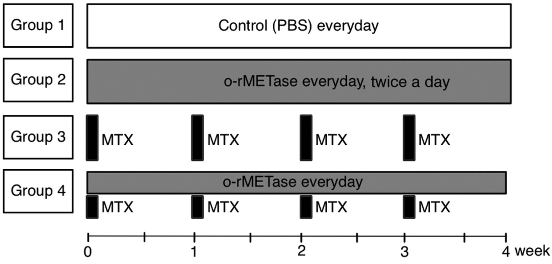
Treatment schema.
Hematoxylin and eosin (H&E) staining. H&E staining was performed according to standard protocols as previously reported (62).
Statistical analyses. We performed statistical analyses using JMP ver. 15.0.0 (SAS Institute, Cary, NC, USA). For the parametric test to compare between groups, Tukey-Kramer HSD was used. Graphs show the mean, and error bars express SD of the mean. A p-value ≤0.05 was defined to indicate statistically significant difference.
Results
Treatment efficacy on the osteosarcoma-PDOX mouse model. There was no significant difference of tumor volume in the osteosarcoma-PDOX between control and o-rMETase alone or MTX alone (p=0.21, 1.00, respectively), in contrast, the combination of o-rMETase and MTX showed significant efficacy, compared to the control (p=0.04). Moreover, the combination therapy showed significant efficacy compared to MTX alone (p=0.04) (Figure 2 and Figure 3).

Efficacy of treatment on the osteosarcoma-PDOX. Relative tumor volume at each time point is shown in line graphs. The relative tumor volume is calculated as the tumor volume at each point divided by the tumor volume at the day of initiation of treatment. n=7 mice/group. *p<0.05. Error bars: ±SD.

Representative photographs from each treatment group of osteosarcoma-PDOX mouse models at the end of treatment. (A) Control administered oral PBS. (B) o-rMETase. (C) MTX. (D) Combination of o-rMETase and MTX. Scale bar: 10 mm.
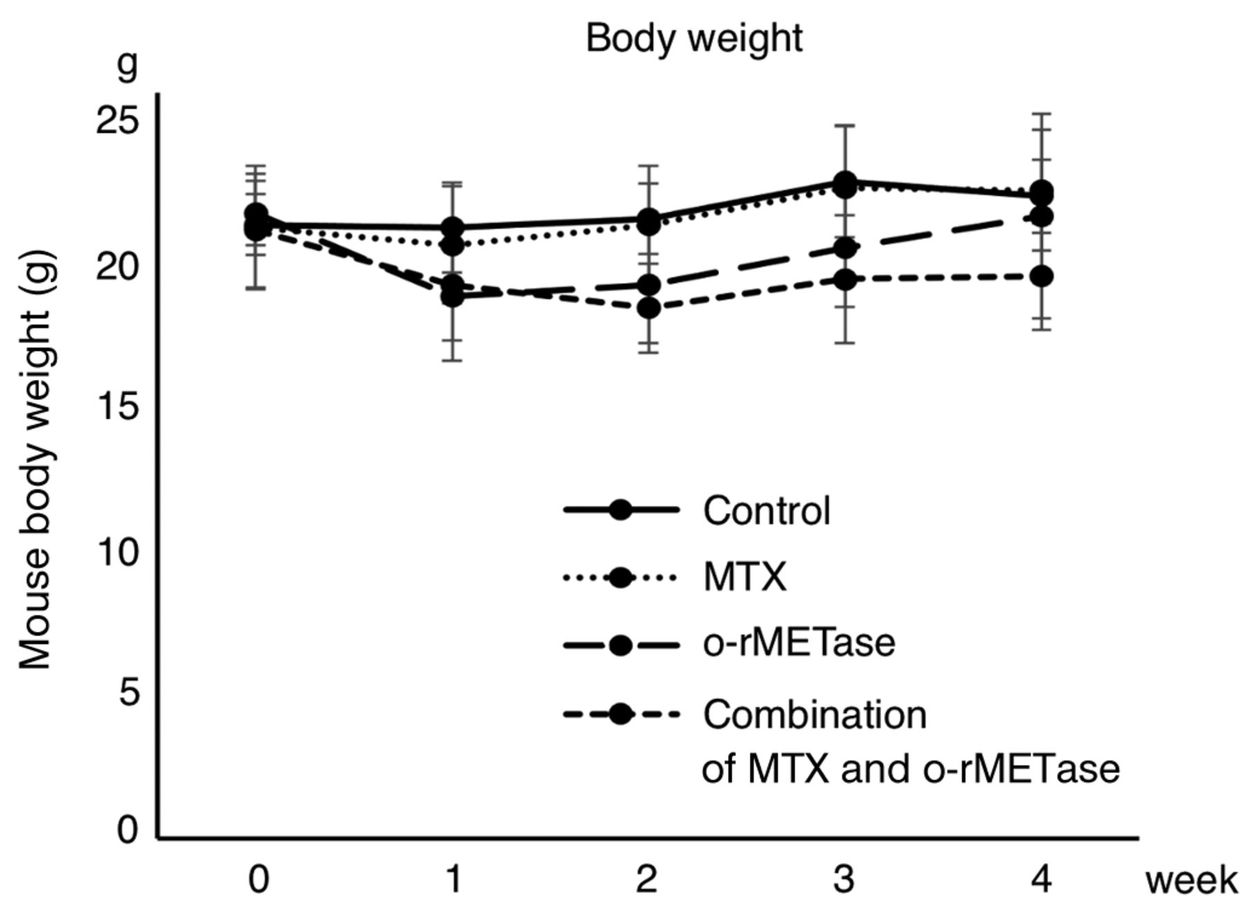
Toxicity of treatment. The body weight of the mice showed no significant reduction in any of the four treatment groups at the end of the experiment (Figure 4). No mice died during the experiment.

Toxicity of treatments on the osteosarcoma-PDOX. Mouse body weight at each point is shown in line graphs. Error bars: ±SD.
Histology of osteosarcoma-PDOX. The tumor tissue of the control osteosarcoma-PDOX comprised high-density spindle-shaped cancer cells. The o-rMETase-alone- or MTX-alone-treated osteosarcoma PDOX tissue resembled the control (Figure 5A-C). In contrast the osteosarcoma PDOX treated with the combination of o-rMETase and MTX had a very low density of cancer-cells (Figure 5D).

Representative photomicrographs of H & E-stained tissue sections of the untreated and treated osteosarcoma-PDOX. (A) Control administered oral PBS. (B) o-rMETase. (C) MTX. (D) Combination of o-rMETase and MTX. Magnification: 200×. Scale bar: 50 μm.
Discussion
Methionine addiction of cancer cells is the result of increased transmethylation reactions, via S-adenosylmethionine (SAM) (24-30). r-METase effectively degrades external methionine (63). The efficacy of orally-administrated r-METase (o-rMETase) was shown previously shown in many types of cancer in PDOX models (18, 21, 22, 35-46, 51-58). We have also reported the efficacy of rMETase on PDOX models of osteosarcoma including o-rMETase (8, 10, 18, 21, 22).
MTX inhibits dihydrofolate reductase (DHFR), resulting in decrease of tetrahydrofolate and methyl-tetrahydrofolate, eventually depleting the levels of endogenously-synthesized methionine and SAM, which is the main methyl donor in the cell (67). Additionally, there is a previous study reporting that low-dose MTX inhibits SAM synthesis directly (61).
In the present study, o-rMETase converted an osteosarcoma PDOX model from MTX-resistant to MTX-sensitive. This might be because MTX inhibits endogenous MET synthesis and o-rMETase restricts the external supply of methionine, resulting in a synergistic methionine-restriction effect on the osteosarcoma PDOX. The combination of o-rMETase and MTX thus has potential clinical promise
Acknowledgements
This study was funded in part by the Robert M. Hoffman Foundation for Cancer Research. This article is dedicated to the memory of A. R. Moossa, MD, Sun Lee, MD, Professor Li Jiaxi and Masaki Kitajima, MD., and Joseph R. Bertino, MD.
Footnotes
Authors’ Contributions
YA, YT and RMH were involved in study conception and design. YA was involved in acquisition of data. YA, YT, JY, KH NM, YK and RMH analyzed and interpreted data. YA, YT and RMH wrote the manuscript. All Authors reviewed and approved the manuscript.
Conflicts of Interest
The Authors have no conflicts of interest to declare in relation to this study. AntiCancer Inc. uses PDOX models for contract research. QH is an employee of AntiCancer Inc. YA, YT, JY, KH, NM, YT, and RMH are or were unsalaried associates of AntiCancer Inc.
- Received November 22, 2021.
- Revision received December 30, 2021.
- Accepted December 31, 2021.
- Copyright © 2022 International Institute of Anticancer Research (Dr. George J. Delinasios), All rights reserved.
References
In this issue


Jump to section
Related Articles
Cited By...
- Extensive Synergy Between Recombinant Methioninase and Eribulin Against Fibrosarcoma Cells But Not Normal Fibroblasts
- Recombinant Methioninase Decreased the Effective Dose of Irinotecan by 15-fold Against Colon Cancer Cells: A Strategy for Effective Low-toxicity Treatment of Colon Cancer
- Non-invasively Imageable Tibia-tumor-fragment Implantation Experimental-bone-metastasis Mouse Model of GFP-expressing Prostate Cancer