
Main menu

  • Home
  • Current Issue
  • Archive
  • Info for
    • Authors
    • Editorial Policies
    • Subscribers
    • Advertisers
    • Editorial Board
    • Special Issues
  • Journal Metrics
  • Other Publications
    • In Vivo
    • Cancer Genomics & Proteomics
    • Cancer Diagnosis & Prognosis
  • More
    • IIAR
    • Conferences
    • 2008 Nobel Laureates
  • About Us
    • General Policy
    • Contact
  • Other Publications
    • Anticancer Research
    • In Vivo
    • Cancer Genomics & Proteomics

User menu

  • Register
  • Subscribe
  • My alerts
  • Log in
  • My Cart

Search

  • Advanced search
Anticancer Research
  • Other Publications
    • Anticancer Research
    • In Vivo
    • Cancer Genomics & Proteomics
  • Register
  • Subscribe
  • My alerts
  • Log in
  • My Cart
Anticancer Research

Advanced Search

  • Home
  • Current Issue
  • Archive
  • Info for
    • Authors
    • Editorial Policies
    • Subscribers
    • Advertisers
    • Editorial Board
    • Special Issues
  • Journal Metrics
  • Other Publications
    • In Vivo
    • Cancer Genomics & Proteomics
    • Cancer Diagnosis & Prognosis
  • More
    • IIAR
    • Conferences
    • 2008 Nobel Laureates
  • About Us
    • General Policy
    • Contact
  • Visit us on Facebook
  • Follow us on Linkedin
Review ArticleReviews

Pre-treatment Apparent Diffusion Coefficient Does Not Predict Therapy Response to Radiochemotherapy in Cervical Cancer: A Systematic Review and Meta-analysis

HANS-JONAS MEYER, ANDREAS WIENKE and ALEXEY SUROV
Anticancer Research March 2021, 41 (3) 1163-1170; DOI: https://doi.org/10.21873/anticanres.14873
HANS-JONAS MEYER
1Department of Diagnostic and Interventional Radiology, University of Leipzig, Leipzig, Germany;
  • Find this author on Google Scholar
  • Find this author on PubMed
  • Search for this author on this site
ANDREAS WIENKE
2Institute of Medical Epidemiology, Biostatistics, and Informatics, Martin-Luther-University Halle-Wittenberg, Halle (Saale), Germany;
  • Find this author on Google Scholar
  • Find this author on PubMed
  • Search for this author on this site
ALEXEY SUROV
3Department of Radiology and Nuclear Medicine, University of Magdeburg, Magdeburg, Germany
  • Find this author on Google Scholar
  • Find this author on PubMed
  • Search for this author on this site
  • Article
  • Figures & Data
  • Info & Metrics
  • PDF
Loading

Abstract

Background/Aim: Magnetic resonance imaging is used for staging purposes in cervical cancer (CC). Diffusion-weighted imaging and the apparent diffusion coefficient (ADC) are associated with tumor microstructure. The present analysis sought to compare pre-treatment ADC values to predict treatment outcome of radiochemotherapy for CC based upon a large patient sample. Materials and Methods: MEDLINE library and SCOPUS databases were assessed for suitable articles up to May 2020. The primary endpoint was the mean ADC value of CC according to the treatment response to radiochemotherapy. In total, 16 studies were included in the analysis. Results: For the response group, 416 patients were included in the analysis (72.5%) and for the no-response group 158 patients were included (27.5%). The mean ADC value of patients with CC with treatment response was 0.87×10⁻³ mm2/s (95% confidence interval=0.81-0.94×10⁻³ mm2/s), and for the patients with no response was 0.92×10⁻³ mm2/s (95% confidence interval=0.85-0.98×10⁻³ mm2/s). Conclusion: Pre-treatment ADC values alone cannot be used to reliably predict treatment response to radiochemotherapy in CC.

Key Words:
  • Meta-analysis
  • systematic review
  • cervical cancer
  • DWI
  • ADC
  • review

Magnetic resonance imaging (MRI) is clinically of great importance for the diagnosis of cervical cancer (CC) (1-4). It has a high sensitivity and can detect tumor infiltration of adjacent structures. Moreover, due to the addition of diffusion-weighted imaging (DWI), information regarding tumor characteristics such as cellularity can be obtained (5-7). DWI is a MRI sequence quantifying the Brownian water movement in tissues, which is inversely correlated with cell density in tumors (5-7).

It has been shown that apparent diffusion coefficient (ADC) values differ significantly between adenocarcinoma and squamous cell carcinoma due to different tumor microstructure (8). In addition, correlations with epidermal growth factor and p53 expression were also reported (7, 9). These data indicate that ADC values can also reflect tumor biology in a more complex way beyond cellularity alone (10).

There is no doubt regarding the benefit of neoadjuvant and definitive chemotherapy in CC (11). A clear survival benefit was shown for treatment responders, which is also shown by the fact that treatment response is an independent prognostic factor in CC (12). However, response rates range from approximately 50% to 92%, whereas the optimal pathological response is lower than 20% (11, 12).

To date, there are no imaging or histopathology modalities which can reliably predict treatment outcome of chemotherapy (11). Yet this approach would be clinically relevant for selecting patients who would benefit from the therapy and those who would not. Thereby, therapy-related toxicity could be reduced if ADC values were able to predict treatment outcome. Previously, preliminary studies indicated that ADC might be used as predictor of therapy response (3, 4).

The present analysis sought to systematically review studies of ADC values in CC according to treatment outcome after radiochemotherapy and to carry out a meta-analysis to elucidate whether ADC values differ significantly according to treatment response.

Materials and Methods

Literature search and inclusion criteria. MEDLINE library and SCOPUS databases were screened for studies investigating ADC values in CC up to May 2020. The following terms for the literature search were used: “Diffusion weighted imaging or diffusion-weighted imaging or DWI or Apparent diffusion coefficient or ADC AND cervical cancer OR cervical carcinoma”. Secondary references were screened and included, when suitable.

The primary endpoint was defined as the ADC values in patients with CC in regard to treatment outcome.

Studies were included when they met the following criteria: (i) Histopathologically confirmed CC, (ii) MRI with DWI before any form of treatment, and (iii) mean ADC values were reported with standard deviation.

The following were exclusion criteria: (i) Case reports, (ii) reviews, (iii) studies without data of pre-treatment DWI, (iv) articles not in English language, and (v) xenograft or animal model experimental study design.

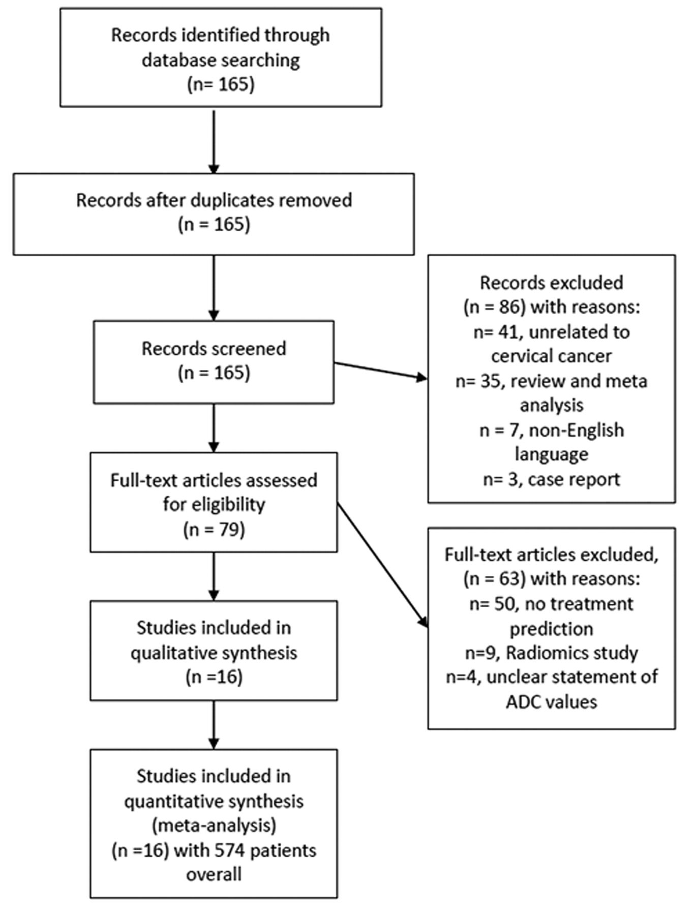
Article acquisition was performed according to Preferred Reporting Items for Systematic Reviews and Meta-Analyses (PRISMA) statement (13) (Figure 1). In total, 16 articles met the inclusion criteria and were included in the present analysis (14-29).

Figure 1.
  • Download figure
  • Open in new tab
  • Download powerpoint
Figure 1.

PRISMA flow chart gives an overview of article acquisition. Overall, 16 studies with 574 patients overall were included in the analysis. ADC: Apparent diffusion coefficient.

Quality assessment. The methodological quality of the included studies was independently assessed by two co-authors (A.S. and H.J.M.) using Quality Assessment of Diagnostic Accuracy Studies (QUADAS-2) (30).

Statistical analysis. The analysis was carried out with RevMan 5.3 (2014; Cochrane Collaboration, Copenhagen, Denmark). Heterogeneity of the results was estimated by the inconsistency index I2 (31, 32). DerSimonian and Laird random-effect models using inverse-variance weights were calculated without any form of correction (33).

Results

Figure 2 gives an overview of the methodological quality of the included studies. Overall, the risk of bias was considered low. Patient selection was regarded as overall well-defined within the respective methodology; yet some studies did not clearly define the inclusion criteria sufficiently enough, which can result in some form of potential bias. The index test defined as the MRI with DWI was clearly reported within the studies and should not be considered a source of potential bias. The DWI measurement can be considered as reliable throughout the studies. For flow and timing, there were no serious concerns to report, as in most studies no delay between imaging and treatment procedures were noticed.

Figure 2.
  • Download figure
  • Open in new tab
  • Download powerpoint
Figure 2.

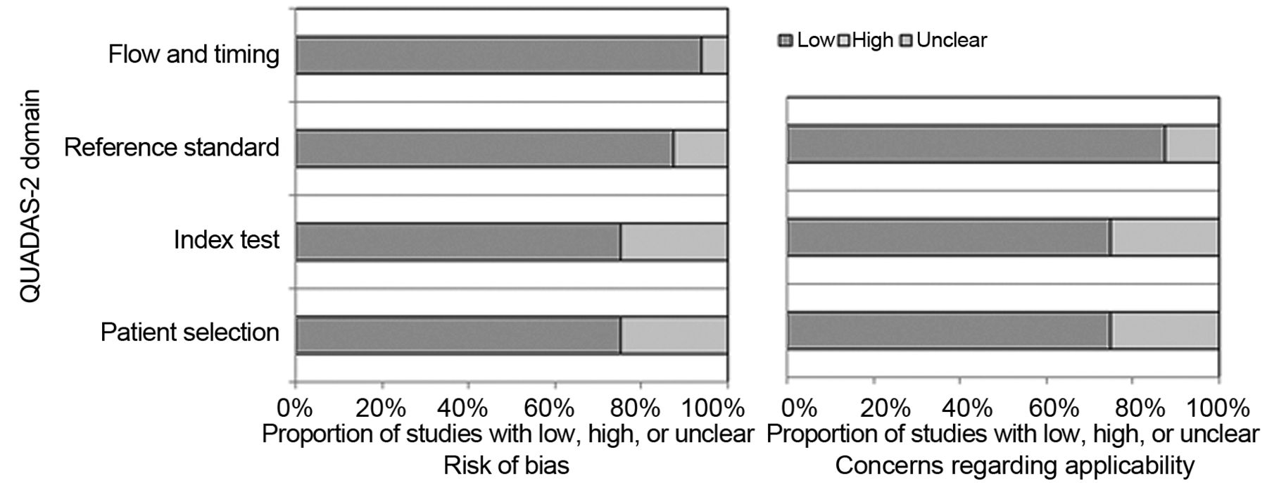
QUADAS-2 quality assessment of the included studies. Most studies showed an overall low potential for sources of bias.

The 16 studies included comprised a total of 574 patients with CC. Table I provides an overview of the included studies.

View this table:
  • View inline
  • View popup
Table I.

Data retrieved from the included studies.

Of the included studies, 10 (62.5%) were of prospective and six (37.5%) of retrospective design. Different 1.5T MRI scanners were used in five (31.3%) studies, 3T scanners in 10 (62.5%) studies, and one study (14) had an unclear scanner technique (6.2%). Regarding b-values, most studies used high b-values of 0 and 800 s/mm2 or higher in 14 studies (87.5%). In two studies (12.5%), no statement of b-values used was given (14, 16). In six studies (37.5%), squamous cell carcinomas and other histological entities were investigated, whereas in 10 studies (62.5%), only squamous cell carcinomas were included. Therefore, most patients had squamous cell carcinomas (n=537, 94.6%) and only 37 patients (6.4%) had other histological entities with 17 patients with adenocarcinomas comprising the second most common entity.

For tumor stages, most studies included different tumor stages ranging from International Federation of Obstetrics and Gynecology-stage IB to IV. Only two studies did not report the exact proportion of tumor stages of their patient sample but only the boundaries of included tumor stages (16, 21).

Different treatment regimens were performed in the studies. Concurrent radiochemotherapy was performed in eight studies (50%), neoadjuvant chemotherapy (25%) in four studies, definitive radiochemotherapy in two studies (12.5%), neoadjuvant radiochemotherapy in one study (6.25%), and in one study, concurrent radiochemotherapy as well as radiotherapy alone were used (23).

For the responder group, 416 patients were included in the analysis (72.5%) and 158 patients for the nonresponders (27.5%).

The mean ADC value for patients with treatment response was 0.87×10⁻³ mm2/s (95% confidence interval=0.81-0.94×10⁻³ mm2/s; Tau2=0.02, Chi2=1047.32, df=15, I2= 99%), and of the patients with no response was 0.92×10⁻³ mm2/s (95% confidence interval=0.85-0.98×10⁻³ mm2/s; Tau2=0.01, Chi2=355.61, df=11, I2=97%) (Figure 3). The high I2 value indicates substantial heterogeneity between the studies. Figure 4 shows these results as a box plot. The ADC values of the groups significantly overlapped with no clear threshold value to distinguish between them.

Figure 3.
  • Download figure
  • Open in new tab
  • Download powerpoint
Figure 3.

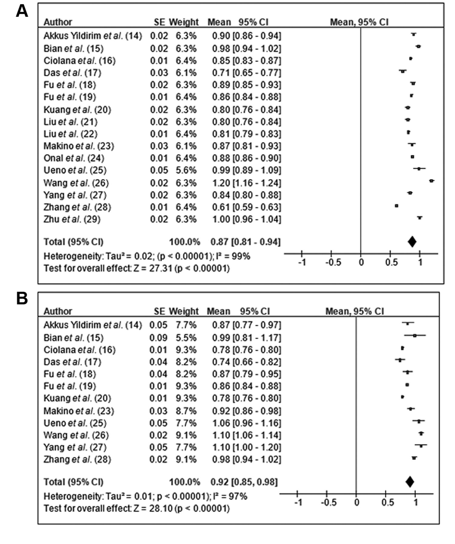
Forrest plots of the mean apparent diffusion coefficient (ADC) values of responders (A) and nonresponders (B) in patients with cervical carcinoma. The mean ADC value of patients with treatment response was 0.87×10⁻³ mm2/s (95% confidence interval=0.81-0.94×10⁻³ mm2/s), that of patients with no response was 0.92×10⁻³ mm2/s (95% confidence interval=0.85-0.98×10⁻³ mm2/s).

Figure 4.
  • Download figure
  • Open in new tab
  • Download powerpoint
Figure 4.

Box plots of the mean apparent diffusion coefficient (ADC) values according to treatment response. The ADC values for groups overlapped significantly, with no clear threshold value to distinguish between them.

Discussion

The present study sought to elucidate whether ADC values of DWI can predict treatment outcome of radiochemotherapy in CC. In short, there were no significant differences between patients with treatment response and those without response according to ADC values.

On the one hand, significantly lower ADC values of patients with treatment response were reported (27, 28), but on the other, ADC values were significantly higher for treatment responders (26). Of note, others did not identify significant differences for ADC values according to treatment response (14, 16, 25). Thus, the present analysis harmonizes the published incongruent results of previous single-centre studies based upon a large sample size.

It is known that radiochemotherapy significantly improves 5-year overall disease-free survival (hazard ratio=0.78) and 5-year overall survival (hazard ratio=0.81) compared to radiotherapy alone (11). There is no doubt that an optimal treatment response is an independent prognostic factor in CC (12). However, the objective response rates range from approximately 50% to 92%, whereas an optimal pathological response is only found in fewer than 20% of patients (11).

In the present analysis, different treatment regimens were included, comprising neoadjuvant chemotherapy and concurrent radiochemotherapy, which might have resulted in the identified heterogeneity of the results. Yet the pre-treatment ADC value was investigated in the present study, which should be predictive for any form of treatment as a valuable biomarker in clinical routine.

It was extensively reported that ADC values are associated with cellularity, cell density and tumor microstructure (5, 7, 9, 10). Moreover, ADC values are also correlated with clinically significant histopathological features, comprising proliferative potential and expression of tumor-suppressor gene TP53 (7, 9, 10). Yet there is still an ongoing debate as to which features of tumor biology are reliably reflected by non-invasive imaging and which not.

Regarding prediction of treatment response by ADC values, there have been promising results for several tumor entities, including head and neck cancer, rectal cancer, and soft-tissue sarcoma (34-36). One can assume that for studies which showed that treatment responders had lower ADC values, a higher cellularity and a higher tumor aggressiveness of the tumor might have resulted in a better treatment response, whereas patients with a higher pre-treatment ADC value might have had lower cellularity and a less aggressive tumor, which resulted in a less effective treatment. On the other hand, there might be the possibility that patients with a more aggressive tumor also have a worse treatment response, as was shown by Wang et al. (26). The relatively high heterogeneity of the underlying data has to be considered. Yet one key finding of this study is that ADC values pooled from the published results indicate that ADC values alone are not able to reliably predict treatment response in CC.

As well as the analysis of pre-treatment ADC values, which might be able to discriminate more aggressive tumors from more benign tumors, the increase of ADC values during treatment seems to be of relevance. This might show tumor regression induced by therapy. Yet in a recent meta-analysis, it was also shown that pre-treatment ADC values were not capable of predicting therapy response in patients with breast cancer in a neoadjuvant setting (37). In short, it is crucial to investigate for which tumor entities DWI and ADC values are useful for treatment prediction and for which ones these cannot be used.

For CC, the treatment response is mainly determined by conventional MRI and morphological measurement of the tumor, considering possible shrinkage (2). Beyond this, a significant T2-weighted signal loss indicates post-treatment fibrosis of the cervix as a result of radiochemotherapy, whereas a decrease in uptake of contrast medium in contrast-enhanced sequences indicates regression of vital tumor tissue, which are both important signs for treatment response (2). However, this assessment can only be made after the treatment and no pre-treatment prediction can be made with conventional imaging.

Notably, there are only few histopathological parameters which were investigated for treatment prediction. Only the proliferative index Ki67 and survivin expression remained statistically significant in a multivariate analysis based on 117 squamous cell cervical carcinomas undergoing neoadjuvant chemotherapy and were associated with treatment response (38).

Presumably, the reported differences in the investigated studies were influenced by different scanner technique, measurement of ADC values, and composition of the patient samples. For example, it is a known fact that adenocarcinoma tends to have different ADC values from squamous cell carcinomas (8).

The present analysis suffers from the following limitations. Firstly, the analysis is based upon results of the literature. There might be some publication bias due to the trend of positive or significant reported results. Secondly, only articles in English language were included. Thirdly, only studies employing clinically DWI sequence using two b-values were included. Yet novel DWI techniques such as intravoxel-incoherent motion and diffusion-kurtosis imaging might perform better for treatment prediction in CC (28, 39, 40). However, no meta-analysis can be performed including these currently with so few articles published. Fourthly, there was substantial inhomogeneity identified in the analysis.

In conclusion, pre-treatment ADC values alone cannot be used to reliably predict treatment response to radiochemo-therapy in CC.

Footnotes

  • This article is freely accessible online.

  • ↵* All Authors contributed equally to this work

  • Authors’ Contributions

    All Authors contributed equally to this work.

  • Conflicts of Interest

    All Authors confirm that there are no conflicts of interest to be declared.

  • Received January 17, 2021.
  • Revision received February 13, 2021.
  • Accepted February 15, 2021.
  • Copyright © 2021 International Institute of Anticancer Research (Dr. George J. Delinasios), All rights reserved.

References

  1. ↵
    1. Mansoori B,
    2. Khatri G,
    3. Rivera-Colón G,
    4. Albuquerque K,
    5. Lea J and
    6. Pinho DF
    : Multimodality imaging of uterine cervical malignancies. AJR Am J Roentgenol 215(2): 292-304, 2020. PMID: 32551909. DOI: 10.2214/AJR.19.21941
    OpenUrlCrossRefPubMed
  2. ↵
    1. Merz J,
    2. Bossart M,
    3. Bamberg F and
    4. Eisenblaetter M
    : Revised FIGO staging for cervical cancer – a new role for MRI. Rofo 192(10): 937-944, 2020. PMID: 32731266. DOI: 10.1055/a-1198-5729
    OpenUrlCrossRefPubMed
  3. ↵
    1. Balcacer P,
    2. Shergill A and
    3. Litkouhi B
    : MRI of cervical cancer with a surgical perspective: staging, prognostic implications and pitfalls. Abdom Radiol (NY) 44(7): 2557-2571, 2019. PMID: 30903231. DOI: 10.1007/s00261-019-01984-7
    OpenUrlCrossRefPubMed
  4. ↵
    1. Balleyguier C,
    2. Sala E,
    3. Da Cunha T,
    4. Bergman A,
    5. Brkljacic B,
    6. Danza F,
    7. Forstner R,
    8. Hamm B,
    9. Kubik-Huch R,
    10. Lopez C,
    11. Manfredi R,
    12. McHugo J,
    13. Oleaga L,
    14. Togashi K and
    15. Kinkel K
    : Staging of uterine cervical cancer with MRI: guidelines of the European Society of Urogenital Radiology. Eur Radiol 21(5): 1102-1110, 2011. PMID: 21063710. DOI: 10.1007/s00330-010-1998-x
    OpenUrlCrossRefPubMed
  5. ↵
    1. Surov A,
    2. Meyer HJ and
    3. Wienke A
    : Correlation between apparent diffusion coefficient (ADC) and cellularity is different in several tumors: a meta-analysis. Oncotarget 8(35): 59492-59499, 2017. PMID: 28938652. DOI: 10.18632/oncotarget.17752
    OpenUrlCrossRefPubMed
    1. Padhani AR,
    2. Liu G,
    3. Koh DM,
    4. Chenevert TL,
    5. Thoeny HC,
    6. Takahara T,
    7. Dzik-Jurasz A,
    8. Ross BD,
    9. Van Cauteren M,
    10. Collins D,
    11. Hammoud DA,
    12. Rustin GJ,
    13. Taouli B and
    14. Choyke PL
    : Diffusion-weighted magnetic resonance imaging as a cancer biomarker: consensus and recommendations. Neoplasia 11(2): 102-125, 2009. PMID: 19186405. DOI: 10.1593/neo.81328
    OpenUrlCrossRefPubMed
  6. ↵
    1. Surov A,
    2. Meyer HJ,
    3. Schob S,
    4. Höhn AK,
    5. Bremicker K,
    6. Exner M,
    7. Stumpp P and
    8. Purz S
    : Parameters of simultaneous 18F-FDG-PET/MRI predict tumor stage and several histopathological features in uterine cervical cancer. Oncotarget 8(17): 28285-28296, 2017. PMID: 28423698. DOI: 10.18632/oncotarget.16043
    OpenUrlCrossRefPubMed
  7. ↵
    1. Lin Y,
    2. Li H,
    3. Chen Z,
    4. Ni P,
    5. Zhong Q,
    6. Huang H and
    7. Sandrasegaran K
    : Correlation of histogram analysis of apparent diffusion coefficient with uterine cervical pathologic finding. AJR Am J Roentgenol 204(5): 1125-1131, 2015. PMID: 25905952. DOI: 10.2214/AJR.14.13350
    OpenUrlCrossRefPubMed
  8. ↵
    1. Schob S,
    2. Meyer HJ,
    3. Pazaitis N,
    4. Schramm D,
    5. Bremicker K,
    6. Exner M,
    7. Höhn AK,
    8. Garnov N and
    9. Surov A
    : ADC Histogram Analysis of Cervical Cancer Aids Detecting Lymphatic Metastases-a Preliminary Study. Mol Imaging Biol 19(6): 953-962, 2017. PMID: 28315203. DOI: 10.1007/s11307-017-1073-y
    OpenUrlCrossRefPubMed
  9. ↵
    1. Meyer HJ,
    2. Gundermann P,
    3. Höhn AK,
    4. Hamerla G and
    5. Surov A
    : Associations between whole tumor histogram analysis parameters derived from ADC maps and expression of EGFR, VEGF, Hif 1-alpha, Her-2 and Histone 3 in uterine cervical cancer. Magn Reson Imaging 57: 68-74, 2019. PMID: 30367998. DOI: 10.1016/j.mri.2018.10.016
    OpenUrlCrossRefPubMed
  10. ↵
    1. Gadducci A and
    2. Cosio S
    : Neoadjuvant chemotherapy in locally advanced cervical cancer: review of the literature and perspectives of clinical research. Anticancer Res 40(9): 4819-4828, 2020. PMID: 32878770. DOI: 10.21873/anticanres.14485
    OpenUrlAbstract/FREE Full Text
  11. ↵
    1. Gadducci A,
    2. Sartori E,
    3. Maggino T,
    4. Zola P,
    5. Cosio S,
    6. Zizioli V,
    7. Lapresa M,
    8. Piovano E and
    9. Landoni F
    : Pathological response on surgical samples is an independent prognostic variable for patients with Stage Ib2-IIb cervical cancer treated with neoadjuvant chemotherapy and radical hysterectomy: an Italian multicenter retrospective study (CTF Study). Gynecol Oncol 131(3): 640-644, 2013. PMID: 24096111. DOI: 10.1016/j.ygyno.2013.09.029
    OpenUrlCrossRefPubMed
  12. ↵
    1. Moher D,
    2. Liberati A,
    3. Tetzlaff J,
    4. Altman DG and PRISMA Group.
    : Preferred reporting items for systematic reviews and meta-analyses: the PRISMA statement. PLoS Med 6(7): e1000097, 2009. PMID: 19621072. DOI: 10.1371/journal.pmed.1000097
    OpenUrlCrossRefPubMed
  13. ↵
    1. Akkus Yildirim B,
    2. Onal C,
    3. Erbay G,
    4. Cem Guler O,
    5. Karadeli E,
    6. Reyhan M and
    7. Koc Z
    : Prognostic values of ADCmean and SUVmax of the primary tumour in cervical cancer patients treated with definitive chemoradiotherapy. J Obstet Gynaecol 39(2): 224-230, 2019. PMID: 30354907. DOI: 10.1080/01443615.2018.1492528
    OpenUrlCrossRefPubMed
    1. Bian H,
    2. Liu F,
    3. Chen S,
    4. Li G,
    5. Song Y,
    6. Sun M and
    7. Dong H
    : Intravoxel incoherent motion diffusion-weighted imaging evaluated the response to concurrent chemoradiotherapy in patients with cervical cancer. Medicine (Baltimore) 98(46): e17943, 2019. PMID: 31725650. DOI: 10.1097/MD.0000000000017943
    OpenUrlCrossRefPubMed
  14. ↵
    1. Ciolina M,
    2. Vinci V,
    3. Villani L,
    4. Gigli S,
    5. Saldari M,
    6. Panici PB,
    7. Perniola G,
    8. Laghi A,
    9. Catalano C and
    10. Manganaro L
    : Texture analysis versus conventional MRI prognostic factors in predicting tumor response to neoadjuvant chemotherapy in patients with locally advanced cancer of the uterine cervix. Radiol Med 124(10): 955-964, 2019. PMID: 31254220. DOI: 10.1007/s11547-019-01055-3
    OpenUrlCrossRefPubMed
    1. Das S,
    2. Chandramohan A,
    3. Reddy JK,
    4. Mukhopadhyay S,
    5. Kumar RM,
    6. Isiah R,
    7. John S,
    8. Oommen R and
    9. Jeyaseelan V
    : Role of conventional and diffusion weighted MRI in predicting treatment response after low dose radiation and chemotherapy in locally advanced carcinoma cervix. Radiother Oncol 117(2): 288-293, 2015. PMID: 26475249. DOI: 10.1016/j.radonc.2015.10.006
    OpenUrlCrossRefPubMed
    1. Fu C,
    2. Bian D,
    3. Liu F,
    4. Feng X,
    5. Du W and
    6. Wang X
    : The value of diffusion-weighted magnetic resonance imaging in assessing the response of locally advanced cervical cancer to neoadjuvant chemotherapy. Int J Gynecol Cancer 22(6): 1037-1043, 2012. PMID: 22683941. DOI: 10.1097/IGC.0b013e31825736d7
    OpenUrlAbstract/FREE Full Text
    1. Fu C,
    2. Feng X,
    3. Bian D,
    4. Zhao Y,
    5. Fang X,
    6. Du W,
    7. Wang L and
    8. Wang X
    : Simultaneous changes of magnetic resonance diffusion-weighted imaging and pathological microstructure in locally advanced cervical cancer caused by neoadjuvant chemotherapy. J Magn Reson Imaging 42(2): 427-435, 2015. PMID: 25328994. DOI: 10.1002/jmri.24779
    OpenUrlCrossRefPubMed
    1. Kuang F,
    2. Yan Z,
    3. Wang J and
    4. Rao Z
    : The value of diffusion-weighted MRI to evaluate the response to radiochemotherapy for cervical cancer. Magn Reson Imaging 32(4): 342-349, 2014. PMID: 24512795. DOI: 10.1016/j.mri.2013.12.007
    OpenUrlCrossRefPubMed
  15. ↵
    1. Liu Y,
    2. Bai R,
    3. Sun H,
    4. Liu H,
    5. Zhao X and
    6. Li Y
    : Diffusion-weighted imaging in predicting and monitoring the response of uterine cervical cancer to combined chemoradiation. Clin Radiol 64(11): 1067-1074, 2009. PMID: 19822239. DOI: 10.1016/j.crad.2009.07.010
    OpenUrlCrossRefPubMed
    1. Liu Y,
    2. Sun H,
    3. Bai R and
    4. Ye Z
    : Time-window of early detection of response to concurrent chemoradiation in cervical cancer by using diffusion-weighted MR imaging: a pilot study. Radiat Oncol 10: 185, 2015. PMID: 26337954. DOI: 10.1186/s13014-015-0493-6
    OpenUrlCrossRefPubMed
  16. ↵
    1. Makino H,
    2. Kato H,
    3. Furui T,
    4. Morishige K and
    5. Kanematsu M
    : Predictive value of diffusion-weighted magnetic resonance imaging during chemoradiotherapy for uterine cervical cancer. J Obstet Gynaecol Res 40(4): 1098-1104, 2014. PMID: 24320754. DOI: 10.1111/jog.12276
    OpenUrlCrossRefPubMed
    1. Onal C,
    2. Erbay G and
    3. Guler OC
    : Treatment response evaluation using the mean apparent diffusion coefficient in cervical cancer patients treated with definitive chemoradiotherapy. J Magn Reson Imaging 44(4): 1010-1019, 2016. PMID: 26919800. DOI: 10.1002/jmri.25215
    OpenUrlCrossRefPubMed
  17. ↵
    1. Ueno Y,
    2. Lisbona R,
    3. Tamada T,
    4. Alaref A,
    5. Sugimura K and
    6. Reinhold C
    : Comparison of FDG PET metabolic tumour volume versus ADC histogram: prognostic value of tumour treatment response and survival in patients with locally advanced uterine cervical cancer. Br J Radiol 90(1075): 20170035, 2017. PMID: 28508679. DOI: 10.1259/bjr.20170035
    OpenUrlCrossRefPubMed
  18. ↵
    1. Wang YC,
    2. Hu DY,
    3. Hu XM,
    4. Shen YQ,
    5. Meng XY,
    6. Tang H and
    7. Li Z
    : Assessing the early response of advanced cervical cancer to neoadjuvant chemotherapy using intravoxel incoherent motion diffusion-weighted magnetic resonance imaging: A pilot study. Chin Med J (Engl) 129(6): 665-671, 2016. PMID: 26960369. DOI: 10.4103/0366-6999.177995
    OpenUrlCrossRefPubMed
  19. ↵
    1. Yang W,
    2. Qiang JW,
    3. Tian HP,
    4. Chen B,
    5. Wang AJ and
    6. Zhao JG
    : Multi-parametric MRI in cervical cancer: early prediction of response to concurrent chemoradiotherapy in combination with clinical prognostic factors. Eur Radiol 28(1): 437-445, 2018. PMID: 28779395. DOI: 10.1007/s00330-017-4989-3
    OpenUrlCrossRefPubMed
  20. ↵
    1. Zhang H,
    2. Zhou Y,
    3. Li J,
    4. Zhang P,
    5. Li Z and
    6. Guo J
    : The value of DWI in predicting the response to synchronous radiochemotherapy for advanced cervical carcinoma: comparison among three mathematical models. Cancer Imaging 20(1): 8, 2020. PMID: 31937371. DOI: 10.1186/s40644-019-0285-6
    OpenUrlCrossRefPubMed
  21. ↵
    1. Zhu L,
    2. Zhu L,
    3. Shi H,
    4. Wang H,
    5. Yan J,
    6. Liu B,
    7. Chen W,
    8. He J,
    9. Zhou Z,
    10. Yang X and
    11. Liu T
    : Evaluating early response of cervical cancer under concurrent chemo-radiotherapy by intravoxel incoherent motion MR imaging. BMC Cancer 16: 79, 2016. PMID: 26860361. DOI: 10.1186/s12885-016-2116-5
    OpenUrlCrossRefPubMed
  22. ↵
    1. Whiting PF,
    2. Rutjes AW,
    3. Westwood ME,
    4. Mallett S,
    5. Deeks JJ,
    6. Reitsma JB,
    7. Leeflang MM,
    8. Sterne JA,
    9. Bossuyt PM and QUADAS-2 Group.
    : QUADAS-2: a revised tool for the quality assessment of diagnostic accuracy studies. Ann Intern Med 155(8): 529-536, 2011. PMID: 22007046. DOI: 10.7326/0003-4819-155-8-201110180-00009
    OpenUrlCrossRefPubMed
  23. ↵
    1. Leeflang MM,
    2. Deeks JJ,
    3. Gatsonis C,
    4. Bossuyt PM and Cochrane Diagnostic Test Accuracy Working Group.
    : Systematic reviews of diagnostic test accuracy. Ann Intern Med 149(12): 889-897, 2008. PMID: 19075208. DOI: 10.7326/0003-4819-149-12-200812160-00008
    OpenUrlCrossRefPubMed
  24. ↵
    1. Zamora J,
    2. Abraira V,
    3. Muriel A,
    4. Khan K and
    5. Coomarasamy A
    : Meta-DiSc: a software for meta-analysis of test accuracy data. BMC Med Res Methodol 6: 31, 2006. PMID: 16836745. DOI: 10.1186/1471-2288-6-31
    OpenUrlCrossRefPubMed
  25. ↵
    1. DerSimonian R and
    2. Laird N
    : Meta-analysis in clinical trials. Control Clin Trials 7(3): 177-188, 1986. PMID: 3802833. DOI: 10.1016/0197-2456(86)90046-2
    OpenUrlCrossRefPubMed
  26. ↵
    1. Chodyla M,
    2. Demircioglu A,
    3. Schaarschmidt BM,
    4. Bertram S,
    5. Bruckmann NM,
    6. Haferkamp J,
    7. Li Y,
    8. Bauer S,
    9. Podleska LE,
    10. Rischpler C,
    11. Forsting M,
    12. Herrmann K,
    13. Umutlu L and
    14. Grueneisen J
    : Evaluation of 18F-FDG PET and DWI datasets for the prediction of therapy response of soft tissues sarcomas under neoadjuvant isolated limb perfusion. J Nucl Med, 2020. PMID: 32737246. DOI: 10.2967/jnumed.120.248260
    OpenUrlAbstract/FREE Full Text
    1. Palmisano A,
    2. Di Chiara A,
    3. Esposito A,
    4. Rancoita PMV,
    5. Fiorino C,
    6. Passoni P,
    7. Albarello L,
    8. Rosati R,
    9. Del Maschio A and
    10. De Cobelli F
    : MRI prediction of pathological response in locally advanced rectal cancer: when apparent diffusion coefficient radiomics meets conventional volumetry. Clin Radiol 75(10): 798.e1-798.e11, 2020. PMID: 32712007. DOI: 10.1016/j.crad.2020.06.023
    OpenUrlCrossRefPubMed
  27. ↵
    1. Ravanelli M,
    2. Grammatica A,
    3. Maddalo M,
    4. Ramanzin M,
    5. Agazzi GM,
    6. Tononcelli E,
    7. Battocchio S,
    8. Bossi P,
    9. Vezzoli M,
    10. Maroldi R and
    11. Farina D
    : Pretreatment DWI with histogram analysis of the ADC in predicting the outcome of advanced oropharyngeal cancer with known human papillomavirus status treated with chemoradiation. AJNR Am J Neuroradiol 41(8): 1473-1479, 2020. PMID: 32732272. DOI: 10.3174/ajnr.A6695
    OpenUrlAbstract/FREE Full Text
  28. ↵
    1. Surov A,
    2. Wienke A and
    3. Meyer HJ
    : Pretreatment apparent diffusion coefficient does not predict therapy response to neoadjuvant chemotherapy in breast cancer. Breast 53: 59-67, 2020. PMID: 32652460. DOI: 10.1016/j.breast.2020.06.001
    OpenUrlCrossRefPubMed
  29. ↵
    1. Zhang Y,
    2. Yan H,
    3. Li R,
    4. Guo Y and
    5. Zheng R
    : High expression of surviving predicts poor prognosis in cervical squamous cell carcinoma treated with paclitaxel and carboplatin. Medicine (Baltimore) 98(20): e15607, 2019. PMID: 31096466. DOI: 10.1097/MD.0000000000015607
    OpenUrlCrossRefPubMed
  30. ↵
    1. Gao S,
    2. Du S,
    3. Lu Z,
    4. Xin J,
    5. Gao S and
    6. Sun H
    : Multiparametric PET/MR (PET and MR-IVIM) for the evaluation of early treatment response and prediction of tumor recurrence in patients with locally advanced cervical cancer. Eur Radiol 30(2): 1191-1201, 2020. PMID: 31493211. DOI: 10.1007/s00330-019-06428-w
    OpenUrlCrossRefPubMed
  31. ↵
    1. Zhang Q,
    2. Yu X,
    3. Ouyang H,
    4. Zhang J,
    5. Chen S,
    6. Xie L and
    7. Zhao X
    : Whole-tumor texture model based on diffusion kurtosis imaging for assessing cervical cancer: a preliminary study. Eur Radiol, 2021. PMID: 33464399. DOI: 10.1007/s00330-020-07612-z
    OpenUrlCrossRef
PreviousNext
Back to top

In this issue

Anticancer Research
Vol. 41, Issue 3
March 2021
  • Table of Contents
  • Table of Contents (PDF)
  • Index by author
  • Back Matter (PDF)
  • Ed Board (PDF)
  • Front Matter (PDF)
Print
Download PDF
Article Alerts
Sign In to Email Alerts with your Email Address
Email Article

Thank you for your interest in spreading the word on Anticancer Research.

NOTE: We only request your email address so that the person you are recommending the page to knows that you wanted them to see it, and that it is not junk mail. We do not capture any email address.

Enter multiple addresses on separate lines or separate them with commas.
Pre-treatment Apparent Diffusion Coefficient Does Not Predict Therapy Response to Radiochemotherapy in Cervical Cancer: A Systematic Review and Meta-analysis
(Your Name) has sent you a message from Anticancer Research
(Your Name) thought you would like to see the Anticancer Research web site.
CAPTCHA
This question is for testing whether or not you are a human visitor and to prevent automated spam submissions.
11 + 0 =
Solve this simple math problem and enter the result. E.g. for 1+3, enter 4.
Citation Tools
Pre-treatment Apparent Diffusion Coefficient Does Not Predict Therapy Response to Radiochemotherapy in Cervical Cancer: A Systematic Review and Meta-analysis
HANS-JONAS MEYER, ANDREAS WIENKE, ALEXEY SUROV
Anticancer Research Mar 2021, 41 (3) 1163-1170; DOI: 10.21873/anticanres.14873

Citation Manager Formats

  • BibTeX
  • Bookends
  • EasyBib
  • EndNote (tagged)
  • EndNote 8 (xml)
  • Medlars
  • Mendeley
  • Papers
  • RefWorks Tagged
  • Ref Manager
  • RIS
  • Zotero
Reprints and Permissions
Share
Pre-treatment Apparent Diffusion Coefficient Does Not Predict Therapy Response to Radiochemotherapy in Cervical Cancer: A Systematic Review and Meta-analysis
HANS-JONAS MEYER, ANDREAS WIENKE, ALEXEY SUROV
Anticancer Research Mar 2021, 41 (3) 1163-1170; DOI: 10.21873/anticanres.14873
Twitter logo Facebook logo Mendeley logo
  • Tweet Widget
  • Facebook Like
  • Google Plus One

Jump to section

  • Article
    • Abstract
    • Materials and Methods
    • Results
    • Discussion
    • Footnotes
    • References
  • Figures & Data
  • Info & Metrics
  • PDF

Related Articles

Cited By...

  • No citing articles found.
  • Google Scholar

More in this TOC Section

  • Cytokine-based Cancer Immunotherapy: Challenges and Opportunities for IL-10
  • Proteolytic Enzyme Therapy in Complementary Oncology: A Systematic Review
  • Multimodal Treatment of Primary Advanced Ovarian Cancer
Show more Reviews

Keywords

  • meta-analysis
  • systematic review
  • cervical cancer
  • DWI
  • ADC
  • review
Anticancer Research

© 2026 Anticancer Research

Powered by HighWire