
Main menu

  • Home
  • Current Issue
  • Archive
  • Info for
    • Authors
    • Editorial Policies
    • Subscribers
    • Advertisers
    • Editorial Board
    • Special Issues
  • Journal Metrics
  • Other Publications
    • In Vivo
    • Cancer Genomics & Proteomics
    • Cancer Diagnosis & Prognosis
  • More
    • IIAR
    • Conferences
    • 2008 Nobel Laureates
  • About Us
    • General Policy
    • Contact
  • Other Publications
    • Anticancer Research
    • In Vivo
    • Cancer Genomics & Proteomics

User menu

  • Register
  • Subscribe
  • My alerts
  • Log in
  • My Cart

Search

  • Advanced search
Anticancer Research
  • Other Publications
    • Anticancer Research
    • In Vivo
    • Cancer Genomics & Proteomics
  • Register
  • Subscribe
  • My alerts
  • Log in
  • My Cart
Anticancer Research

Advanced Search

  • Home
  • Current Issue
  • Archive
  • Info for
    • Authors
    • Editorial Policies
    • Subscribers
    • Advertisers
    • Editorial Board
    • Special Issues
  • Journal Metrics
  • Other Publications
    • In Vivo
    • Cancer Genomics & Proteomics
    • Cancer Diagnosis & Prognosis
  • More
    • IIAR
    • Conferences
    • 2008 Nobel Laureates
  • About Us
    • General Policy
    • Contact
  • Visit us on Facebook
  • Follow us on Linkedin
Research ArticlePROCEEDINGS OF THE 23RD ANNUAL MEETING OF THE SOCIETY OF BIOTHERAPEUTIC APPROACHES

Quercetin Suppresses Proliferation of Liver Cancer Cell Lines In Vitro

TORU HISAKA, HISAMUNE SAKAI, TOSHIHIRO SATO, YUICHI GOTO, YORIKO NOMURA, SHOGO FUKUTOMI, FUMIHIKO FUJITA, TOMOAKI MIZOBE, OSAMU NAKASHIMA, MASAHIKO TANIGAWA, YOSHIKI NAITO, JUN AKIBA, SACHIKO OGASAWARA, KOUTA NAKASHIMA, YOSHITO AKAGI, KOJI OKUDA and HIROHISA YANO
Anticancer Research August 2020, 40 (8) 4695-4700; DOI: https://doi.org/10.21873/anticanres.14469
TORU HISAKA
1Department of Surgery, Faculty of Medicine, Kurume University, Kurume, Japan
  • Find this author on Google Scholar
  • Find this author on PubMed
  • Search for this author on this site
  • For correspondence: thisaka{at}med.kurume-u.ac.jp
HISAMUNE SAKAI
1Department of Surgery, Faculty of Medicine, Kurume University, Kurume, Japan
  • Find this author on Google Scholar
  • Find this author on PubMed
  • Search for this author on this site
TOSHIHIRO SATO
1Department of Surgery, Faculty of Medicine, Kurume University, Kurume, Japan
  • Find this author on Google Scholar
  • Find this author on PubMed
  • Search for this author on this site
YUICHI GOTO
1Department of Surgery, Faculty of Medicine, Kurume University, Kurume, Japan
  • Find this author on Google Scholar
  • Find this author on PubMed
  • Search for this author on this site
YORIKO NOMURA
1Department of Surgery, Faculty of Medicine, Kurume University, Kurume, Japan
  • Find this author on Google Scholar
  • Find this author on PubMed
  • Search for this author on this site
SHOGO FUKUTOMI
1Department of Surgery, Faculty of Medicine, Kurume University, Kurume, Japan
  • Find this author on Google Scholar
  • Find this author on PubMed
  • Search for this author on this site
FUMIHIKO FUJITA
1Department of Surgery, Faculty of Medicine, Kurume University, Kurume, Japan
  • Find this author on Google Scholar
  • Find this author on PubMed
  • Search for this author on this site
TOMOAKI MIZOBE
1Department of Surgery, Faculty of Medicine, Kurume University, Kurume, Japan
  • Find this author on Google Scholar
  • Find this author on PubMed
  • Search for this author on this site
OSAMU NAKASHIMA
2Department of Clinical Laboratory Medicine, Kurume University Hospital, Kurume, Japan
  • Find this author on Google Scholar
  • Find this author on PubMed
  • Search for this author on this site
MASAHIKO TANIGAWA
3Department of Diagnostic Pathology, Kurume University Hospital, Kurume, Japan
4Department of Pathology, Faculty of Medicine, Kurume University, Kurume, Japan
  • Find this author on Google Scholar
  • Find this author on PubMed
  • Search for this author on this site
YOSHIKI NAITO
3Department of Diagnostic Pathology, Kurume University Hospital, Kurume, Japan
  • Find this author on Google Scholar
  • Find this author on PubMed
  • Search for this author on this site
JUN AKIBA
3Department of Diagnostic Pathology, Kurume University Hospital, Kurume, Japan
  • Find this author on Google Scholar
  • Find this author on PubMed
  • Search for this author on this site
SACHIKO OGASAWARA
4Department of Pathology, Faculty of Medicine, Kurume University, Kurume, Japan
  • Find this author on Google Scholar
  • Find this author on PubMed
  • Search for this author on this site
KOUTA NAKASHIMA
4Department of Pathology, Faculty of Medicine, Kurume University, Kurume, Japan
  • Find this author on Google Scholar
  • Find this author on PubMed
  • Search for this author on this site
YOSHITO AKAGI
1Department of Surgery, Faculty of Medicine, Kurume University, Kurume, Japan
  • Find this author on Google Scholar
  • Find this author on PubMed
  • Search for this author on this site
KOJI OKUDA
1Department of Surgery, Faculty of Medicine, Kurume University, Kurume, Japan
  • Find this author on Google Scholar
  • Find this author on PubMed
  • Search for this author on this site
HIROHISA YANO
4Department of Pathology, Faculty of Medicine, Kurume University, Kurume, Japan
  • Find this author on Google Scholar
  • Find this author on PubMed
  • Search for this author on this site
  • Article
  • Figures & Data
  • Info & Metrics
  • PDF
Loading

Abstract

Background/Aim: We investigated the anti-proliferative effect of quercetin on liver cancer cell lines. Materials and Methods: Thirteen liver cancer cell lines were cultured followed by treatment with varying concentrations of quercetin (0-100 μM) or quercetin and 5-FU, and the cell viability was analysed by the MTT assay. Flow cytometry was also used to examine cell cycle progression after treatment with quercetin. Results: The addition of quercetin resulted in a dose- and time-dependent suppression of cell proliferation. In some cell lines, treatment with quercetin and 5-FU caused an additional or synergistic effect. Most cell lines displayed cell cycle arrest at different phases of the cell cycle. Conclusion: Quercetin inhibits the proliferation of liver cancer cells via induction of apoptosis and cell cycle arrest.

  • Quercetin
  • antiproliferation effect
  • apoptosis
  • hepatocellular carcinoma

Hepatocellular carcinoma (HCC) is the most frequent primary cancer, and an important medical problem (1). Many HCC patients have a history of chronic hepatitis or liver cirrhosis caused by Hepatitis B virus (HBV) or Hepatitis C virus (HCV) infection (2-4). Treatments for poor liver function associated with hepatitis are limited and outcomes are poor. Even with the most successful treatments, the 5-year recurrence rate of HCC is very high (1). Treating and preventing recurrence is critical for improving survival rates.

Quercetin is a type of flavonoid contained in many plants, and possesses antioxidant, anti-inflammatory, and immunological capabilities (5). Quercetin can suppress cell proliferation and induce apoptosis in human cancers, including breast, lung, oral, and prostate; however, there is no report on its effect on liver cancer (6-9). In the current study, we examined the antitumor effects of quercetin on 13 HCC cell lines in vitro.

Materials and Methods

Cell lines and cell culture. This study utilized 11 HCC cell lines (KIM-1, KYN-1, KYN-2, KYN-3, HAK-1A, HAK-1B, HAK-2, HAK-3, HAK-4, HAK-5, and HAK-6), and 2 human combined hepatocellular and cholangiocarcinoma (CHC) cell lines (KMCH-1 and KMCH-2). The cell lines were originally established in the Department of Pathology, Kurume University Faculty of Medicine, and each of them retains the morphological and functional features of the original tumor as previously described (10-18). Each cell line was grown in Dulbecco's modified Eagle medium (Nissui Seiyaku, Co., Japan) supplemented with 2.5% heat-inactivated (56°C, 30 min) fetal bovine serum (Bioserum, Victoria, Australia), 100 U/ml penicillin, 100 μg/ml streptomycin (GIBCO BRL/Life Technologies, Inc., Gaithersburg, MD, USA) and 12 mmol/l sodium bicarbonate, in a 5% CO2 humidified atmosphere at 37°C.

Effects of quercetin on the proliferation of hepatocellular carcinoma and combined hepatocellular and cholangiocarcinoma cell lines in vitro. The effects of quercetin with and without 5-FU on proliferation were examined using 3-(4,5-dimethylthiazol-2yl-yl)-2,5-diphenyl tetrazolium bromide (MTT) assay kit (Chemicon, Temecula, CA, USA) as previously described (10). Quercetin was obtained from Sigma Chemical, and 5-fluorouracil (5-FU) was purchased from Kyowa Hakko (Tokyo, Japan). Briefly, cells (1.5-8×10³ cells per well) were seeded on 96-well plates (Falcon, Becton Dickinson Labware, Tokyo, Japan), cultured for 24 h, and then culture medium was replaced with a new one containing quercetin (0, 12.5, 25, 50, 100 μM) with or without 5-FU (0, 10, 100 μM). After treatment of cells for 24, 48 or 72 h, the number of viable cells was examined.

Quantitative analysis of apoptosis induced by quercetin in vitro. Cells cultured with or without quercetin for 72 h were stained with Annexin V-EGFP (enhanced green fluorescent protein) and analysed by using an Apoptosis detection kit (Medical and Biological Laboratories Co., Ltd. Nagoya, Japan) according to the manufacturer's protocol. After staining, the cells were analyzed using a FACScan (Becton Dickinson Immunocytometry Systems Franklin Lakes, NJ, USA), and annexin V-EGFP-positive apoptotic cell rate was determined.

Figure 1.
  • Download figure
  • Open in new tab
  • Download powerpoint
Figure 1.

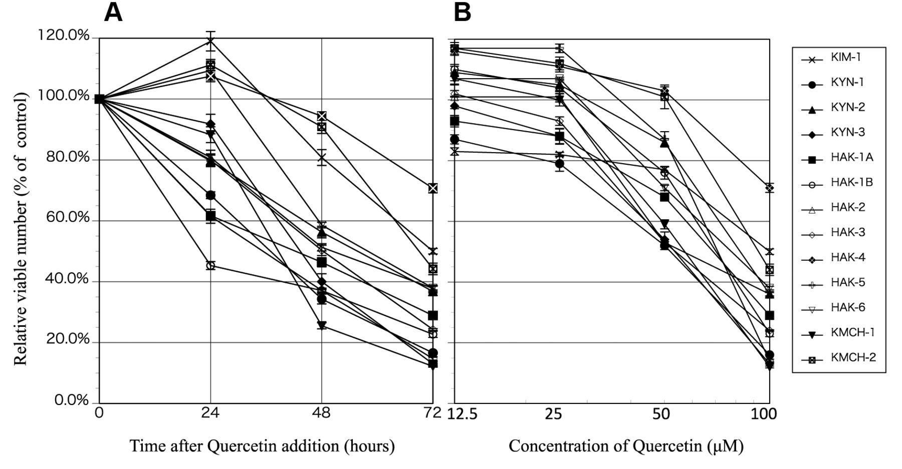
Effect of quercetin on the proliferation of liver cancer cell lines. (A) Relative viable cell number (% of the control) was reduced in a time-dependent manner after the addition of 100 μM of quercetin. (B) 72 h after adding 12.5, 25, 50 or 100 μM of quercetin cell proliferation was suppressed in a dose-dependent manner but in varying degrees. The suppression was statistically significant (p<0.01-0.0001) in the range of 12.5-100 μM of quercetin in all cell lines. Eight samples were used in each experiment (n=8). The experiment was repeated at least three times for each cell line to confirm reproducibility of the data, and almost identical results were obtained. Error bar represents SE. Mean absorbance of the control cells (cells cultured without quercetin) was determined as 100%, and the mean of the other groups was expressed as a ratio.

Cell cycle analysis. Cell lines were cultured with or without quercetin (25 or 100 μM) for 48 h, labeled with 10 μmol/l BrdU for 30 min, fixed in 70% cold ethanol at 4°C overnight, stained with anti-BrdU and propidium iodide (Sigma Chemical Co.), and analyzed using a FACScan. Staining was performed using the modified technique described elsewhere (19). Briefly, fixed cells were washed with PBS, subjected to double-strand DNA denaturation treatment with 2 mol/l HCl at room temperature for 30 min, neutralized with 0.1 mol/l Na2B4O7, washed twice with PBS containing 0.5% Tween 20, and 0.5% BSA (PBS-T), incubated for 30 min at room temperature with 20 μl anti-BrdU antibody, and washed with PBS-T. Then, cells were incubated for 30 min at room temperature with 4 μl fluorescein isothiocyanate–conjugated goat anti-mouse immunoglobulin, and washed with PBS-T. DNA was counterstained with 5 μg/ml of propidium iodide for at least 20 min before flow cytometric analysis. Percentage of cells in the G0/G1, S, and G2/M phases of the cell cycle was calculated from a dot or contour plot obtained after flow cytometric analyses of double-stained cells.

Statistics. Significance in differences was estimated by using the Student's t test (two-tailed).

Results

In all 13 cell lines, the viable cell number reduced to around 20 to 70%, after 72 h of treatment with 100 μM of quercetin. In four of the cell lines (KIM-1, HAK4, HAK-6, KMCH-1), the cell proliferation reached a plateau 24 h after quercetin addition, and there was a reduction in their relative viable cell numbers thereafter. In the other 9 cell lines, there were varying degrees of cell growth inhibition over the course of 72 h since quercetin addition (Figure 1A). All cell lines showed a dose-dependent decrease in the relative viable cell count 72 h after quercetin addition (Figure 1B). The relative viable cell count for cells cultured with 100 μM of quercetin ranged between 12 to 70%, with a mean of 31.4±17%. In 10 cell lines (KYN-1, KYN-2, KYN-3, HAk-1A, HAK-1B, HAK-2, HAk-3, HAK5, HAK-6, KMCH-1, KMCH-2), the relative viable cell number reduced to below 50%. Quantitative analysis of Annexin V-EGFP-positive apoptotic cells revealed that the appearance of apoptosis was significantly higher in the cells treated with quercetin compared to untreated cell lines (Table I).

View this table:
  • View inline
  • View popup
  • Download powerpoint
Table I.

Quantitative analysis of apoptosis induced by quercetin in 13 liver cancer cell lines.

Figure 2.
  • Download figure
  • Open in new tab
  • Download powerpoint
Figure 2.

Effect of quercetin on the cell cycle distribution of 13 liver cancer cell lines. Cells were treated with 0-100 μM quercetin for 48 h and analyzed by flow cytometry. Cell cycle distribution is shown as a histogram. Most liver cancer cell lines underwent cell cycle arrest, but the phase of arrest varied from cell line to cell line (A: G0/G1 phase arrest, B: G2/M phase arrest, C: S phase arrest).

We examined the effect of quercetin on the cell cycle of liver cancer cell lines. Cell cycle arrest was observed in 12 out of 13 cell lines. Following the addition of quercetin, a dose-dependent G0/G1 phase cell cycle arrest was observed in 4 cell lines (HAK-3, HAK-6, KMCH-1, KMCH-2), in the G2/M phase in 6 cell lines (KYN-2, KYN-3, HAK-1B, HAK-2, HAK-4, HAK-5), and in the S phase in 2 cell lines (KIM-1, KYN-1) (Figure 2).

Figure 3.
  • Download figure
  • Open in new tab
  • Download powerpoint
Figure 3.

Effects of quercetin or quercetin+5-FU on the growth of 13 cell line aftertreatment for 72 h. The experiment was repeated at least three times for each cell line to confirm reproducibility of the data, and almost identical results were obtained. Error bar represents SE.

The combination treatment of quercetin and 5-FU produced additional or synergic effects in 9 cell lines (KIM-1, KYN-2, KYN-3, HAK-1B, HAK-2, HAK-5, HAK-6, KMCH-1, KMCH-2) (Figure 3).

Discussion

Quercetin induced a dose-dependent and time -dependent antiproliferative effect in most of the 13 cell lines, and a certain degree of apoptosis in all 13 cell lines. The liver cancer cell lines used in this study had been established from different types of liver cancers, including well to poorly differentiated HCC, sarcomatous HCC, and combined hepatocellular and cholangiocarcinoma. The fact that potent growth inhibition was observed in cell lines of different histology should be emphasized. Although in vitro antiproliferative findings may not exactly resemble those in clinical applications, these findings indicate that quercetin may be efficacious in the treatment of various types of HCC. Experimental reports thus far indicate that quercetin activates caspases, which are involved in the induction of apoptosis, and has an inhibitory effect on the PI3K/Akt and ERK pathways (20). It is possible that the liver cancer cell lines used in this study may have undergone apoptosis and cell growth inhibition by similar mechanisms.

Even though our results on the cell cycle arrest induced by quercetin indicated that most liver cancer cell lines underwent cell cycle arrest, the arrest phase varied from cell line to cell line. Of the 13 liver cancer cell lines studied, 4 cell lines underwent cell cycle arrest in the G0/G1 phase, 2 cell lines in the S phase, and 6 cell lines in the G2/M phase. This is similar to cell cycle arrest by quercetin as reported previously, where the arrest has been shown to take place at different points of the cell cycle (21-23). Quercetin may be able to induce G0/G1 phase, G2/M phase and S phase cell cycle arrest, depending on the type of liver cancer cell line. Further research is needed to determine the exact mechanism.

Treatment with the combination of quercetin and 5-FU led to enhanced growth inhibition in some cell lines, compared to when quercetin was administered on its own. This result was similar to previous reports by Dai et al. demonstrating that 5-FU enhanced quercetin's inhibition of cell growth in vitro and in vivo (24). As the combined use of quercetin and 5-FU showed additional or synergistic effects relative to the use of quercetin alone, this result suggests that the combined use may inhibit the growth of liver cancer at a greater extent. In conclusion, the present study revealed that quercetin suppressed the proliferation of liver cancer cells via induction of apoptosis and cell cycle arrest. Further investigations are needed to clarify the signalling pathways involved in quercetin-induced apoptosis and proliferation inhibition in liver cancer cells.

Acknowledgements

The Authors are grateful to Ms Akemi Fujiyoshi, Ms Mika Hanada and Ms Atsuko Matsuo for their assistance in the experiments.

Footnotes

  • Authors' Contributions

    Toru Hisaka designed the study, and wrote the initial draft of the manuscript. Toru Hisaka and Hisamune Sakai contributed to analysis and interpretation of data, and assisted in the preparation of the manuscript. All other Authors have contributed to data collection and interpretation, and critically reviewed the manuscript. All Authors approved the final version of the manuscript, and agreed to be accountable for all aspects of the work in ensuring that questions related to the accuracy or integrity of any part of the work are appropriately investigated and resolved.

  • This article is freely accessible online.

  • Conflicts of Interest

    The Authors declare no conflicts of interest associated with this manuscript.

  • Received May 23, 2020.
  • Revision received June 14, 2020.
  • Accepted June 15, 2020.
  • Copyright© 2020, International Institute of Anticancer Research (Dr. George J. Delinasios), All rights reserved

References

  1. ↵
    1. Forner A,
    2. Reig M,
    3. Bruix J
    : Hepatocellular carcinoma. Lancet 391(31): 1301-1314, 2019. PMID: 29307467. DOI: 10.1016/S0140-6736(18)30010-2
    OpenUrl
  2. ↵
    1. Bréchot C,
    2. Jaffredo F,
    3. Lagorce D,
    4. Gerken G,
    5. Büchenfelde KM,
    6. Papakonstoninou A,
    7. Hadziyannis S,
    8. Romeo R,
    9. Colombo M,
    10. Rodes J,
    11. Bruix J,
    12. Williams R,
    13. Naoumov N
    : Impact of HBV, HCV and GBV-C/HGV on hepatocellular carcinoma in Europe: results of a European concerted action. J Hepatol 29(2): 173-183, 1998. PMID: 9722197. DOI: 10.1016/s0168-8278(98)80001-9.
    OpenUrlCrossRefPubMed
    1. Di Bisceglie AM,
    2. Order SE,
    3. Klein JL,
    4. Waggoner JG,
    5. Sjogren MH,
    6. Kuo G,
    7. Houghton M,
    8. Choo QL,
    9. Hoofnagle JH
    : The role of chronic viral hepatitis in hepatocellular carcinoma in the United States. Am J Gastroenterol 86(3): 335-338, 1991. PMID: 1847790.
    OpenUrlPubMed
  3. ↵
    1. Shiratori Y,
    2. Shiina S,
    3. Imamura M,
    4. Kato N,
    5. Kanai F,
    6. Okudaira T,
    7. Teratani T,
    8. Tohgo G,
    9. Toda N,
    10. Ohashi M
    : Characteristic difference of hepatocellular carcinoma between hepatitis B- and C-viral infection in Japan. Hepatology 22(4 Pt 1): 1027-1033, 1995. PMID: 7557847. DOI: 10.1016/0270-9139(95)90605-3.
    OpenUrlCrossRefPubMed
  4. ↵
    1. Li Y,
    2. Yao J,
    3. Han C,
    4. Yang J,
    5. Chaudhry MT,
    6. Wang S,
    7. Liu H,
    8. Yin Y
    : Quercetin, inflammation and immunity. Nutrients 8(3): 167, 2016. PMID: 26999194. DOI: 10.3390/nu8030167
    OpenUrl
  5. ↵
    1. Singhal RL,
    2. Yeh YA,
    3. Praja N,
    4. Olah E,
    5. Sledge GW Jr..,
    6. Weber G
    : Quercetin down-regulates signal transduction in human breast carcinoma cells. Biochem Biophys Res Commun 208(1): 425-431, 1995. PMID: 7887960. DOI: 10.1006/bbrc.1995.1355
    OpenUrlCrossRefPubMed
    1. Nguyen TT,
    2. Tran E,
    3. Nguyen TH,
    4. Do PT,
    5. Huynh TH,
    6. Huynh H
    : The Role of Activated MEK-ERK pathway in quercetin-induced growth inhibition and apoptosis in A549 lung cancer cells. Carcinogenesis 25(5): 647-659, 2004. PMID: 14688022. DOI: 10.1093/carcin/bgh052
    OpenUrlCrossRefPubMed
    1. Zhang W,
    2. Yin G,
    3. Dai J,
    4. Sun YU,
    5. Hoffman RM,
    6. Yang Z,
    7. Fan Y
    : Chemoprevention by quercetin of oral squamous cell carcinoma by suppression of the NF-κB signaling pathway in DMBA-treated hamsters. Anticancer Res 37(8): 4041-4049, 2017. PMID: 28739686. DOI: 10.21873/anticanres.11789
    OpenUrlAbstract/FREE Full Text
  6. ↵
    1. Mokbel K,
    2. Wazir U,
    3. Mokbel K
    : Chemoprevention of prostate cancer by natural agents: evidence from molecular and epidemiological studies. Anticancer Res 39(10): 5231-5259, 2019. PMID: 31570421. DOI: 10.21873/anticanres.13720
    OpenUrlAbstract/FREE Full Text
  7. ↵
    1. Yano H,
    2. Iemura A,
    3. Haramaki M,
    4. Ogasawara S,
    5. Takayama A,
    6. Akiba J,
    7. Kojiro M
    : Interferon alfa receptor expression and growth inhibition by interferon alfa in human liver cancer cell lines. Hepatology 29(6): 1708-1717, 1999. PMID: 10347112. DOI: 10.1002/hep.510290624
    OpenUrlCrossRefPubMed
    1. Murakami T
    : Establishment and characterization of human hepatoma cell line (KIM-1). Acta hepatol Jpn 25: 532-539, 1984.
    OpenUrl
    1. Yano H,
    2. Kojiro M,
    3. Nakashima T
    : A new human hepatocellular carcinoma cell line (KYN-1) with a transformation to adenocarcinoma. In Vitro Cell Dev Biol 22(11): 637-646, 1986. PMID: 2430933. DOI: 10.1007/bf02623477
    OpenUrlPubMed
    1. Yano H,
    2. Maruiwa M,
    3. Murakami T,
    4. Fukuda K,
    5. Ito Y,
    6. Sugihara S,
    7. Kojiro M
    : A new human pleomorphic hepatocellular carcinoma cell line, KYN-2. Acta Hepatol Jpn 38: 953-966, 1988.
    OpenUrl
    1. Murakami T,
    2. Maruiwa M,
    3. Fukuda K,
    4. Kojiro M,
    5. Tanaka M,
    6. Tanikawa K
    : Characterization of new human hepatoma cell line (KYN-3) derived from the ascites of the hepatoma patient [Abstract]. Proceedings of Japanese Cancer Association. Jpn J Cancer Res 292, 1988.
    1. Yano H,
    2. Iemura A,
    3. Fukuda K,
    4. Mizoguchi A,
    5. Haramaki M,
    6. Kojiro M
    : Establishment of two distinct human hepatocellular carcinoma cell lines from a single nodule showing clonal dedifferentiation of cancer cells. Hepatology 18(2): 320-327, 1993. PMID: 8393423.
    OpenUrlCrossRefPubMed
    1. Haramaki M,
    2. Yano H,
    3. Iemura A,
    4. Momosaki S,
    5. Ogasawara S,
    6. Inoue M,
    7. Yamaguchi R,
    8. Kusaba A,
    9. Utsunomiya I,
    10. Kojiro M
    : A new human hepatocellular carcinoma cell line (HAK-2) forms various structures in collagen gel matrices. Hum Cell 10(3): 183-192, 1997. PMID: 9436038.
    OpenUrlPubMed
    1. Murakami T,
    2. Yano H,
    3. Maruiwa M,
    4. Sugihara S,
    5. Kojiro M
    : Establishment and Characterization of a human combined hepatocholangiocarcinoma cell line and its heterologous transplantation in nude mice. Hepatology 7(3): 551-556, 1987. PMID: 3032760. DOI: 10.1002/hep.1840070322
    OpenUrlCrossRefPubMed
  8. ↵
    1. Yano H,
    2. Iemura A,
    3. Haramaki M,
    4. Momosaki S,
    5. Ogasawara S,
    6. Higaki K,
    7. Kojiro M
    : A human combined hepatocellular and cholangiocarcinoma cell line (KMCH-2) that shows the features of hepatocellular carcinoma or cholangiocarcinoma under different growth conditions. J Hepatol 24(4): 413-422, 1996. PMID: 8738727. DOI: 10.1016/s0168-8278(96)80161-9
    OpenUrlPubMed
  9. ↵
    1. Yano H,
    2. Mizoguchi A,
    3. Fukuda K,
    4. Haramaki M,
    5. Ogasawara S,
    6. Momosaki S,
    7. Kojiro M
    : The herbal medicine Sho-Saiko-To inhibits proliferation of cancer cell lines by inducing apoptosis and arrest at the G0/G1 phase. Cancer Res 54(2): 448-454, 1994. PMID: 8275481.
    OpenUrlAbstract/FREE Full Text
  10. ↵
    1. Granado-Serrano AB,
    2. Martín MA,
    3. Bravo L,
    4. Goya L,
    5. Ramos S
    : Quercetin induces apoptosis via caspase activation, regulation of Bcl-2, and inhibition of PI-3-kinase/Akt and ERK pathways in a human hepatoma cell line (HepG2). J Nutr 136(11): 2715-2721, 2019. PMID: 17056790. DOI: 10.1093/jn/136.11.2715
    OpenUrl
  11. ↵
    1. Srisa-Nga K,
    2. Mankhetkorn S,
    3. Okonogi S,
    4. Khonkarn R
    : Delivery of superparamagnetic polymeric micelles loaded with quercetin to hepatocellular carcinoma cells. J Pharm Sci 108(2): 996-1006, 2019. PMID: 30121312. DOI: 10.1016/j.xphs.2018.08.008
    OpenUrl
    1. Pi J,
    2. Li B,
    3. Tu L,
    4. Zhu H,
    5. Jin H,
    6. Yang F,
    7. Bai H,
    8. Cai H,
    9. Cai J
    : Investigation of Quercetin-induced HepG2 cell apoptosis-associated cellular biophysical alterations by atomic force microscopy. Scanning 38(2): 100-112, 2016. PMID: 26179807. DOI: 10.1002/sca.21245
    OpenUrl
  12. ↵
    1. Sudan S,
    2. Vasantha Rupasinghe Hp
    : Antiproliferative activity of long chain acylated esters of quercetin-3-O-glucoside in hepatocellular carcinoma HepG2 cells. Exp Biol Med 240(11): 1452-1464, 2015. PMID: 25681471. DOI: 10.1177/1535370215570828
    OpenUrlCrossRefPubMed
  13. ↵
    1. Dai W,
    2. Gao Q,
    3. Qiu J,
    4. Yuan J,
    5. Wu G,
    6. Shen G
    : Quercetin induces apoptosis and enhances 5-fu therapeutic efficacy in hepatocellular carcinoma. Tumour Biol 37(5): 6307-6313, 2016. PMID: 26628295. DOI: 10.1007/s13277-015-4501-0
    OpenUrl
PreviousNext
Back to top

In this issue

Anticancer Research
Vol. 40, Issue 8
August 2020
  • Table of Contents
  • Table of Contents (PDF)
  • Index by author
  • Back Matter (PDF)
  • Ed Board (PDF)
  • Front Matter (PDF)
Print
Download PDF
Article Alerts
Sign In to Email Alerts with your Email Address
Email Article

Thank you for your interest in spreading the word on Anticancer Research.

NOTE: We only request your email address so that the person you are recommending the page to knows that you wanted them to see it, and that it is not junk mail. We do not capture any email address.

Enter multiple addresses on separate lines or separate them with commas.
Quercetin Suppresses Proliferation of Liver Cancer Cell Lines In Vitro
(Your Name) has sent you a message from Anticancer Research
(Your Name) thought you would like to see the Anticancer Research web site.
CAPTCHA
This question is for testing whether or not you are a human visitor and to prevent automated spam submissions.
5 + 3 =
Solve this simple math problem and enter the result. E.g. for 1+3, enter 4.
Citation Tools
Quercetin Suppresses Proliferation of Liver Cancer Cell Lines In Vitro
TORU HISAKA, HISAMUNE SAKAI, TOSHIHIRO SATO, YUICHI GOTO, YORIKO NOMURA, SHOGO FUKUTOMI, FUMIHIKO FUJITA, TOMOAKI MIZOBE, OSAMU NAKASHIMA, MASAHIKO TANIGAWA, YOSHIKI NAITO, JUN AKIBA, SACHIKO OGASAWARA, KOUTA NAKASHIMA, YOSHITO AKAGI, KOJI OKUDA, HIROHISA YANO
Anticancer Research Aug 2020, 40 (8) 4695-4700; DOI: 10.21873/anticanres.14469

Citation Manager Formats

  • BibTeX
  • Bookends
  • EasyBib
  • EndNote (tagged)
  • EndNote 8 (xml)
  • Medlars
  • Mendeley
  • Papers
  • RefWorks Tagged
  • Ref Manager
  • RIS
  • Zotero
Reprints and Permissions
Share
Quercetin Suppresses Proliferation of Liver Cancer Cell Lines In Vitro
TORU HISAKA, HISAMUNE SAKAI, TOSHIHIRO SATO, YUICHI GOTO, YORIKO NOMURA, SHOGO FUKUTOMI, FUMIHIKO FUJITA, TOMOAKI MIZOBE, OSAMU NAKASHIMA, MASAHIKO TANIGAWA, YOSHIKI NAITO, JUN AKIBA, SACHIKO OGASAWARA, KOUTA NAKASHIMA, YOSHITO AKAGI, KOJI OKUDA, HIROHISA YANO
Anticancer Research Aug 2020, 40 (8) 4695-4700; DOI: 10.21873/anticanres.14469
Twitter logo Facebook logo Mendeley logo
  • Tweet Widget
  • Facebook Like
  • Google Plus One

Jump to section

  • Article
    • Abstract
    • Materials and Methods
    • Results
    • Discussion
    • Acknowledgements
    • Footnotes
    • References
  • Figures & Data
  • Info & Metrics
  • PDF

Related Articles

Cited By...

  • No citing articles found.
  • Google Scholar

More in this TOC Section

PROCEEDINGS OF THE 23RD ANNUAL MEETING OF THE SOCIETY OF BIOTHERAPEUTIC APPROACHES

  • Mutant KRAS Promotes NKG2D+ T Cell Infiltration and CD155 Dependent Immune Evasion
  • The Impact of Histological Variant on Oncological Outcomes in Patients With Urothelial Carcinoma of the Bladder Treated With Radical Cystectomy
  • Clinical Outcomes of Surgical Resection for Brain Metastases from Non-small Cell Lung Cancer
Show more PROCEEDINGS OF THE 23RD ANNUAL MEETING OF THE SOCIETY OF BIOTHERAPEUTIC APPROACHES

Experimental Studies

  • The Glucose and Glutamine Requirements of Cancer and Normal Cells Do Not Distinguish Them, in Contrast to Their Methionine Requirement, Suggesting the Warburg Effect Is Not a Cancer Paradigm
  • Integrative Analysis Combining Machine Learning and Functional Experiments Uncovers ISG15 As a Key Determinant of Cisplatin Resistance in Gastric Cancer
  • Allow Aloe to Do the Work: Aloe vera Constrains Growth of Bladder Cancer Cells and Modulates Expression of Key Costimulatory Molecules
Show more Experimental Studies

Keywords

  • quercetin
  • antiproliferation effect
  • apoptosis
  • Hepatocellular carcinoma
Anticancer Research

© 2026 Anticancer Research

Powered by HighWire