


Abstract
Background/Aim: Triple-negative breast cancer (TNBC) is a highly aggressive form of breast cancer (BC) and lacks targeted therapy and alternate therapeutic combinations. There is a necessity to increase disease-free survival in patients particularly within the first 5 years of diagnosis. 2,3-dichloro-5,8-dimethoxy-1,4-naphthoquinone (Z285), a novel 1,4 naphthoquinone analog, has been shown to have cytotoxic activity in BC cell lines and in combination with 4-hydroxytamoxifen (4-OHT). A known metabolite of tamoxifen, was postulated to decrease cell proliferation. Thus, this study investigates the use of Z285 and 4-OHT alone or in combination as a novel therapeutic alternative for TNBC. Materials and Methods: Cell proliferation assays were performed on MDA-MB-231, Hs578T, MCF7 and HCC1806 cell lines at varying time points with Z285 and 4-OHT alone and in combination. Furthermore, ROS activity was measured to determine the changes in oxidative stress caused by both drugs. Results: The results showed dose- and time-dependent decreases in proliferation for all cell lines when treated with Z285, 4-OHT and their combination. Combinatorial analysis performed at 72 h using Synergyfinder® showed additive effects in MCF7, HCC1806 and Hs578T and an antagonistic response in MDA-MB-231. Z285 caused a significant increase in ROS production in three cell lines after 8 h, but HCC1806 showed no change in effect. Conclusion: These promising results suggest the independent ability of each compound as a stand-alone chemotherapeutic agent, or in combinatorial therapy for the treatment of TNBC.
Triple-negative breast cancer (TNBC) lacks the expression of estrogen receptor α (ERα), progesterone receptor (PR) and human epidermal growth factor receptor 2 (HER2). The loss of these therapeutic targets limits the treatment options to chemotherapy, surgery, and radiation. This subtype accounts for 10-15% of diagnosed breast cancers (BC) but approximately 35% of metastatic BC-related deaths are attributed to TNBC (1). It is an aggressive phenotype and disproportionately effects African American (AA) women where the diagnosis of this disease is twice as likely than in European American (EA) women (2). In the AA population, there is also a higher incidence in premenopausal women with this disease (3, 4). Studies of Sub-Saharan and West African women indicate increased frequency and younger age of diagnoses of TNBC when compared to AA women (5). The five- and ten-year survival rates are lower for TNBC patients when compared to other breast cancer subtypes thus novel therapies are needed to increase survival especially in stage IV (6-8).
Over the years, several therapeutic targets have been purported in vitro and in vivo to be effective in TNBC treatment such as EGFR, PI3K/mTOR, PARP, Src and RAS/RAF/MEK inhibitors but in clinical trials many have failed to be efficacious (9, 10). Recently, a new class of drugs has been approved that target BRCA1/2-mutated TNBC patients. This class of drugs, PARP inhibitors, demonstrate synthetic lethality (11, 12). However, this class of drugs only targets approximately 15% of diagnosed TNBC (13). Topoisomerase I antibody drug conjugate, sacituzumab govitecan-hizy, has recently been approved for metastatic TNBC (14-16). In general, monotherapy for cancer treatment has been proven to be ineffective due to heterogeneity of cells within a tumor and development of drug resistance (17). Thus, novel compounds and innovative therapeutic combinations are needed for the treatment of breast cancer, particularly TNBC (18, 19).
Z285 is a member of the 1,4 naphthoquinones class of compounds that have been shown to have anticancer, antibacterial, and antimalarial function (20-22). Similarly, Z285 has demonstrated cytotoxic effects in androgen-dependent and -independent prostate cancer cell lines as well as BC cell lines (23, 24). 4-OHT is an active metabolite of tamoxifen, a drug routinely used as BC prophylaxis as well as treatment of ERα-positive BC (25, 26). Therefore, this study examines the use of 2,3-dichloro-5,8-dimethoxy-1,4-naphthoquinone (Z285) and 4-hydroxytamoxifen (4-OHT) alone or in combination as a novel therapeutic alternative for TNBC.
Materials and Methods
Materials. CellTiter 96® AQueous One Solution Cell Proliferation Assay was obtained from Promega (Madison, WI, USA) and DMSO was purchased from Sigma-Aldrich (St. Louis, MO, USA). 4-hydroxytamoxifen was acquired from Fisher Scientific (Hanover Park, IL, USA) and PBS from Gibco (Gaithersburg, MD, USA). 2,3-Dichloro-5,8-dimethoxy-1,4-naphthoquinone was synthesized in house (23). CM-H2DCFDA was purchased from Invitrogen (Carlsbad, CA, USA). All drugs were made up as 10 mM stock dissolved in DMSO.
Cell culture. Stock cultures of the human ERα-positive BC (MCF7) and TNBC (HCC1806 obtained from AA patient and MDA-MB 231, Hs578T both taken from EA patients) cell lines were obtained from the American Type Culture Collection (ATCC) (Rockville, MD, USA). The cells were grown in 75 cm3 flasks in RPMI-1640 from ATCC (Rockville, MD, USA) medium supplemented with 10% fetal bovine serum (FBS) obtained from ATCC (Rockville, MD, USA) and 1% penicillin/streptomycin from Gibco (Gaithersburg, MD, USA) and incubated in humidified atmosphere 5% CO2 at 37°C. Upon reaching 80-90% confluency, the cells were trypsinized, and quantified with T10 cell counter from Bio-Rad (Hercules, CA, USA). For all experiments, cells were starved by plating in phenol free RPMI 1640, 1% charcoal stripped FBS and 1% penicillin/streptomycin for 24 h to achieve cell cycle synchronization and limit estrogenic exposure. Drugs were diluted to appropriate concentrations in this media.
Cell proliferation. All cell lines were plated at 8×10³ cells/well in 96-well plate. Cells were allowed to attach overnight in growth media. The media was aspirated, and cells were further incubated in starvation media for 24 h. Relevant concentrations of the compounds were diluted in starvation media.
Single drug: Cells were treated in a concentration- and time-dependent manner with Z285 and 4-OHT at 0.1, 0.2, 0.5, 1, 2, 5, 10, 20 and 50 μM for 24, 72 and 120 h, respectively.
Combinatorial drug treatment: Cells were treated with Z285 for final concentrations of 2 or 5 μM and 1, 3, 6, 12, 15 μM of 4-OHT for 24, 72 and 120 h.
At the end of the respective time periods, 20 μl MTS was added to all the wells for 2 h and read at 495 nm using Perkin Elmer Wallac Victor 3 1420 plate reader (Waltham, MA, USA).
Data analysis: Graphpad v8 was used to calculate the IC50 for each cell line with each compound and its combination through nonlinear regression analysis. Combinatorial synergy study. Cells were treated with Z285 with final concentrations of 1, 2, 4, 8, 16 μM and with 4-OHT 3, 6, 12, 24, 48 μM for 72 h. After 72 h, 20 μL of MTS was added to the wells for 2 hours and read at 495 nm using Perkin Elmer Wallac Victor 3 1420 plate reader (Waltham, MA, USA).
Data analysis: Synergistic analysis was performed using Synergyfinder®, utilizing the Bliss Independence Reference model.
ROS assay. According to the manufacturers guidelines (Invitrogen), media was aspirated, cells were then washed with room temperature PBS and incubated in 10 μM CM-H2DCFA for 45 min. The cells were additionally washed twice in PBS followed by treatment with Z285 at final concentrations of 2, 4, 8, and 16 μM and at 4-OHT 3, 6, 12 and 24 μM and their combination for 8 h. Plate was read at 492-495 nm excitation and 517-527 nm emission using Biotek Cytation 3 imaging reader (Winooski, VT, USA).
Data analysis: One-way Anova followed by post-hoc Dunnetts test compared each mean to the control (untreated cells incubated with dye) was used to determine significance.
Results
In this study, three TNBC cell lines and one ERα expressing cell line were evaluated to determine the efficacy of Z285 and 4OHT alone and in combination on cell proliferation.
Z285 treatment. The cell line which was most susceptible to Z285 was Hs578T at 24 and 120 h with an IC50 of 5.21 and 1.63 μM, respectively. At 72 h, HCC1806 showed the most sensitivity with an IC50 of 4.26 μM as shown in Table I. MCF7 was least susceptible to Z285 at 72 h and 120 h with IC50 values of 9.43 and 5.50 μM, whereas HCC1806 showed the least sensitivity at 24 h with, respectively; IC50 of 22 μM as shown in Figure 1A-C.

Cell viability assay of Z285 alone and 4-OHT alone treated cells at 24, 72 and 120 h. (A) 24 h Z285, (B) 72 h Z285, (C) 120 h Z285, (D) 24 h 4-OHT, (E) 72 h 4-OHT,(F) 120 h 4-OHT. After starving cells for 24 h, A-D were treated with 0.1-50 μM of Z285 and D-F were treated with 0.1-50 μM of 4-OHT. IC50 was calculated by non-linear regression for each cell line at every time point. Each group analysis was performed in triplicate.
IC50 values of cells treated with Z285 alone at 24, 72 and 120 h.
4-OHT treatment. MDA-MB-231 exhibited the highest susceptibility at 14.8, 4.83 and 2.65 μM for 24, 72 and 120 h, respectively as shown in Figure 1D-F. Whereas, the IC50 for HCC1806 decreased from 16.2 to 6.83 μM and Hs578T showed a reduction in the IC50 from 12.6 to 7.16 μM between 24 h and 120 h. Interestingly, MCF7 was least sensitive to 4-OHT at every time point reaching an IC50 of approximately 20 μM at 24 h and decreasing to 8.5 μM by 120 h as shown in Table II.
IC50 values of cells treated with 4-OHT alone at 24, 72 and 120 h.
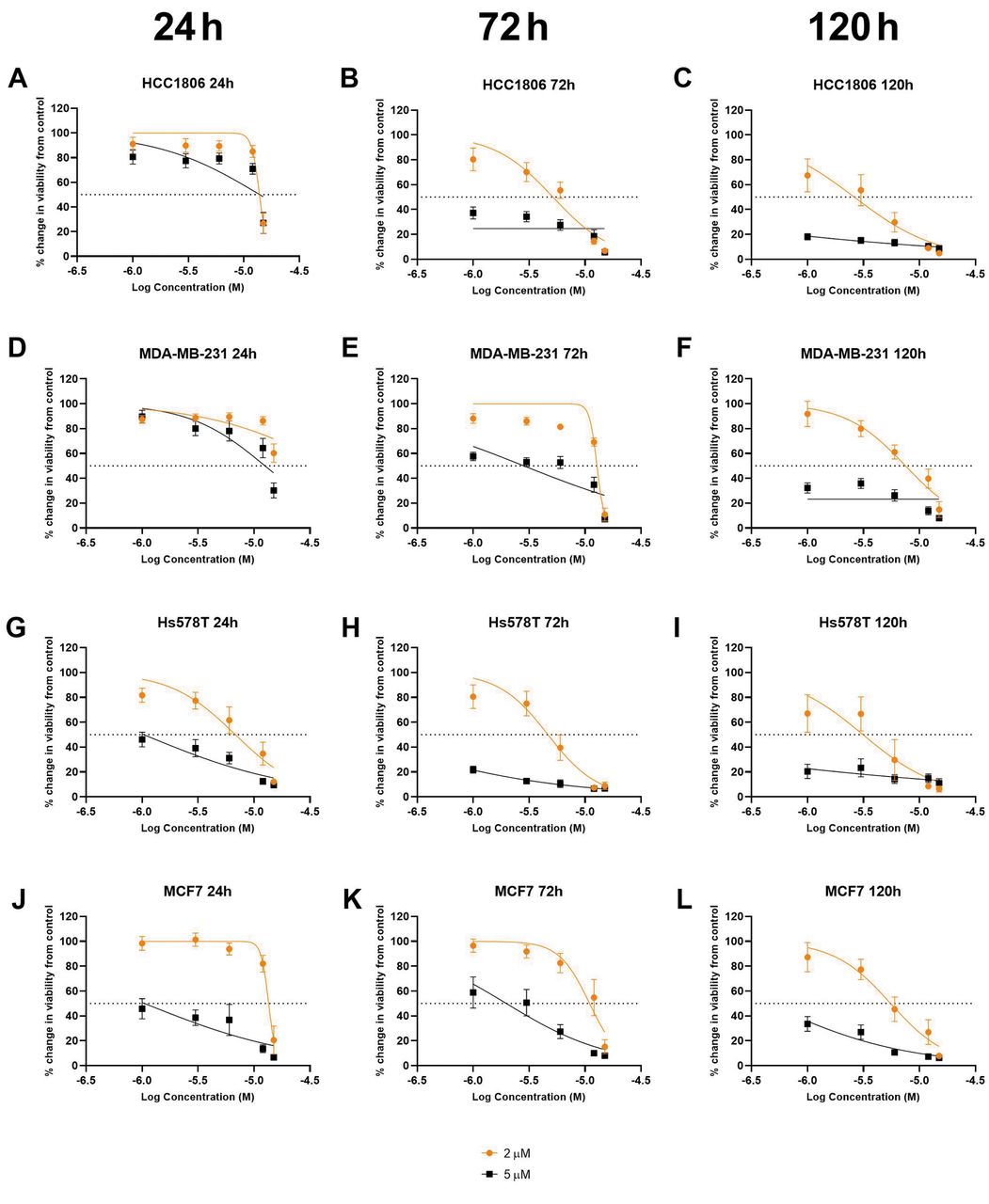
Z285 and 4-OHT combination treatment. Each cell line demonstrated a reduction in cell proliferation following combination treatment. Z285 was given at 2 and 5 μM in combination with 4-OHT at concentrations between 1 and 30 μM (Figure 2). When compared to the IC50 of 4-OHT alone, combination treatment with Z285 and 4-OHT showed decrease in the IC50 for HCC1806, Hs578T and MCF7 at concentrations of Z285 2 and 5 μM and 5 μM Z285 for MDA-MB-231 (Table III). HCC1806 showed a 14% decrease in IC50 for at both concentrations of Z285 at 24 h. A further 39% reduction was observed at 2 μM of Z285 at 72 h and a 62 and 99% at 120 h was shown at 2 and 5 μM Z285 when compared to 4-OHT-treated cells, respectively. MDA-MB-231 demonstrated a 15 and 41% decrease in IC50 at 24 and 72 h respectively after 5 μM Z285 and 4-OHT combination when compared to 4-OHT alone. In Hs578T, the IC50 decreased by 48, 42 and 56% for 2 μM combination at 24, 72 and 120 h, respectively. Furthermore, a 92, 98 and 99% decrease in IC50 was observed with 5 μM combination at 24, 72 and 120 h, respectively. MCF7 showed approximately 33% decrease in IC50 for all time points in 2 μM combination while at 5 μM combination a 95% decrease in IC50 was seen at all times. It should be noted that the combination treatment at 2 μM for MDA-MB-231 showed an increase in the IC50 values when compared with 4-OHT by itself there was a 225, 165 and 188% increase in IC50 at 24, 72 and 120 h, respectively.

Cell viability assay of cells treated with Z285 and 4-OHT at 24, 72 and 120 h. After starving cells for 24 h. All cells were treated with either 2 or 5 μM of Z285 and 1-15 μM of 4-OHT combined. Column 1 is 24 h, Column 2 is 72 h and column 3 is 120 h (A-C) are HCC1806, (D-F) are MDA-MB-231, (G-I) are Hs578T and (J-L) are MCF7. Non-linear regression is perfored on every cell line at every time point. Each group analysis was performed in triplicate.
Percentage change from 4-OHT treated cells alone.
Combinatorial analysis. Analysis of drug combination by Synergyfinder® indicated Bliss δ scores of 0.9, 4.9, and 5.6 for HCC1806, MCF7 and Hs578T respectively, thereby showing an additive effect for decreased cell proliferation. MDA-MB-231 showed a score of -10 indicating an antagonistic effect (Figure 3). Bliss independence reference model in this software compares the observed versus predicted inhibition response and where less than -10 is considered antagonistic, between -10 and 10 is considered to be additive and above 10 is synergistic.

Combinatorial analysis of cell lines treated with Z285 and 4-OHT. (A) HCC1806, (B) MDA-MB-231, (C) Hs578T, (D) MCF7. Synergyfinder using Bliss Independence reference model determined that an additive effect was produced with δ scores of HCC1806, Hs578T and MCF7 and MDA-MB-231 treated cells resulted with an antagonsitic effect.
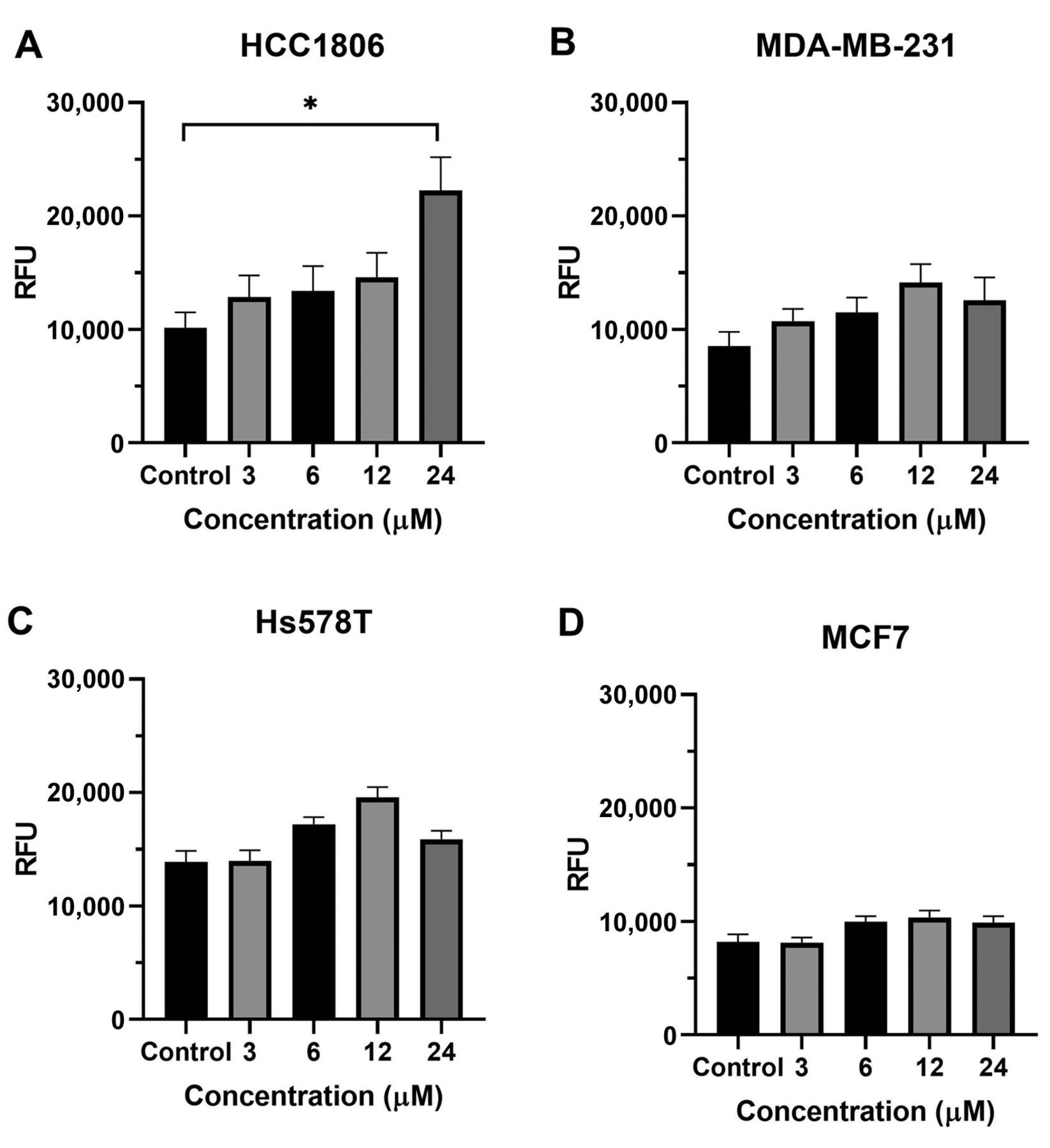
ROS generation. Z285 demonstrated a significant increase in oxidative stress in Hs78T, MCF7 and MDA-MB-231 at 8 and 16 μM after 8 h exposure. Hs578T demonstrated the most significant increases in ROS with p-value ≤0.0001 at both 8 and 16 μM (Figure 4C). MDA-MB-231 and MCF7 showed p-values of ≤0.01 and ≤0.0001 at 8 and 16 μM, shown respectively in Figure 4B and D. HCC1806 showed a trend increase in ROS levels without significance. HCC1806 treated with 4-OHT produced a significant increase in ROS levels at 24 μM with a p-value of ≤0.05. The other cell lines treated with 4-OHT showed no significant changes in ROS production (Figure 5A-D).

ROS assay of cells treated with Z285. (A) HCC1806, (B) MDA-MB-231, (C) Hs578T, (D) MCF7. Cells were washed with PBS after starving for 24h. A total of 10 μM CM-H2DCFA was added for 45 min, then the cells were washed with PBS twice before treatments were added. All cell lines were treated with Z285 at 2, 4, 8, and 16 μM for 8 h. One-way ANOVA followed by post-hoc Dunnetts test comparing each mean to the control (untreated cells with incubated with dye) was used to determine significance. Each group was performed in triplicate. **p≤0.01, ****p≤0.0001.
ROS assay of cells treated with 4-OHT. (A) HCC1806, (B) MDA-MB-231, (C) Hs578T, (D) MCF7. Cells were washed with PBS after starving for 24 h. A total of 10 μM CM-H2DCFA was added for 45 min, then the cells were washed with PBS twice before treatments were added. A-D were treated with 3, 6, 12 and 24 μM for 8 h. One-way ANOVA followed by post-hoc Dunnetts test comparing each mean to the control (untreated cells with incubated with dye) was used to determine significance. Each group analysis was performed in triplicate, *p≤0.05.
When Z285 and 4-OHT were combined, MDA-MB-231 and MCF7 showed some combinations with significant increases in ROS production. In comparison to the MDA-MB-231 control, 16 μM Z285 and 6 μM 4-OHT combination caused an increase in ROS with a statistical significance of p≤0.01. MCF7 demonstrated significant increases in ROS levels at 4 μM Z285 and 6 μM, 8 μM Z285 and 3 μM 4-OHT and 8 μM Z285 and 6 μM 4-OHT with p-values of ≤0.05, ≤0.01, ≤0.05, respectively. No statistical changes in ROS was observed in HCC1806 and MCF7 (Figure 6).

ROS assay of combination treatment with Z285 and 4-OHT. (A) HCC1806, (B) MDA-MB-231, (C) Hs578T, (D) MCF7. Cells were washed with PBS after starving for 24 h. 10 μM CM-H2DCFA was added for 45 min, then the cells were washed with PBS twice before treatments were added. A-D were treated with a combintion of Z285 at 2, 4, 8, and 16 μM and 3, 6, 12 and 24 μM for 8 h. One-way ANOVA followed by post-hoc Dunnetts test comparing each mean to the control (untreated cells with incubated with dye) was used to determine significance. Each group was performed in triplicate. *p≤0.05, **p≤0.01.
Discussion
TNBC loss of ERα, PR and HER2 receptors make it difficult to treat due to lack of receptor targets. Though treatment options for TNBC have improved in recent years with targeted therapy such as PARP and topoisomerase I, there is still a significant number of patients who are unresponsive to these drugs (27, 28). Data show a 73% five-survival rate of TNBC to a 96% five-survival rate for non-TNBC counterparts. This is based on the grade of tumor, stage of cancer, age of patient, and overall health status. Stage IV diagnoses of TNBC has a general survival rate as low as 26.7% (29, 30).
This disease disproportionately effects premenopausal AA women, with twice as many AA women being diagnosed with this subtype when compared to EA women. Up to 40% of BC diagnoses in premenopausal AA women can be attributed to TNBC (31). In comparison to EA women, AA women demonstrate enhanced genetic risk factors like BRCA1, Aurora A and B, EZH2 and p53 mutations; as well as, increased rates of obesity and lower socioeconomic factors (32). TNBC in young women is more likely to be of a more aggressive subtype, and is more likely to present at an advanced stage, either because of its biological aggressive nature or because of delayed diagnosis (33). Young age was seen as an independent prognostic factor. According to these findings, patients diagnosed with BC at ≤35 years of age had a worse prognosis compared to premenopausal women above this age (34). Therefore, it is essential that innovative therapies are presented to increase the survivability of diagnosed patients.
This study investigated the efficacy of Z285 and 4-OHT as possible therapy for TNBC. Z285, is a 1,4 naphthoquinone with many therapeutic effects including antibacterial, antifungal, anti-inflammatory, antiviral and antitumor, specifically in prostate and breast cancer cell lines (35). In our previous study, it was shown that this compound causes inhibition of topoisomerase I, an enzyme responsible for producing single-strand breakage and relegation by unwinding supercoiled DNA to allow DNA replication and transcription. Thus inhibition of this enzyme can lead to apoptosis in BC cells (24, 36). Also, it significantly increases retinoblastoma, a tumor suppressor, levels in these cells. The cell proliferation inhibitory effects of Z285 has been previously observed in TNBC, ERα positive and androgen-dependent and - independent prostate cancer cell lines. The compound was observed to cause cell-cycle arrest in the S phase in all BC cell lines as well as androgen-independent prostate cancer cell lines (23, 24).
In the present investigation, four BC cell lines were used including HCC1806, Hs578T, MDA-MB-231and MCF7. HCC1806 was used specifically to evaluate an AA TNBC cell line in comparison to EA cells. As described by Lehmann et al., TNBC can be subdivided into seven categories: basal-like 1 (BL1), basal-like (BL2), Mesenchymal (M), Mesenchymal stem-like (MSL), immunomodulatory (IM), luminal androgen receptor (LAR) and unstable (UNS) (9). BL1 and BL2 are described as basal-like with BL1 exhibiting greater DNA damage response and cell-cycle gene expression and BL2 is enriched in growth factor signaling and myoepithelial markers. Whereas LAR, expresses a 9-fold increase of AR expression compared to other subtypes and has luminal gene expression. M shows increased gene expression of epithelial-mesenchymal-transition and growth factor pathways. MSL though similar to M shows reduced expression of proliferative genes. IM has immune signaling transduction pathways most likely due to a mixture of tumor cells and infiltrating lymphocytes. UNS tumors do not fall into any of the aforementioned categories. Thus, HCC1806 is classified as BL2 and MDA-MB-231 and Hs578T are categorized as MSL (9).
The evaluation did corroborate prior results as well as demonstrate the compounds ability to decrease cell proliferation in the multiple TNBC cell lines. Prior data indicate topoisomerase I inhibition as a mechanism for inhibition of cell viability, it is important to note that other mechanisms can be suggested for this compound’s action especially as the androgen dependent cells were not arrested in the S-phase similar to the other cell lines. Further, topoisomerase II is responsible for recombination, the separation of daughter chromosomes, and proper chromosome structure, condensation, and decondensation and inhibition of this enzyme is associated with other compounds in this class (37).
It may be suggested that BC cells treated with Z285 in the current study causes an increased generation of ROS resulting in alterations in cell signaling leading to cell damage thereby causing decreased cell proliferation. This cellular damage could be caused by hydroxyl radicals binding to cysteine-rich proteins and lipids, resulting in lipid peroxidation of cellular membrane and leading to apoptosis (38-42).
Concentration-response of the cell lines with 4-OHT produced interesting results with MCF7 in that they were the least sensitive to the drug as compared to the TNBC cell lines. These cell lines do not express the ERα target commonly associated with this drug so they should not be more susceptible to 4-OHT. Studies by Lin et al., (39) and Yaacob and Ismail (40) corroborated the MCF data from the current study in that high concentrations above 10 μM of 4-OHT failed to produce a 50% reduction in cell proliferation in MCF7 cells.
Tamoxifen and its active metabolites, endoxifen and 4-OHT are ligands for ERα; as well as, ERβ, GPER1, Estrogen Related Receptor β and Estrogen Related Receptor γ (43-45). 4-OHT has a similar relative binding affinities (RBA) to both ERα and Erβ (46) when compared to tamoxifen. On its own, 4-OHT decreased proliferation in MCF7 as expected but it also decreased proliferation in all three TNBC cell lines with greater sensitivity. Studies by Manna and Holz showed that 5-10% of ERα-negative cells are susceptible to tamoxifen (47). This can be attributed to the varying levels of expression of ERβ in these cell lines (48-51). 4-OHT is an agonist for ERβ inhibiting cell proliferation, migration, and invasion in TNBC cell lines (52). ERs have ER/ligand independent activity that can lead to activation of cytoplasmic proteins or phosphorylation of transcription factors (53).
Synergyfinder® using Bliss independence reference model was used to identify the relationship between the two compounds when combined (54). The effects of the drug combination showed an additive relationship in decreasing cell proliferation in HCC1806, Hs578T and MCF7. The additive relationship demonstrated in these cell lines maybe due to the changes in ROS production or DNA damage caused by Z285 thus increasing the susceptibility of the cells to 4-OHT. Though this combination demonstrates additivity in most of these cell lines, the methods of cell death may vary.
MDA-MB-231 demonstrated an antagonistic response with the compound combination. Lin et al., also observed some antagonistic responses when treating this cell line with shikonin and 4-OHT, shikonin being a naphthoquinone (55). This response may indicate a functional independence of the two compounds on cell proliferation. MDA-MB-231 cells have hypermethylation of CpG island in the promoter region of ESR1 thus effectively silencing the gene (56). Moreover, it has been proposed that the loss of ERα expression is due to the hyperactivation of MAPK (57, 58). Therefore, ROS production may modulate MAPK thus altering ERα expression in the other cell lines, but this would be ineffective in MDA-MB-231 (59).
CM-H2DCFDA indirectly reacts with H2O2 to produce a fluorescent molecule that is used to measure ROS levels (60). The increase in oxidative stress after 8 h corresponds to the decrease in cell proliferation seen at 24 h with HCC1806 being the least responsive and Hs578T showing increased susceptibility to Z285. It has been shown that 1,4 naphthoquinone derivatives can increase ROS levels and modulate the three major MAPK pathways ERK, JNK and p38 as well as the PI3K/AKT and JAK/STAT3 (61-64). These cellular stressors can cause a reduction in cell proliferation and an induction of apoptosis. In the current study, treatment with 4-OHT produced an increased trend in ROS production, whereas, at high concentration there was a slight decrease in ROS levels in MDA-MB-231 and Hs578T. Bekele et al., reported that incubation of MCF7 at 24h shows a significant increase in ROS (65). Therefore, a longer incubation period maybe required to achieve statistically significant increases in ROS production in the other cell lines. The combination treatment of the two compounds demonstrated an increase trend in the ROS production in HCC1806 and Hs578T.
The additive effect observed in the synergy analysis maybe due to other mechanisms independent of increased ROS generation. Kawiak et al., suggested that glucose regulated protein 78 (GRP78) down-regulation and Bcl-2-interacting killer (Bik) up-regulation by plumbagin was shown to increase the sensitivity of MCF7 and T47D to tamoxifen. Bik is a proapoptotic protein and increased Bik forms a complex with Bcl-2 on the endoplasmic reticulum activating apoptotic process (66). In addition, the activation of c-jun by JNK signaling is needed for 4-OHT induced cell death and 1,4 napthoquinones have demonstrated an increase in JNK activation and thus potentially enhancing the effect when the compounds are combined (67, 68).
Future studies with an expanded number of AA TNBC cell lines would be needed to improve comparison between the two ethnicities. Also, expansion into increased molecular subtypes of TNBC cell lines (i.e. LAR and BL1) would further the knowledge of the efficacy of Z285 and 4OH-T combination for all TNBC. This information may be translated into a molecular subytpe-specific therapeutic modality for TNBC.
Conclusion
The present study demonstrated a beneficial relationship between Z285 and 4-OHT. However, the mechanisms that are associated with the additive effect have not been fully elucidated. Therefore, combination of these two compounds may be an alternative therapy for TNBC patients who are unresponsive to other treatments.
Acknowledgements
This work was supported by funding from the Charles and Mary Latham Foundation Fund. The Authors would like to thank Dr. O. Bakare from the Department of Chemistry at Howard University for generously providing Z285.
Footnotes
Authors’ Contributions
Experimental design was performed by AGJR and RLC. Experiments and statistical analysis were performed by AGJR. AGJR, YMK and RLC were responsible for manuscript writing and editing.
This article is freely accessible online.
Conflicts of Interest
The Authors have no conflicts of interest to declare.
- Received October 1, 2020.
- Revision received October 26, 2020.
- Accepted November 1, 2020.
- Copyright © 2020 International Institute of Anticancer Research (Dr. George J. Delinasios), All rights reserved.


