
Main menu

  • Home
  • Current Issue
  • Archive
  • Info for
    • Authors
    • Subscribers
    • Advertisers
    • Editorial Board
  • Other Publications
    • In Vivo
    • Cancer Genomics & Proteomics
    • Cancer Diagnosis & Prognosis
  • More
    • IIAR
    • Conferences
    • 2008 Nobel Laureates
  • About Us
    • General Policy
    • Contact
  • Other Publications
    • Anticancer Research
    • In Vivo
    • Cancer Genomics & Proteomics

User menu

  • Register
  • Subscribe
  • My alerts
  • Log in
  • My Cart

Search

  • Advanced search
Anticancer Research
  • Other Publications
    • Anticancer Research
    • In Vivo
    • Cancer Genomics & Proteomics
  • Register
  • Subscribe
  • My alerts
  • Log in
  • My Cart
Anticancer Research

Advanced Search

  • Home
  • Current Issue
  • Archive
  • Info for
    • Authors
    • Subscribers
    • Advertisers
    • Editorial Board
  • Other Publications
    • In Vivo
    • Cancer Genomics & Proteomics
    • Cancer Diagnosis & Prognosis
  • More
    • IIAR
    • Conferences
    • 2008 Nobel Laureates
  • About Us
    • General Policy
    • Contact
  • Visit us on Facebook
  • Follow us on Linkedin
Research ArticleExperimental Studies

Color-coded Imaging of the Fate of Cancer-cell-derived Exosomes During Pancreatic Cancer Metastases in a Nude-mouse Model

TOMOYUKI SATAKE, ATSUSHI SUETSUGU, MIKI NAKAMURA, TAKAHIRO KUNISADA, SHIGETOYO SAJI, HISATAKA MORIWAKI, MASAHITO SHIMIZU and ROBERT M. HOFFMAN
Anticancer Research August 2019, 39 (8) 4055-4060; DOI: https://doi.org/10.21873/anticanres.13561
TOMOYUKI SATAKE
1Gifu University Graduate School of Medicine, Gifu, Japan
  • Find this author on Google Scholar
  • Find this author on PubMed
  • Search for this author on this site
ATSUSHI SUETSUGU
1Gifu University Graduate School of Medicine, Gifu, Japan
2AntiCancer, Inc., San Diego, CA, U.S.A.
3Department of Surgery, University of California, San Diego, CA, U.S.A.
  • Find this author on Google Scholar
  • Find this author on PubMed
  • Search for this author on this site
  • For correspondence: asue@gifu-u.ac.jp all@anticancer.com
MIKI NAKAMURA
1Gifu University Graduate School of Medicine, Gifu, Japan
  • Find this author on Google Scholar
  • Find this author on PubMed
  • Search for this author on this site
TAKAHIRO KUNISADA
1Gifu University Graduate School of Medicine, Gifu, Japan
  • Find this author on Google Scholar
  • Find this author on PubMed
  • Search for this author on this site
SHIGETOYO SAJI
1Gifu University Graduate School of Medicine, Gifu, Japan
  • Find this author on Google Scholar
  • Find this author on PubMed
  • Search for this author on this site
HISATAKA MORIWAKI
1Gifu University Graduate School of Medicine, Gifu, Japan
  • Find this author on Google Scholar
  • Find this author on PubMed
  • Search for this author on this site
MASAHITO SHIMIZU
1Gifu University Graduate School of Medicine, Gifu, Japan
  • Find this author on Google Scholar
  • Find this author on PubMed
  • Search for this author on this site
ROBERT M. HOFFMAN
2AntiCancer, Inc., San Diego, CA, U.S.A.
3Department of Surgery, University of California, San Diego, CA, U.S.A.
  • Find this author on Google Scholar
  • Find this author on PubMed
  • Search for this author on this site
  • For correspondence: asue@gifu-u.ac.jp all@anticancer.com
  • Article
  • Figures & Data
  • Info & Metrics
  • PDF
Loading

Abstract

Background/Aim: Tumor-derived exosomes play important roles in tumor metastases. In this report, we observed the fate of tumor-derived exosomes in pancreatic cancer metastatic nude-mouse models using color-coded imaging. Materials and Methods: Mia-PaCa-2 human pancreatic cancer cells expressing red fluorescent protein (RFP) were transduced by exosome-specific pCT-CD63-green fluorescent protein (GFP) and injected in the spleen of nude mice. Results: Four weeks after injection of these cells into the spleen, liver metastases developed and tumor-derived exosomes were observed within the metastatic cancer cells and in Kupffer cells. Furthermore, tumor-derived exosomes diffused to bone marrow and lung cells, especially macrophages, without any metastases present. Conclusion: In the present study, we visualized the distribution of cancer-derived exosomes for the first time at the cellular level, in a pancreatic-cancer metastatic model.

  • Pancreatic cancer
  • exosomes
  • tumor-derived exosomes
  • macrophage
  • green fluorescent protein
  • red fluorescent protein
  • color-coded imaging
  • nude mice

Exosomes are small membrane vesicles of approximate 30-150 nm in diameter (1). Tetraspanins, such as CD9, CD63, and CD81, are enriched in the membrane of exosomes and serve as exosomal markers (2). Exosomes contain bioactive molecules including DNA, mRNA, microRNA, and proteins (3-5). These components have a role in cell-to-cell communication. Exosomes have an important function in metastasis (6-8). Exosomes derived from tumors and also cancer-associated fibroblasts (CAFs) enhance metastasis. They may also affect angiogenesis, epithelial to mesenchymal transition (EMT), and immune modulation (9-12). We previously reported that breast-cancer-derived exosomes were incorporated into tumor-associated cells and contributed to developing a metastatic niche visualized by using color-coded imaging (13). Macrophages also have an important role in metastasis. For instance, tumor-associated macrophages (TAMs) have been shown to promote tumor growth in the tumor microenvironment (TME). Tumor-derived exosomes have been shown to induce polarization of macrophages, which enhanced tumor metastasis (14-18).

In the present report, we visualized the distribution of tumor-derived exosomes in a pancreatic cancer metastatic mouse model, and observed that tumor-derived exosomes were present in Kupffer cells in the liver, bone marrow and lung without any metastases.

Materials and Methods

Cell line and culture conditions. Mia-PaCa-2 human pancreatic adenocarcinoma cells, were engineered to stably express red fluorescent protein (RFP) as previously reported (19). The cells were maintained in RPMI 1640 medium (Gibco-BRL, Grand island, NY, USA) supplemented with 10% heat-inactivated fetal bovine serum (FBS) and 1% penicillin and streptomycin (Gibco-BRL). The cells were cultured in a humidified atmosphere containing 5% CO2 at 37°C.

Lentiviral exosome labeling. Mia-PaCa-2 cells were transduced with lentiviral vector, pCT-CD63-green fluorescent protein (GFP) (System Biosciences, CA, USA), which contains the tetraspanin CD63 gene fused to GFP for tracking exosomes (20).

Mice. All experiments were conducted in accordance with the institutional guidelines of Gifu University, Gifu, Japan, and approved by the animal research committee and the committee on living modified organisms of Gifu University (approval number 26-37). In order to minimize any suffering of the animals, anesthesia and analgesics were used for all surgical experiments. Animals were anesthetized by subcutaneous injection of a 0.02 ml solution of 20 mg/kg ketamine. The response of animals during surgery was monitored to ensure adequate depth of anesthesia. Animals were housed in a barrier facility on a high-efficiency particulate arrestance (HEPA)-filtered rack under standard conditions of 12-h light/dark cycles. Mice were fed with an autoclaved laboratory rodent diet.

Pancreatic adenocarcinoma metastasis model. Balb-c/nu-nu mice (8 weeks old) were used as hosts. Mia-PaCa-2 cells, expressing RFP and transduced with pCT-CD63-GFP, were harvested by trypsinization, washed three times with cold serum-free medium, and then resuspended in serum-free RPMI 1640 medium. Mia-PaCa-2 cells (2.0×10⁶) were injected into the spleen of mice. Four weeks later, all the mice were sacrificed and had primary tumors as well as liver metastases. Liver metastases as well as lung and bone marrow were cultured and observed for tumor-derived exosomes by fluorescence imaging.

Tumor imaging. The SZX microscope and FV1000 confocal microscope (Olympus Corp. Tokyo, Japan) were used for imaging.

Results and Discussion

In order to track the distribution of tumor-derived exosomes, pCT-CD63-GFP (System Biosciences), which contains the tetraspanin CD63 gene fused to GFP was used to transduce Mia-PaCa-2 human pancreatic adenocarcinoma cells expressing RFP. Tumor-derived exosomes were observed with a confocal microscope using color-coded imaging (Figure 1).

Mia-PaCa-2 human pancreatic cancer cells expressing exosome-specific pCT-CD63-GFP were injected into the spleen of Balb-c/nu-nu mice. Four weeks later, all mice were sacrificed. Primary tumor in the spleen (Figure 2A) and multiple metastases in the liver were imaged (Figure 2B). Metastases were not observed in the lung (Figure 2C). Mia-PaCa-2 cells in the metastases contained GFP-expressing exosomes (Figure 3A).

Liver metastases as well as lung and bone marrow were cultured to observe tumor-derived exosomes. Interestingly, although there were no cancer cells expressing RFP present, tumor-derived exosomes were observed in bone marrow cells, especially in macrophages (Figure 4B). Kupffer cells derived from the liver contained tumor-derived exosomes (Figure 3B). In addition, tumor-derived exosomes were occasionally observed in lung macrophages without any metastases (Figure 4A).

Tumor-derived exosomes develop a pre-metastatic niche and can determine metastasis (21). Tumor-derived exosomes also have immune suppressive effects and promote metastasis (22, 23). On the other hand, tumor-derived exosomes from poorly metastatic cells may induce immune surveillance in the pre-metastatic niche (24). These tumor promoting or suppressing mechanisms of exosomes may be related to bone marrow derived cells, including macrophages. Macrophages are polarized between M1 and M2 phenotypes. M2 macrophages have tumor-promoting effects, while M1 macrophages have tumor-suppressive effects (15, 17). In addition, myeloid-derived suppressor cells (MDSC) suppress anti-tumor immunity and promote tumor progression and metastases (25, 26). These immune-suppressive functions are related to tumor-derived exosomes. The present report is the first to demonstrate tumor-derived exosomes in both metastatic and non-metastatic organs in vivo in a tumor-bearing animal.

Figure 1.
  • Download figure
  • Open in new tab
  • Download powerpoint
Figure 1.

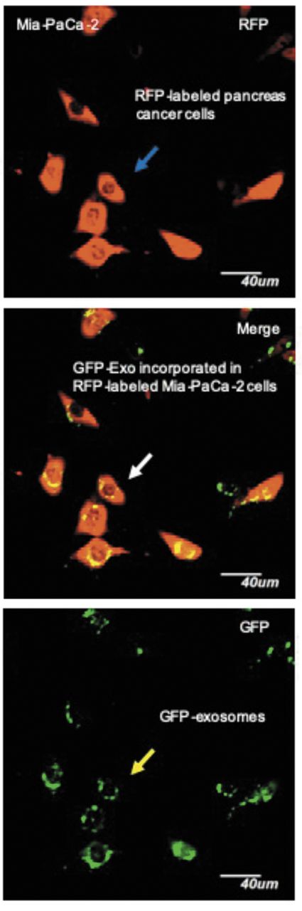
RFP expressing Mia-PaCa-2 cells were transduced with pCT-CD63-GFP for tracking exosomes. Blue arrow indicates RFP-labeled pancreatic cancer cells. Yellow arrow indicates GFP-exosomes. White arrow indicates GFP-exosome incorporated in RFP-labeled Mia-PaCa-2 cells. (Bar=40 μm).

Figure 2.
  • Download figure
  • Open in new tab
  • Download powerpoint
Figure 2.

Pancreatic cancer experimental metastases model established with red fluorescent protein (RFP) expressing Mia-PaCa-2 cells with green fluorescent protein (GFP) expressing exosomes. Primary tumor in spleen (white arrow) and metastatic tumors in liver (blue arrows). No metastases in lung.

Further experiments are needed to determine how tumor-derived exosomes promote or suppress metastases, and characterize how macrophages are educated by tumor-derived exosomes.

Figure 3.
  • Download figure
  • Open in new tab
  • Download powerpoint
Figure 3.

Mia-PaCa-2 cells in liver metastasis contained GFP-expressing exosomes. (A) RFP-labeled Mia-Paca-2 cells producing GFP-labelled exosomes in liver metastases (yellow arrows). GFP expressing exosomes are present in metastasis (Bar=30 μm). (B) Blue arrows indicate GFP-exosomes incorporated in RFP-labeled Mia-Paca-2 cells. Yellow arrows indicate macrophages in liver containing tumor-derived exosomes (Bar=30 μm).

Figure 4.
  • Download figure
  • Open in new tab
  • Download powerpoint
Figure 4.

Distribution of tumor-derived exosomes. Yellow arrows indicate macrophages in lung (A) (Bar=30 μm) and bone marrow (B) (Bar=50 μm) containing tumor-derived exosomes.

Footnotes

  • Authors' Contributions

    T.S., A.S., and R.M.H. conceived and planned the experiments. T.S., M.N., and A.S. carried out the experiments. T.S., M.N., A.S., and R.M.H. contributed to the interpretation of the results. All Authors provided critical feedback and helped shape the research and manuscript.

  • This article is freely accessible online.

  • Conflicts of Interest

    None of the Authors have any conflicts of interest with regard to this study.

  • Received May 14, 2019.
  • Revision received June 14, 2019.
  • Accepted June 18, 2019.
  • Copyright© 2019, International Institute of Anticancer Research (Dr. George J. Delinasios), All rights reserved

References

  1. ↵
    1. Kowal J,
    2. Tkach M,
    3. Théry C
    : Biogenesis and secretion of exosomes. Curr Opin Cell Biol 29: 116-125, 2014. PMID: 24959705. DOI: 10.1016/j.ceb.2014.05.004
    OpenUrlCrossRefPubMed
  2. ↵
    1. Andreu Z,
    2. Yáñez-Mó M
    : Tetraspanins in extracellular vesicle formation and function. Front Immunol 5: 442, 2014. PMID: 25278937. DOI: 10.3389/fimmu.2014.00442
    OpenUrlCrossRefPubMed
  3. ↵
    1. Choi DS,
    2. Kim DK,
    3. Kim YK,
    4. Gho YS
    : Proteomics, transcriptomics and lipidomics of exosomes and ectosomes. Proteomics 13: 1554-1571, 2013. PMID: 23401200. DOI: 10. 1002/pmic.201200329
    OpenUrlCrossRefPubMed
    1. Thakur BK,
    2. Zhang H,
    3. Becker A,
    4. Matei I,
    5. Huang Y,
    6. Costa-Silva B,
    7. Zheng Y,
    8. Hoshino A,
    9. Brazier H,
    10. Xiang J,
    11. Williams C,
    12. Rodriguez-Barrueco R,
    13. Silva JM,
    14. Zhang W,
    15. Hearn S,
    16. Elemento O,
    17. Paknejad N,
    18. Manova-Todorova K,
    19. Welte K,
    20. Bromberg J,
    21. Peinado H,
    22. Lyden D
    : Double-stranded DNA in exosomes: a novel biomarker in cancer detection. Cell Res 24: 766-769, 2014. PMID: 24710597. DOI: 10.1038/cr.2014.44
    OpenUrlCrossRefPubMed
  4. ↵
    1. Valadi H,
    2. Ekström K,
    3. Bossios A,
    4. Sjöstrand M,
    5. Lee JJ,
    6. Lötvall JO
    : Exosome-mediated transfer of mRNAs and microRNAs is a novel mechanism of genetic exchange between cells. Nat Cell Biol 9: 654-659, 2007. PMID: 17486113. DOI: 10.1038/ncb1596
    OpenUrlCrossRefPubMed
  5. ↵
    1. Becker A,
    2. Thakur BK,
    3. Weiss JM,
    4. Kim HS,
    5. Peinado H,
    6. Lyden D
    : Extracellular vesicles in cancer: cell-to-cell mediators of metastasis. Cancer Cell 30: 836-848, 2016. PMID: 27960084. DOI: 10.1016/j.ccell.2016.10.009
    OpenUrlCrossRefPubMed
    1. Peinado H,
    2. Lavotshkin S,
    3. Lyden D
    : The secreted factors responsible for pre-metastatic niche formation: Old sayings and new thoughts. Semin. Cancer Biol 21: 139-146, 2011. PMID: 21251983. DOI: 10.1016/j.semcancer.2011.01.002
    OpenUrl
  6. ↵
    1. Kahlert C,
    2. Kalluri R
    : Exosomes in tumor microenvironment influence cancer progression and metastasis. J Mol Med 91: 431-437, 2013. PMID: 23519402. DOI: 10.1007/s00109-013-1020-6
    OpenUrlCrossRefPubMed
  7. ↵
    1. Umezu T,
    2. Ohyashiki K,
    3. Kuroda M,
    4. Ohyashiki JH
    : Leukemia cell to endothelial cell communication via exosomal miRNAs. Oncogene 32: 2747-2755, 2013. PMID: 22797057. DOI: 10.1038/onc.2012.295
    OpenUrlCrossRefPubMed
    1. Luga V,
    2. Zhang L,
    3. Viloria-Petit A.M,
    4. Ogunjimi AA,
    5. Inanlou MR,
    6. Chiu E,
    7. Buchanan M,
    8. Hosein AN,
    9. Basik M,
    10. Wrana JL
    : Exosomes mediate stromal mobilization of autocrine Wnt-PCP signaling in breast cancer cell migration. Cell 151: 1542-1556, 2012. PMID: 23260141. DOI: 10.1016/j.cell.2012.11.024
    OpenUrlCrossRefPubMed
    1. Donnarumma E,
    2. Fiore D,
    3. Nappa M,
    4. Roscigno G,
    5. Adamo A,
    6. Iaboni M,
    7. Russo V,
    8. Affinito A,
    9. Puoti I,
    10. Quintavalle C,
    11. Rienzo A,
    12. Piscuoglio S,
    13. Thomas R,
    14. Condorelli G
    : Cancer-associated fibroblasts release exosomal microRNAs that dictate an aggressive phenotype in breast cancer. Oncotarget 8: 19592-19608, 2017. PMID: 28121625. DOI: 10.18632/oncotarget. 14752
    OpenUrlCrossRefPubMed
  8. ↵
    1. Peinado H,
    2. Alečković M,
    3. Lavotshkin S,
    4. Matei I,
    5. Costa-Silva B,
    6. Moreno-Bueno G,
    7. Hergueta-Redondo M,
    8. Williams C,
    9. García-Santos G,
    10. Ghajar CM,
    11. et al
    : Melanoma exosomes educate bone marrow progenitor cells toward a pro-metastatic phenotype through MET. Nat Med 18: 883-891, 2012. PMID: 22635005. DOI: 10.1038/nm.2753
    OpenUrlCrossRefPubMed
  9. ↵
    1. Suetsugu A,
    2. Honma K,
    3. Saji S,
    4. Moriwaki H,
    5. Hoffman RM
    : Imaging exosome transfer from breast cancer cells to stroma at metastatic sites in orthotopic nude-mouse models. Adv Drug Deliv Rev 65: 383-390, 2013. PMID: 22921594. DOI: 10.1016/j.addr.2012.08.007
    OpenUrlCrossRefPubMed
  10. ↵
    1. Wynn TA,
    2. Chawla A,
    3. Pollard JW
    : Macrophage biology in development, homeostasis and disease. Nature 496: 445-455, 2013. PMID: 23619691. DOI: 10.1038/nature12034
    OpenUrlCrossRefPubMed
  11. ↵
    1. Martinez FO,
    2. Sica A,
    3. Mantovani A,
    4. Locati M
    : Macrophage activation and polarization. Front Biosci 13: 453-461, 2008. PMID: 17981560.
    OpenUrlCrossRefPubMed
    1. Xue J,
    2. Schmidt SV,
    3. Sander J,
    4. Draffehn A,
    5. Krebs W,
    6. Quester I,
    7. De Nardo D,
    8. Gohel TD,
    9. Emde M,
    10. Schmidleithner L,
    11. Ganesan H,
    12. Nino-Castro A,
    13. Mallmann MR,
    14. Labzin L,
    15. Theis H,
    16. Kraut M,
    17. Beyer M,
    18. Latz E,
    19. Freeman TC,
    20. Ulas T,
    21. Schultze JL
    : Transcriptome-based network analysis reveals a spectrum model of human macrophage activation. Immunity 40: 274-288, 2014. PMID: 24530056. DOI: 10.1016/j.immuni.2014.01.006
    OpenUrlCrossRefPubMed
  12. ↵
    1. Mantovani A,
    2. Sica A,
    3. Sozzani S,
    4. Allavena P,
    5. Vecchi A,
    6. Locati M
    : The chemokine system in diverse forms of macrophage activation and polarization. Trends Immunol 25: 677-686, 2004. PMID: 15530839. DOI: 10.1016/j.it.2004.09.015
    OpenUrlCrossRefPubMed
  13. ↵
    1. Quatromoni JG,
    2. Eruslanov E
    : Tumor-associated macrophages: function, phenotype, and link to prognosis in human lung cancer. Am J Transl Res 4: 376-389, 2012. PMID: 23145206.
    OpenUrlPubMed
  14. ↵
    1. Katz MH,
    2. Takimoto S,
    3. Spivack D,
    4. Moossa AR,
    5. Hoffman RM,
    6. Bouvet M
    : An imageable highly metastatic orthotopic red fluorescent protein model of pancreatic cancer. Clin Exp Metastasis 21: 7-12, 2004. PMID: 15065597.
    OpenUrlCrossRefPubMed
  15. ↵
    1. Garcia NA,
    2. Ontoria-Oviedo I,
    3. González-King H,
    4. Diez-Juan A,
    5. Sepúlveda P
    : Glucose starvation in cardiomyocytes enhances exosome secretion and promotes angiogenesis in endothelial cells. PLoS One 10: e0138849, 2015. PMID: 26393803. DOI: 10.1371/journal.pone.0138849
    OpenUrlCrossRefPubMed
  16. ↵
    1. Hoshino A,
    2. Costa-Silva B,
    3. Shen TL,
    4. Rodrigues G,
    5. Hashimoto A,
    6. Tesic Mark M,
    7. Molina H,
    8. Kohsaka S,
    9. Di Giannatale A,
    10. Ceder S,
    11. Singh S,
    12. Williams C,
    13. Soplop N,
    14. Uryu K,
    15. Pharmer L,
    16. King T,
    17. Bojmar L,
    18. Davies AE,
    19. Ararso Y,
    20. Zhang T,
    21. Zhang H,
    22. Hernandez J,
    23. Weiss JM,
    24. Dumont-Cole VD,
    25. Kramer K,
    26. Wexler LH,
    27. Narendran A,
    28. Schwartz GK,
    29. Healey JH,
    30. Sandstrom P,
    31. Labori KJ,
    32. Kure EH,
    33. Grandgenett PM,
    34. Hollingsworth MA,
    35. de Sousa M,
    36. Kaur S,
    37. Jain M,
    38. Mallya K,
    39. Batra SK,
    40. Jarnagin WR,
    41. Brady MS,
    42. Fodstad O,
    43. Muller V,
    44. Pantel K,
    45. Minn AJ,
    46. Bissell MJ,
    47. Garcia BA,
    48. Kang Y,
    49. Rajasekhar VK,
    50. Ghajar CM,
    51. Matei I,
    52. Peinado H,
    53. Bromberg J,
    54. Lyden D
    : Tumour exosome integrins determine organotropic metastasis. Nature 527: 329-335, 2015. PMID: 26524530. DOI: 10.1038/nature15756
    OpenUrlCrossRefPubMed
  17. ↵
    1. Xiang X,
    2. Poliakov A,
    3. Liu C,
    4. Liu Y,
    5. Deng Z,
    6. Wang J,
    7. Cheng Z,
    8. Shah SV,
    9. Wang GJ,
    10. Zhang L,
    11. Grizzle WE,
    12. Mobley J,
    13. Zhang HG
    : Induction of myeloid-derived suppressor cells by tumor exosomes. Int J Cancer 124: 2621-2633, 2009. PMID: 19235923. DOI: 10.1002/ijc.24249
    OpenUrlCrossRefPubMed
  18. ↵
    1. Wen SW,
    2. Sceneay J,
    3. Lima LG,
    4. Wong CS,
    5. Becker M,
    6. Krumeich S,
    7. Lobb RJ,
    8. Castillo V,
    9. Wong KN,
    10. Ellis S,
    11. Parker BS,
    12. Möller A
    : The biodistribution and immune suppressive effects of breast cancer–derived exosomes. Cancer Res 76: 6816-6827, 2016. PMID: 27760789. DOI: 10.1158/0008-5472.CAN-16-0868
    OpenUrlAbstract/FREE Full Text
  19. ↵
    1. Plebanek MP,
    2. Angeloni NL,
    3. Vinokour E,
    4. Li J,
    5. Henkin A,
    6. Martinez-Marin D,
    7. Filleur S,
    8. Bhowmick R,
    9. Henkin J,
    10. Miller SD,
    11. Ifergan I,
    12. Lee Y,
    13. Osman I,
    14. Thaxton CS,
    15. Volpert OV
    : Pre-metastatic cancer exosomes induce immune surveillance by patrolling monocytes at the metastatic niche. Nat Commun 8: 1319, 2017. PMID: 29105655. DOI: 10.1038/s41467-017-01433-3
    OpenUrlCrossRefPubMed
  20. ↵
    1. Nagaraj S,
    2. Gabrilovich DI
    : Myeloid-derived suppressor cells. Adv Exp Med Biol 601: 213-223, 2007. PMID: 17713008. DOI: 10.1007/978-0-387-72005-0_22
    OpenUrlCrossRefPubMed
  21. ↵
    1. Wang J,
    2. De Veirman K,
    3. De Beule N,
    4. Maes K,
    5. De Bruyne E,
    6. Van Valckenborgh E,
    7. Vanderkerken K,
    8. Menu E
    : The bone marrow microenvironment enhances multiple myeloma progression by exosome-mediated activation of myeloid-derived suppressor cells. Oncotarget 6: 43992-44004, 2015. PMID: 26556857. DOI: 10.18632/oncotarget.6083
    OpenUrl
PreviousNext
Back to top

In this issue

Anticancer Research: 39 (8)
Anticancer Research
Vol. 39, Issue 8
August 2019
  • Table of Contents
  • Table of Contents (PDF)
  • Index by author
  • Back Matter (PDF)
  • Ed Board (PDF)
  • Front Matter (PDF)
Print
Download PDF
Article Alerts
Sign In to Email Alerts with your Email Address
Email Article

Thank you for your interest in spreading the word on Anticancer Research.

NOTE: We only request your email address so that the person you are recommending the page to knows that you wanted them to see it, and that it is not junk mail. We do not capture any email address.

Enter multiple addresses on separate lines or separate them with commas.
Color-coded Imaging of the Fate of Cancer-cell-derived Exosomes During Pancreatic Cancer Metastases in a Nude-mouse Model
(Your Name) has sent you a message from Anticancer Research
(Your Name) thought you would like to see the Anticancer Research web site.
CAPTCHA
This question is for testing whether or not you are a human visitor and to prevent automated spam submissions.
3 + 13 =
Solve this simple math problem and enter the result. E.g. for 1+3, enter 4.
Citation Tools
Color-coded Imaging of the Fate of Cancer-cell-derived Exosomes During Pancreatic Cancer Metastases in a Nude-mouse Model
TOMOYUKI SATAKE, ATSUSHI SUETSUGU, MIKI NAKAMURA, TAKAHIRO KUNISADA, SHIGETOYO SAJI, HISATAKA MORIWAKI, MASAHITO SHIMIZU, ROBERT M. HOFFMAN
Anticancer Research Aug 2019, 39 (8) 4055-4060; DOI: 10.21873/anticanres.13561

Citation Manager Formats

  • BibTeX
  • Bookends
  • EasyBib
  • EndNote (tagged)
  • EndNote 8 (xml)
  • Medlars
  • Mendeley
  • Papers
  • RefWorks Tagged
  • Ref Manager
  • RIS
  • Zotero
Reprints and Permissions
Share
Color-coded Imaging of the Fate of Cancer-cell-derived Exosomes During Pancreatic Cancer Metastases in a Nude-mouse Model
TOMOYUKI SATAKE, ATSUSHI SUETSUGU, MIKI NAKAMURA, TAKAHIRO KUNISADA, SHIGETOYO SAJI, HISATAKA MORIWAKI, MASAHITO SHIMIZU, ROBERT M. HOFFMAN
Anticancer Research Aug 2019, 39 (8) 4055-4060; DOI: 10.21873/anticanres.13561
del.icio.us logo Digg logo Reddit logo Twitter logo CiteULike logo Facebook logo Google logo Mendeley logo
  • Tweet Widget
  • Facebook Like
  • Google Plus One

Jump to section

  • Article
    • Abstract
    • Materials and Methods
    • Results and Discussion
    • Footnotes
    • References
  • Figures & Data
  • Info & Metrics
  • PDF

Related Articles

  • No related articles found.
  • PubMed
  • Google Scholar

Cited By...

  • Exosome Transfer Between Pancreatic-cancer Cells Is Associated With Metastasis in a Nude-mouse Model
  • Google Scholar

More in this TOC Section

  • 5-Azacytidine (5-aza) Induces p53-associated Cell Death Through Inhibition of DNA Methyltransferase Activity in Hep3B and HT-29 Cells
  • Prognostic Value of WNT1, NOTCH1, PDGFRβ, and CXCR4 in Oral Squamous Cell Carcinoma
  • Hypoxia-adapted Multiple Myeloma Stem Cells Resist γδ-T-Cell-mediated Killing by Modulating the Mevalonate Pathway
Show more Experimental Studies

Similar Articles

Keywords

  • pancreatic cancer
  • exosomes
  • tumor-derived exosomes
  • macrophage
  • green fluorescent protein
  • red fluorescent protein
  • color-coded imaging
  • nude mice
Anticancer Research

© 2023 Anticancer Research

Powered by HighWire