
Main menu

  • Home
  • Current Issue
  • Archive
  • Info for
    • Authors
    • Subscribers
    • Advertisers
    • Editorial Board
  • Other Publications
    • In Vivo
    • Cancer Genomics & Proteomics
    • Cancer Diagnosis & Prognosis
  • More
    • IIAR
    • Conferences
    • 2008 Nobel Laureates
  • About Us
    • General Policy
    • Contact
  • Other Publications
    • Anticancer Research
    • In Vivo
    • Cancer Genomics & Proteomics

User menu

  • Register
  • Subscribe
  • My alerts
  • Log in
  • My Cart

Search

  • Advanced search
Anticancer Research
  • Other Publications
    • Anticancer Research
    • In Vivo
    • Cancer Genomics & Proteomics
  • Register
  • Subscribe
  • My alerts
  • Log in
  • My Cart
Anticancer Research

Advanced Search

  • Home
  • Current Issue
  • Archive
  • Info for
    • Authors
    • Subscribers
    • Advertisers
    • Editorial Board
  • Other Publications
    • In Vivo
    • Cancer Genomics & Proteomics
    • Cancer Diagnosis & Prognosis
  • More
    • IIAR
    • Conferences
    • 2008 Nobel Laureates
  • About Us
    • General Policy
    • Contact
  • Visit us on Facebook
  • Follow us on Linkedin
Research ArticleExperimental Studies

Nuclear Accumulation of β-Catenin in Cancer Stem Cell Radioresistance and Stemness in Human Colon Cancer

HIDEKAZU TANAKA, MASAYA KAWAGUCHI, SHINICHI SHODA, TOSHIHARU MIYOSHI, RYOTA IWASAKI, FUMINORI HYODO, TAKASHI MORI, AKIRA HARA, HIROYUKI TOMITA and MASAYUKI MATSUO
Anticancer Research December 2019, 39 (12) 6575-6583; DOI: https://doi.org/10.21873/anticanres.13873
HIDEKAZU TANAKA
1Department of Radiology, Gifu University Graduate School of Medicine, Gifu, Japan
2Department of Radiation Oncology, Yamaguchi University Graduate School of Medicine, Ube, Japan
  • Find this author on Google Scholar
  • Find this author on PubMed
  • Search for this author on this site
MASAYA KAWAGUCHI
1Department of Radiology, Gifu University Graduate School of Medicine, Gifu, Japan
3Department of Tumor Pathology, Gifu University Graduate School of Medicine, Gifu, Japan
  • Find this author on Google Scholar
  • Find this author on PubMed
  • Search for this author on this site
SHINICHI SHODA
1Department of Radiology, Gifu University Graduate School of Medicine, Gifu, Japan
4Department of Radiology Service, Gifu University Hospital, Gifu, Japan
  • Find this author on Google Scholar
  • Find this author on PubMed
  • Search for this author on this site
TOSHIHARU MIYOSHI
4Department of Radiology Service, Gifu University Hospital, Gifu, Japan
  • Find this author on Google Scholar
  • Find this author on PubMed
  • Search for this author on this site
RYOTA IWASAKI
5Animal Medical Center, Gifu University, Gifu, Japan
  • Find this author on Google Scholar
  • Find this author on PubMed
  • Search for this author on this site
FUMINORI HYODO
1Department of Radiology, Gifu University Graduate School of Medicine, Gifu, Japan
6Department of Frontier Science for Imaging, Gifu University Graduate School of Medicine, Gifu, Japan
  • Find this author on Google Scholar
  • Find this author on PubMed
  • Search for this author on this site
TAKASHI MORI
7Department of Veterinary Clinical Oncology, Faculty of Applied Biological Sciences, Gifu University, Gifu, Japan
8Center for Highly Advanced Integration of Nano and Life Science, Gifu University, Gifu, Japan
  • Find this author on Google Scholar
  • Find this author on PubMed
  • Search for this author on this site
AKIRA HARA
3Department of Tumor Pathology, Gifu University Graduate School of Medicine, Gifu, Japan
  • Find this author on Google Scholar
  • Find this author on PubMed
  • Search for this author on this site
HIROYUKI TOMITA
3Department of Tumor Pathology, Gifu University Graduate School of Medicine, Gifu, Japan
  • Find this author on Google Scholar
  • Find this author on PubMed
  • Search for this author on this site
  • For correspondence: h_tomita@gifu-u.ac.jp
MASAYUKI MATSUO
1Department of Radiology, Gifu University Graduate School of Medicine, Gifu, Japan
  • Find this author on Google Scholar
  • Find this author on PubMed
  • Search for this author on this site
  • Article
  • Figures & Data
  • Info & Metrics
  • PDF
Loading

Abstract

Background/Aim: The aim of this study was to examine whether the Wnt/β-catenin signal activation is a cause of radioresistance in colon cancer by assessing the β-catenin localization and its correlation with cancer stem cells (CSCs). Materials and Methods: The nuclear levels of β-catenin, the hallmark of Wnt activation, were analyzed in HCT116 and SW480 cells by immunohistochemistry, before and after irradiation. Further, we assessed CSC populations by staining for aldehyde dehydrogenase-1 (ALDH1) and CD44. Results: β-catenin was localized predominantly in the nucleus and plasma membrane in SW480 and HCT116 cells, respectively. Compared to HCT116 cells, SW480 cells displayed higher Wnt activation. At 24 h after irradiation, most of the DSBs in SW480 cells were repaired, but were still present in HCT116 cells. Additionally, compared to HCT116 cells, a significantly higher proportion of SW480 cells were ALDH1- and CD44-positive. Conclusion: Colon cancers with nuclear β-catenin accumulation demonstrated greater radio-resistance with a higher number of CSCs.

  • Wnt/β-catenin signaling pathway
  • cancer stem cell
  • radioresistance
  • colorectal cancer
  • β-catenin

There are two Wnt/β-catenin signaling pathways: the canonical pathway and the non-canonical pathway (1, 2). β-catenin is the key molecule in the canonical Wnt pathway. In Wnt-off state, β-catenin in the cytoplasm is within a protein complex composed of adenomatous polyposis coli (APC), Axin, glycogen synthase kinase 3β (GSK3β), and casein kinase 1α (CK1α), and phosphorylated by GSK3β and CK1α. Then, β-catenin is ubiquitinated and targeted for destruction by the proteasome. As a result, the amount of β-catenin is low. Conversely, in the Wnt-on state, Wnt ligands bind to the receptor complex composed of a Frizzled (Fz) receptor and low-density lipoprotein receptor-related proteins (LRP5/6). This receptor recruits Disheveled (Dvl) to the plasma membrane. Dvl forms a complex with Axin, Fz, and LRP5/6 and acts as a direct competitive inhibitor of GSK3β. Thus, β-catenin escapes phosphorylation and accumulates in the cytoplasm. The stabilized β-catenin translocates into the nucleus, where it serves as a transcriptional co-activator and influences gene transcription. Thus, β-catenin localization to the nucleus reflects Wnt activity (3-5).

The canonical Wnt/β-catenin signaling pathway plays an important role in cell proliferation, growth, and differentiation (6, 7). Mutations in Wnt signaling pathway components cause many diseases, including cancer. Furthermore, dysregulation of the Wnt signaling pathway is associated with the expansion of stem cell and progenitor cell lineages and is also highly associated with carcinogenesis (8-10). Most colorectal cancer (CRC) cases are caused by mutations in Wnt signaling pathway components (11-13). Originally, a mutation in the APC gene was associated with familial adenomatous polyposis (FAP). Approximately 1% of FAP cases progress to CRC (14, 15). Furthermore, 85% of sporadic intestinal neoplasia cases have mutations in APC, whereas activating mutations in β-catenin are found in approximately 50% of CRC tumors without APC mutations (16, 17). The Wnt signaling pathway is related to both carcinogenesis and treatment resistance (18). Several reports have evaluated the correlation between the Wnt signaling pathway and resistance to chemotherapy (19, 20). Several attempts to discover new drugs targeting the Wnt signaling pathway and to improve chemo-resistance have been made (21-23). Although the relationship between the Wnt signaling pathway and chemoresistance has been well studied, their relationship with radioresistance remains unclear.

The Wnt signaling pathway is responsible for designating cancer stem cells (CSCs) (24). The nuclear localization and subsequent activation of T cell factor (TCF) transcription is an important process in CSCs (25). CD44 is a multifunctional cell surface adhesion protein that reacts with extracellular matrix components. Increased CD44 expression leads to enhanced CSC properties in colon cancer cells (26, 27). There are also reports showing that the combination of CD44 and CD133 is correlated with features of CSCs (28). CD44 helps to facilitate cell-cell and cell-matrix interactions through its affinity for hyaluronic acid; it is also involved in cell adhesion and the assembly of growth factors on the cell surface (29). Increased resistance to radiation therapy has been reported in CD44high cells compared to CD44low cells (29-31).

The aldehyde dehydrogenase (ALDH) family of enzymes are cytosolic isoenzymes responsible for oxidizing intracellular aldehydes and retinol to retinoic acid in early stem cell differentiation (32). ALDH1 is expressed in various normal stem and progenitor cells and contributes to cell proliferation and differentiation (27, 33). Some reports have shown that nuclear localization of ALDH1A1 is associated with a dismal prognosis in clinical CRC cases (34, 35). ALDH1 is also correlated with radioresistance (31, 36, 37).

The epithelial cell adhesion molecule (EpCAM) is also known as an epithelial-specific antigen. EpCAM was initially discovered as a predominant antigen in human colon carcinoma; it is a transmembrane glycoprotein that is highly expressed in rapidly proliferating carcinomas (38). Recent data have suggested a more multifaceted role of EpCAM in not only cell-cell adhesion but also cell signaling, migration, proliferation, and differentiation (39). EpCAM is also reported to be a putative CSC marker (40). EpCAM expression is also correlated with radioresistance (41).

This study focused on the localization of β-catenin and analyzed its correlation with radioresistance and cancer stem-like cells.

Materials and Methods

Cell culture. Two human CRC cell lines, HCT116 and SW480 (American Type Culture Collection, Manassas, VA, USA), were maintained in culture using standard protocols. The HCT116 cell line was cultured in McCoy's 5A medium (American Type Culture Collection, Manassas, VA, USA) supplemented with 10% fetal bovine serum and 1% penicillin-streptomycin. The SW480 cell line was cultured in Leibovitz's L-15 medium (American Type Culture Collection, Manassas, VA, USA) supplemented with 10% fetal bovine serum and 1% penicillin-streptomycin.

Irradiation. Photon irradiation was delivered by a Primus system (Siemens Healthcare, Malvern, PA, USA). The photon energy was 4 MV and the dose rate was 250 monitor units per min. Cells were plated into dishes at approximately 50% confluency. Twenty-four h after plating, a 1-cm bolus was put on dishes and the cells in dishes were irradiated.

Clonogenic assay. Each cell line was irradiated in dishes with 0 (control), 2, 4, and 8 Gy, respectively. Subsequently, 100 and 1,000 cells were re-plated. After 14 days of cultivation, the cells were rinsed with phosphate buffer saline. A 3-ml of 6.0% glutaraldehyde and 0.5% crystal violet was then added and incubated for 30 min. The glutaraldehyde crystal violet mixture was then rinsed with water and the plate was allowed to dry in normal air at room temperature. Finally, the colonies were counted.

Cell block. The cell samples were fixed for one h by mixing with 10% buffered formalin. After one h, the samples were centrifuged at 1000 x g for 15 min. The supernatant was discarded and a further fresh 100% and then 70% alcohol was added to the sediment and incubated for one day. On the following day, the sediment containing the cells was processed along with other routine histopathological specimens.

IHC staining. A labeled streptavidin-biotin (LSAB) method was used for immunohistochemical (IHC) staining of deparaffinized 3-μm-thick sections. The sections were placed in citrate buffer (10 mmol-1, pH 6.0) and then autoclaved at a chamber temperature of 121°C for 1 min to retrieve the antigen. They were then rinsed and blocked in 3% hydrogen peroxide. Non-specific binding sites were blocked in 0.01 M phosphate-buffered saline (pH 7.4) containing 2% bovine serum albumin (Wako Pure Chemical, Osaka, Japan) for 40 min. The primary antibodies used were purified mouse anti-β-catenin [mouse immunoglobulin (IgG), 1:100, BD bioscience, CA, USA], anti-gamma H2A.X (phospho S139) antibody (rabbit IgG, 1:100, abcam, CA, UK), anti-ALDH1A1 antibody (ab52492) (rabbit IgG, 1:500, abcam, CA, UK), purified mouse anti-human CD44 (550392) (mouse IgG, 1:25, BD Pharmingen, NJ, USA), and anti-EpCAM antibody (ab71916) (rabbit IgG, 1:50, abcam, Cambridge, UK).

Results

Localization of β-catenin in colorectal cancer (CRC) cell lines. To investigate the localization of β-catenin, the hallmark of Wnt activation, in CRC cell lines, we performed IHC with β-catenin antibody in SW480 and HCT116 CRC cell lines, which have similar protein expression levels in western blot analysis (42, 43). HCT116 cells express wild-type APC and mutant β-catenin, while SW480 cells express mutant APC. SW480 cells had one mutant allele with loss of the other allele (44). β-catenin expression was localized in the nucleus and cytoplasm of SW480 cells and in the membrane of HCT116 cells (Figure 1). The results indicated that SW480 cells had a different Wnt/β-catenin signaling pathway activation compared to HCT116 cells.

Figure 1.
  • Download figure
  • Open in new tab
  • Download powerpoint
Figure 1.

Expression of β-catenin in HCT116 and SW480 cells. In SW480 cells, β-catenin predominantly localized in the nucleus, whereas in HCT116 cells, it was primarily found in the plasma membrane. Arrow indicates cellular membrane. Arrowhead indicates nucleus. Scale bars: 50 μm.

Comparison of DNA repair rates after irradiation. To assess the level of DNA double-strand break (DSB) repair after irradiation, expression of γH2AX, a surrogate marker of DSBs, was evaluated in both SW480 and HCT116 cell lines after 4 Gy irradiation. The number of foci was significantly lower in SW480 cells than that in HCT116 cells at 24 h after irradiation (Figure 2). These results suggested that nuclear β-catenin, the hallmark of Wnt activation, is associated with DSB repair at 24 h after irradiation.

Sensitivity of CRC cells to X-rays assessed by clonogenic assay. To measure the survival of both SW480 and HCT116 cells after irradiation, clonogenic assays were performed following 0, 2, 4, and 8 Gy irradiation (Figure 3). Fourteen days after irradiation, colonies on each dish were counted. At each irradiation dose, the ratio of colony numbers to the control group (0 Gy) was calculated and expressed logarithmically. Figure 3 shows the results of clonogenic assays starting from 1,000 cells. There were significantly more colonies in SW480 cells than in HCT116 cells at 2, 4, and 8 Gy (p=0.0316, <0.0001, and 0.0002, respectively). Similarly, starting from 100 cells, the clonogenic assay showed significantly higher numbers of colonies in SW480 cells than those in HCT116 cells at 2, 4, and 8 Gy (data not shown). No colony formation was observed in the dishes where 100 HCT116 cells plated after 8 Gy irradiation. These results indicate that SW480 cells were more radioresistant compared to HCT116 cells. Taken together, Wnt-activated CRC cells may be radioresistant relatively to non-activated CRC cells.

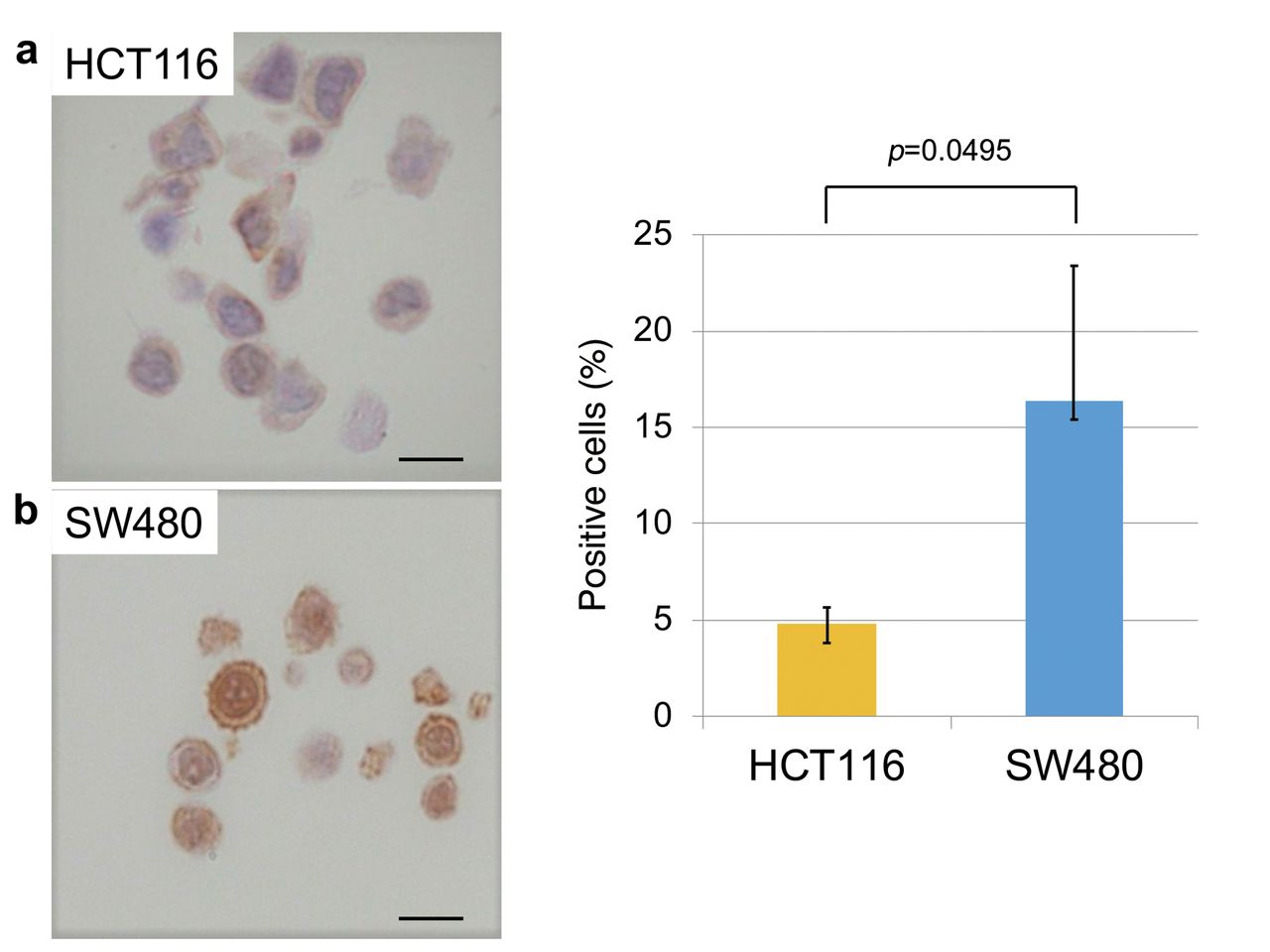
Expression of CSC markers in Wnt-activated and non-activated CRC cells. Next, to evaluate the correlation between putative CSC markers and nuclear β-catenin, IHC for ALDH1, CD44, and EpCAM was performed in each cell line. SW480 cells, which have high β-catenin accumulation in the nucleus, had a significantly higher proportion of ALDH1-positive cells compared to HCT116 cells, which showed almost exclusive membranous staining of β-catenin (Figure 4). Similar to ALDH1 expression, the proportion of CD44-positive SW480 cells was also significantly higher than that in HCT116 cells (Figure 5). In contrast, the proportion of EpCAM-positive SW480 cells was significantly lower than that in HCT116 cells (Figure 6). These results suggest that ALDH1 and CD44 expression may be radioresistant markers as well as nuclear β-catenin expression in CRC cells.

Discussion

When the Wnt/β-catenin signaling pathway is active, β-catenin is not degraded; it accumulates and translocates into the nucleus. Thus, increased β-catenin levels in the nucleus indicate Wnt-active status. In contrast, when Wnt is inactive, β-catenin is degraded by the proteasome and the levels of β-catenin in the cytoplasm are decreased. Translocation into the nucleus does not occur because the cytoplasmic β-catenin level is not high. Therefore, higher β-catenin levels in the plasma membrane rather than the nucleus indicates inactive Wnt. In SW480 cells, β-catenin was predominantly observed in the nucleus, whereas it was primarily observed in the plasma membrane in HCT116 cells. SW480 cells showed higher Wnt/β-catenin signaling pathway activation compared to HCT116 cells.

Figure 2.
  • Download figure
  • Open in new tab
  • Download powerpoint
Figure 2.

γH2AX expression after irradiation. (a) γH2AX foci after 4 Gy irradiation in HCT116 and SW480 cells. At 24 h after irradiation, most of the double-strand breaks (DSBs) in the SW480 cells are repaired, whereas they remain in the HCT116 cells. (b) The number of γH2AX foci in SW480 cells was significantly lower than that in HCT116 cells at 24 h after irradiation (Student's t-test; N=10). Scale bars: 10 μm.

Photon beams have an anti-tumor effect by damaging tumor DNA. The DSB levels were assessed via γH2AX, a surrogate marker of DSBs. At the early time point, there was no difference in DNA damage foci formation between HCT116 and SW480 cells. However, 24 h after irradiation, most of the DSBs in SW480 cells were repaired, but remained in HCT116 cells. The number of γH2AX foci in SW480 cells was significantly lower than that in HCT116 cells at 24 h after irradiation. Wnt signaling is related to DNA damage repair, suggesting increased repair with high Wnt activity (45). This is considered to be one of the causes of resistance to treatment. Moreover, the results of the colony formation assay showed that SW480 cells, with higher Wnt activity, formed higher number of colonies and showed resistance to irradiation compared to HCT116 cells, with lower Wnt activity. This result was similar regardless of the irradiation dose and numbers of seeding cells.

Wnt signaling is also involved in CSC control (46-48). CD44, CD133, ALDH1, Lgr5, EpCAM, etc. are putative markers of CSC in colorectal cancer (26-28, 33, 35, 40, 49). Among them, we evaluated CD44, ALDH1, and EpCAM by IHC. Koh et al. have reported that activation of gastrin by β-catenin is an early event of colon carcinogenesis that promotes tumor development (50). Wnt signaling is enhanced by the secretion of progastrin, a direct target of the oncogenic Tcf/β-catenin pathway (51, 52). Jin et al. have reported an increased proportion of CD44-positive cells following the addition of prosgastrin in a colon cancer cell line, indicating an increase in putative CSC (53). In addition, Giraud et al. have reported that decreased proportion of ALDHhigh cells by suppression of progastrin resulted in decreased tumorigenesis and glycolytic ability and restricted the self-renewal ability of CSC (54). Thus, there may be a relationship between the Wnt signaling and CSC. In this study, SW480 cells with high Wnt activity had a significantly higher proportion of CD44-positive cells than HCT116 cells. A similar result was observed for ALDH1. SW480 cells with high Wnt activity had a significantly lower proportion of EpCAM-positive cells than HCT116 cells. While there are reports that EpCAM may be a marker of CSC, this finding remains controversial. EpCAM expression has been linked to poorer disease-free and overall survival in breast cancer (55). Gosens et al. have observed strong EpCAM staining in the tumor center and a progressive loss at the tumor front associated with poor local and overall recurrence-free survival in colorectal cancer (56). Lugli et al. have reported a correlation between the loss of membranous EpCAM and tumor invasiveness and progression (57). EpCAM overexpression was related to lymph node classification and tumor border configuration (pushing or infiltration) but not to local recurrence. These results suggest that the loss of EpCAM at the tumor border results in reduced adhesion and induces the epithelial-mesenchymal transition, resulting in lymph node and distant metastasis. Pastushenko et al. have analyzed cells that did not express EpCAM and isolated cell populations based on their expression of the mesenchymal-cell receptor proteins CD51, CD61 and CD106. The authors identified two cell populations that were the most likely to metastasize; EpCAM(−), CD106(−), CD51(−), CD61(−) and EpCAM(−), CD106(+), CD51(−), CD61(−) (58, 59).These findings suggest that EpCAM affects metastasis and prognosis but has little direct effect on radiosensitivity.

Figure 3.
  • Download figure
  • Open in new tab
  • Download powerpoint
Figure 3.

Colony formation after irradiation in SW480 and HCT116 cells. (a) Colony formation assay in HCT116 and SW480 cells. (b) Comparison of radioresistance between SW480 and HCT116 cells (Student's t-test; N=3). Scale bars: 10 μm.

Figure 4.
  • Download figure
  • Open in new tab
  • Download powerpoint
Figure 4.

ALDH1 expression in HTC116 and SW480 cells. (a) Expression of ALDH1 in HTC116 and SW480 cells. (b) SW480 cells had a significantly higher proportion of ALDH1-positive cells compared to HCT116 cells (Student's t-test; N=5). Error bars represent the standard deviation of the mean value. Scale bars: 10 μm.

Figure 5.
  • Download figure
  • Open in new tab
  • Download powerpoint
Figure 5.

CD44 expression in HTC116 and SW480 cells. (a) Expression of CD44 in HTC116 and SW480 cells. (b) SW480 cells had a significantly higher proportion of CD44-positive cells compared to HCT116 cells (Student's t-test; N=5). Error bars represent the standard deviation of the mean value. Scale bars: 10 μm.

Figure 6.
  • Download figure
  • Open in new tab
  • Download powerpoint
Figure 6.

EpCAM expression in HCT116 and SW480 cells. Expression of EpCAM in HCT116 and SW480 cells. (b) HCT116 cells had a significantly higher proportion of EpCAM-positive cells compared to SW480 cells (Student's t-test; N=5). Error bars represent the standard deviation of the mean value. Scale bars: 10 μm.

In conclusion, nuclear accumulation of β-catenin is an indicator of active Wnt signaling. Cell lines with higher nuclear β-catenin accumulation showed resistance to radiation therapy compared to cell lines with lower accumulation, as well as a significantly higher positive proportion of putative CSC. These findings suggest that the Wnt/β-catenin signaling pathway is related to radioresistance and stemness of CSC.

Acknowledgements

The paper has been edited and rewritten by an experienced scientific editor, who has improved its grammar and stylistic expression. The Authors thank Enago, Crimson Interactive Pvt, Ltd.

Footnotes

  • ↵* These Authors contributed equally to this study.

  • Authors' Contributions

    HTan, FH, HTom, Tmor, AH, and MM conceived and designed the experiments. HTan, MK, Tmiy, FH, Htom, Tmor, AH, and MM analyzed the data. HTan, MK, SS, RI, Htom, and Tmor wrote the first draft of the manuscript. HTan, MK, SS, Tmiy, RI, FH, Htom, Tmor, AH, and MM contributed to the writing of the manuscript. HTan, MK, SS, Tmiy, RI, FH, Htom, Tmor, AH, and MM agreed with the manuscript results and conclusions. HTan, MK, SS, Tmiy, RI, FH, Htom, Tmor, AH, and MM jointly developed the structure and arguments for the paper. HTan, MK, SS, Tmiy, RI, FH, Htom, Tmor, AH, and MM made critical revisions and approved the final version. HTan, MK, SS, Tmiy, RI, FH, Htom, Tmor, AH, and MM reviewed and approved the final manuscript.

  • Conflicts of Interest

    The Authors declare that they have no competing interests regarding this study.

  • Received September 29, 2019.
  • Revision received November 16, 2019.
  • Accepted November 18, 2019.
  • Copyright© 2019, International Institute of Anticancer Research (Dr. George J. Delinasios), All rights reserved

References

  1. ↵
    1. Xiao YF,
    2. Yong X,
    3. Tang B,
    4. Qin Y,
    5. Zhang JW,
    6. Zhang D,
    7. Xie R,
    8. Yang SM
    : Notch and wnt signaling pathway in cancer: Crucial role and potential therapeutic targets (review). Int J Oncol 48: 437-449, 2016. PMID: 26648421. DOI: 10.3892/ijo.2015.3280
    OpenUrl
  2. ↵
    1. Tabatabai R,
    2. Linhares Y,
    3. Bolos D,
    4. Mita M,
    5. Mita A
    : Targeting the wnt pathway in cancer: A review of novel therapeutics. Target Oncol 12: 623-641, 2017. PMID: 28653295. DOI: 10.1007/s11523-017-0507-4
    OpenUrl
  3. ↵
    1. Wu X,
    2. Tu X,
    3. Joeng KS,
    4. Hilton MJ,
    5. Williams DA,
    6. Long F
    : Rac1 activation controls nuclear localization of beta-catenin during canonical wnt signaling. Cell 133: 340-353, 2008. PMID: 18423204. DOI: 10.1016/j.cell.2008.01.052
    OpenUrlCrossRefPubMed
    1. Zhang N,
    2. Wei P,
    3. Gong A,
    4. Chiu WT,
    5. Lee HT,
    6. Colman H,
    7. Huang H,
    8. Xue J,
    9. Liu M,
    10. Wang Y,
    11. Sawaya R,
    12. Xie K,
    13. Yung WK,
    14. Medema RH,
    15. He X,
    16. Huang S
    : Foxm1 promotes beta-catenin nuclear localization and controls wnt target-gene expression and glioma tumorigenesis. Cancer Cell 20: 427-442, 2011. PMID: 22014570. DOI: 10.1016/j.ccr.2011.08.016
    OpenUrlCrossRefPubMed
  4. ↵
    1. Kim I,
    2. Hur J,
    3. Jeong S
    : Hur represses wnt/beta-catenin-mediated transcriptional activity by promoting cytoplasmic localization of beta-catenin. Biochem Biophys Res Commun 457: 65-70, 2015. PMID: 25534855. DOI: 10.1016/j.bbrc. 2014.12.052
    OpenUrlCrossRefPubMed
  5. ↵
    1. Logan CY,
    2. Nusse R
    : The wnt signaling pathway in development and disease. Annu Rev Cell Dev Biol 20: 781-810, 2004. PMID: 15473860. DOI: 10.1146/annurev.cellbio.20.01 0403.113126
    OpenUrlCrossRefPubMed
  6. ↵
    1. Rosenbluh J,
    2. Wang X,
    3. Hahn WC
    : Genomic insights into wnt/beta-catenin signaling. Trends Pharmacol Sci 35: 103-109, 2014. PMID: 24365576. DOI: 10.1016/j.tips.2013.11.007
    OpenUrlCrossRefPubMed
  7. ↵
    1. Batlle E,
    2. Bacani J,
    3. Begthel H,
    4. Jonkheer S,
    5. Gregorieff A,
    6. van de Born M,
    7. Malats N,
    8. Sancho E,
    9. Boon E,
    10. Pawson T,
    11. Gallinger S,
    12. Pals S,
    13. Clevers H
    : Ephb receptor activity suppresses colorectal cancer progression. Nature 435: 1126-1130, 2005. PMID: 15973414. DOI: 10.1038/nature03626
    OpenUrlCrossRefPubMed
    1. Katoh M
    : Wnt/pcp signaling pathway and human cancer (review). Oncol Rep 14: 1583-1588, 2005. PMID: 16273260. DOI: 10.3892/or.14.6.1583
    OpenUrlPubMed
  8. ↵
    1. Clevers H,
    2. Nusse R
    : Wnt/beta-catenin signaling and disease. Cell 149: 1192-1205, 2012. PMID: 22682243. DOI: 10.1016/j.cell.2012.05.012
    OpenUrlCrossRefPubMed
  9. ↵
    1. Liu W,
    2. Dong X,
    3. Mai M,
    4. Seelan RS,
    5. Taniguchi K,
    6. Krishnadath KK,
    7. Halling KC,
    8. Cunningham JM,
    9. Boardman LA,
    10. Qian C,
    11. Christensen E,
    12. Schmidt SS,
    13. Roche PC,
    14. Smith DI,
    15. Thibodeau SN
    : Mutations in axin2 cause colorectal cancer with defective mismatch repair by activating beta-catenin/tcf signalling. Nat Genet 26: 146-147, 2000. PMID: 11017067. DOI: 10.1038/79859
    OpenUrlCrossRefPubMed
    1. Polakis P
    : Wnt signaling and cancer. Genes Dev 14: 1837-1851, 2000. PMID: 10921899. DOI: 10.1101/gad.14.15.1837
    OpenUrlFREE Full Text
  10. ↵
    1. Holcombe RF,
    2. Marsh JL,
    3. Waterman ML,
    4. Lin F,
    5. Milovanovic T,
    6. Truong T
    : Expression of wnt ligands and frizzled receptors in colonic mucosa and in colon carcinoma. Mol Pathol 55: 220-226, 2002. PMID: 12147710. DOI: 10.1136/mp.55.4.220
    OpenUrlAbstract/FREE Full Text
  11. ↵
    1. Moser AR,
    2. Pitot HC,
    3. Dove WF
    : A dominant mutation that predisposes to multiple intestinal neoplasia in the mouse. Science 247: 322-324, 1990. PMID: 2296722. DOI: 10.1126/science.2296722
    OpenUrlAbstract/FREE Full Text
  12. ↵
    1. Nishisho I,
    2. Nakamura Y,
    3. Miyoshi Y,
    4. Miki Y,
    5. Ando H,
    6. Horii A,
    7. Koyama K,
    8. Utsunomiya J,
    9. Baba S,
    10. Hedge P
    : Mutations of chromosome 5q21 genes in fap and colorectal cancer patients. Science 253: 665-669, 1991. PMID: 1651563. DOI: 10.1126/science.1651563
    OpenUrlAbstract/FREE Full Text
  13. ↵
    1. Kinzler KW,
    2. Vogelstein B
    : Lessons from hereditary colorectal cancer. Cell 87: 159-170, 1996. PMID: 8861899. DOI: 10.1016/s0092-8674(00)81333-1
    OpenUrlCrossRefPubMed
  14. ↵
    1. Sparks AB,
    2. Morin PJ,
    3. Vogelstein B,
    4. Kinzler KW
    : Mutational analysis of the apc/beta-catenin/tcf pathway in colorectal cancer. Cancer Res 58: 1130-1134, 1998. PMID: 9515795.
    OpenUrlAbstract/FREE Full Text
  15. ↵
    1. Huang S,
    2. Fantini D,
    3. Merrill BJ,
    4. Bagchi S,
    5. Guzman G,
    6. Raychaudhuri P
    : Ddb2 is a novel regulator of wnt signaling in colon cancer. Cancer Res 77: 6562-6575, 2017. PMID: 29021137. DOI: 10.1158/0008-5472.CAN-17-1570
    OpenUrlAbstract/FREE Full Text
  16. ↵
    1. Wickstrom M,
    2. Dyberg C,
    3. Milosevic J,
    4. Einvik C,
    5. Calero R,
    6. Sveinbjornsson B,
    7. Sanden E,
    8. Darabi A,
    9. Siesjo P,
    10. Kool M,
    11. Kogner P,
    12. Baryawno N,
    13. Johnsen JI
    : Wnt/beta-catenin pathway regulates mgmt gene expression in cancer and inhibition of wnt signalling prevents chemoresistance. Nat Commun 6: 8904, 2015. PMID: 26603103. DOI: 10.1038/ncomms9904 1
    OpenUrl
  17. ↵
    1. Chen B,
    2. Zhang D,
    3. Kuai J,
    4. Cheng M,
    5. Fang X,
    6. Li G
    : Upregulation of mir-199a/b contributes to cisplatin resistance via wnt/beta-catenin-abcg2 signaling pathway in aldha1(+) colorectal cancer stem cells. Tumour Biol 39: 1010428317715155, 2017. PMID: 28639895. DOI: 10.1177/1010428317715155
    OpenUrl
  18. ↵
    1. Fischer M,
    2. Yen WC,
    3. Kapoun AM,
    4. Wang M,
    5. O'Young G,
    6. Lewicki J,
    7. Gurney A,
    8. Hoey T
    : Anti-dll4 inhibits growth and reduces tumor-initiating cell frequency in colorectal tumors with oncogenic kras mutations. Cancer Res 71: 1520-1525, 2011. PMID: 21193546. DOI: 10.1158/0008-5472.CAN-10-2817
    OpenUrlAbstract/FREE Full Text
    1. Osada T,
    2. Chen M,
    3. Yang XY,
    4. Spasojevic I,
    5. Vandeusen JB,
    6. Hsu D,
    7. Clary BM,
    8. Clay TM,
    9. Chen W,
    10. Morse MA,
    11. Lyerly HK
    : Antihelminth compound niclosamide downregulates wnt signaling and elicits antitumor responses in tumors with activating apc mutations. Cancer Res 71: 4172-4182, 2011. PMID: 21531761. DOI: 10.1158/0008-5472.CAN-10-3978
    OpenUrlAbstract/FREE Full Text
  19. ↵
    1. Zhang Y,
    2. Li Q,
    3. Zhou D,
    4. Chen H
    : Genistein, a soya isoflavone, prevents azoxymethane-induced up-regulation of wnt/beta-catenin signalling and reduces colon pre-neoplasia in rats. Br J Nutr 109: 33-42, 2013. PMID: 22716201. DOI: 10.1017/S0007114512000876
    OpenUrlCrossRefPubMed
  20. ↵
    1. Valkenburg KC,
    2. Graveel CR,
    3. Zylstra-Diegel CR,
    4. Zhong Z,
    5. Williams BO
    : Wnt/beta-catenin signaling in normal and cancer stem cells. Cancers (Basel) 3: 2050-2079, 2011. PMID: 24212796. DOI: 10.3390/cancers3022050
    OpenUrl
  21. ↵
    1. Morgan RG,
    2. Ridsdale J,
    3. Tonks A,
    4. Darley RL
    : Factors affecting the nuclear localization of beta-catenin in normal and malignant tissue. J Cell Biochem 115: 1351-1361, 2014. PMID: 24610469. DOI: 10.1002/jcb.24803
    OpenUrlCrossRefPubMed
  22. ↵
    1. Du L,
    2. Wang H,
    3. He L,
    4. Zhang J,
    5. Ni B,
    6. Wang X,
    7. Jin H,
    8. Cahuzac N,
    9. Mehrpour M,
    10. Lu Y,
    11. Chen Q
    : Cd44 is of functional importance for colorectal cancer stem cells. Clin Cancer Res 14: 6751-6760, 2008. PMID: 18980968. DOI: 10.1158/1078-0432.CCR-08-1034
    OpenUrlAbstract/FREE Full Text
  23. ↵
    1. Hatano Y,
    2. Fukuda S,
    3. Hisamatsu K,
    4. Hirata A,
    5. Hara A,
    6. Tomita H
    : Multifaceted interpretation of colon cancer stem cells. Int J Mol Sci 18, 2017. PMID: 28678194. DOI: 10.3390/ijms18071446
  24. ↵
    1. Wang C,
    2. Xie J,
    3. Guo J,
    4. Manning HC,
    5. Gore JC,
    6. Guo N
    : Evaluation of cd44 and cd133 as cancer stem cell markers for colorectal cancer. Oncol Rep 28: 1301-1308, 2012. PMID: 22895640. DOI: 10.3892/or.2012.1951
    OpenUrlPubMed
  25. ↵
    1. Sahlberg SH,
    2. Spiegelberg D,
    3. Glimelius B,
    4. Stenerlow B,
    5. Nestor M
    : Evaluation of cancer stem cell markers cd133, cd44, cd24: Association with akt isoforms and radiation resistance in colon cancer cells. PLoS One 9: e94621, 2014. PMID: 24760019. DOI: 10.1371/journal.pone.0094621
    OpenUrlCrossRefPubMed
    1. Phillips TM,
    2. McBride WH,
    3. Pajonk F
    : The response of cd24(−/low)/cd44+ breast cancer-initiating cells to radiation. J Natl Cancer Inst 98: 1777-1785, 2006. PMID: 17179479. DOI: 10.1093/jnci/djj495
    OpenUrlCrossRefPubMed
  26. ↵
    1. Croker AK,
    2. Allan AL
    : Inhibition of aldehyde dehydrogenase (aldh) activity reduces chemotherapy and radiation resistance of stem-like aldhhicd44(+) human breast cancer cells. Breast Cancer Res Treat 133: 75-87, 2012. PMID: 21818590. DOI: 10.1007/s10549-011-1692-y
    OpenUrlCrossRefPubMed
  27. ↵
    1. Yoshida A
    : Molecular genetics of human aldehyde dehydrogenase. Pharmacogenetics 2: 139-147, 1992. PMID: 1306115 DOI: 10.1097/00008571-199208000-00001
    OpenUrlCrossRefPubMed
  28. ↵
    1. Huang EH,
    2. Hynes MJ,
    3. Zhang T,
    4. Ginestier C,
    5. Dontu G,
    6. Appelman H,
    7. Fields JZ,
    8. Wicha MS,
    9. Boman BM
    : Aldehyde dehydrogenase 1 is a marker for normal and malignant human colonic stem cells (sc) and tracks sc overpopulation during colon tumorigenesis. Cancer Res 69: 3382-3389, 2009. PMID: 19336570. DOI: 10.1158/0008-5472.CAN-08-4418
    OpenUrlAbstract/FREE Full Text
  29. ↵
    1. Kahlert C,
    2. Gaitzsch E,
    3. Steinert G,
    4. Mogler C,
    5. Herpel E,
    6. Hoffmeister M,
    7. Jansen L,
    8. Benner A,
    9. Brenner H,
    10. Chang-Claude J,
    11. Rahbari N,
    12. Schmidt T,
    13. Klupp F,
    14. Grabe N,
    15. Lahrmann B,
    16. Koch M,
    17. Halama N,
    18. Buchler M,
    19. Weitz J
    : Expression analysis of aldehyde dehydrogenase 1a1 (aldh1a1) in colon and rectal cancer in association with prognosis and response to chemotherapy. Ann Surg Oncol 19: 4193-4201, 2012. PMID: 22878609. DOI: 10.1245/s10434-012-2518-9
    OpenUrlCrossRefPubMed
  30. ↵
    1. Tomita H,
    2. Tanaka K,
    3. Tanaka T,
    4. Hara A
    : Aldehyde dehydrogenase 1a1 in stem cells and cancer. Oncotarget 7: 11018-11032, 2016. PMID: 26783961. DOI: 10.18632/oncotarget.6920
    OpenUrlCrossRefPubMed
  31. ↵
    1. Chen YC,
    2. Chen YW,
    3. Hsu HS,
    4. Tseng LM,
    5. Huang PI,
    6. Lu KH,
    7. Chen DT,
    8. Tai LK,
    9. Yung MC,
    10. Chang SC,
    11. Ku HH,
    12. Chiou SH,
    13. Lo WL
    : Aldehyde dehydrogenase 1 is a putative marker for cancer stem cells in head and neck squamous cancer. Biochem Biophys Res Commun 385: 307-313, 2009. PMID: 19450560. DOI: 10.1016/j.bbrc.2009.05.048
    OpenUrlCrossRefPubMed
  32. ↵
    1. Mihatsch J,
    2. Toulany M,
    3. Bareiss PM,
    4. Grimm S,
    5. Lengerke C,
    6. Kehlbach R,
    7. Rodemann HP
    : Selection of radioresistant tumor cells and presence of aldh1 activity in vitro. Radiother Oncol 99: 300-306, 2011. PMID: 21704411. DOI: 10.1016/j.radonc.2011.06.003
    OpenUrlPubMed
  33. ↵
    1. Ni J,
    2. Cozzi PJ,
    3. Duan W,
    4. Shigdar S,
    5. Graham PH,
    6. John KH,
    7. Li Y
    : Role of the epcam (cd326) in prostate cancer metastasis and progression. Cancer Metastasis Rev 31: 779-791, 2012. PMID: 22718399. DOI: 10.1007/s10555-012-9389-1
    OpenUrlCrossRefPubMed
  34. ↵
    1. Patriarca C,
    2. Macchi RM,
    3. Marschner AK,
    4. Mellstedt H
    : Epithelial cell adhesion molecule expression (cd326) in cancer: A short review. Cancer Treat Rev 38: 68-75, 2012. PMID: 21576002. DOI: 10.1016/j.ctrv.2011.04.002
    OpenUrlCrossRefPubMed
  35. ↵
    1. Dalerba P,
    2. Dylla SJ,
    3. Park IK,
    4. Liu R,
    5. Wang X,
    6. Cho RW,
    7. Hoey T,
    8. Gurney A,
    9. Huang EH,
    10. Simeone DM,
    11. Shelton AA,
    12. Parmiani G,
    13. Castelli C,
    14. Clarke MF
    : Phenotypic characterization of human colorectal cancer stem cells. Proc Natl Acad Sci USA 104: 10158-10163, 2007. PMID: 17548814. DOI: 10.1073/pnas. 0703478104
    OpenUrlAbstract/FREE Full Text
  36. ↵
    1. Ni J,
    2. Cozzi P,
    3. Hao J,
    4. Beretov J,
    5. Chang L,
    6. Duan W,
    7. Shigdar S,
    8. Delprado W,
    9. Graham P,
    10. Bucci J,
    11. Kearsley J,
    12. Li Y
    : Epithelial cell adhesion molecule (epcam) is associated with prostate cancer metastasis and chemo/radioresistance via the pi3k/akt/mtor signaling pathway. Int J Biochem Cell Biol 45: 2736-2748, 2013. PMID: 24076216. DOI: 10.1016/j.biocel.2013.09.008
    OpenUrlCrossRefPubMed
  37. ↵
    1. Hao L,
    2. Zhao Y,
    3. Wang Z,
    4. Yin H,
    5. Zhang X,
    6. He T,
    7. Song S,
    8. Sun S,
    9. Wang B,
    10. Li Z,
    11. Su Q
    : Expression and clinical significance of sall4 and beta-catenin in colorectal cancer. J Mol Histol 47: 117-128, 2016. PMID: 26779651. DOI: 10.1007/s10735-016-9656-5
    OpenUrl
  38. ↵
    1. Qin Y,
    2. Tang B,
    3. Hu CJ,
    4. Xiao YF,
    5. Xie R,
    6. Yong X,
    7. Wu YY,
    8. Dong H,
    9. Yang SM
    : An htert/zeb1 complex directly regulates e-cadherin to promote epithelial-to-mesenchymal transition (emt) in colorectal cancer. Oncotarget 7: 351-361, 2016. PMID: 26540342. DOI: 10.18632/oncotarget.5968
    OpenUrlCrossRefPubMed
  39. ↵
    1. El-Bahrawy M,
    2. Poulsom R,
    3. Rowan AJ,
    4. Tomlinson IT,
    5. Alison MR
    : Characterization of the e-cadherin/catenin complex in colorectal carcinoma cell lines. Int J Exp Pathol 85: 65-74, 2004. PMID: 15154912. DOI: 10.1111/j.0959-9673.2004.0371.x
    OpenUrlCrossRefPubMed
  40. ↵
    1. Karimaian A,
    2. Majidinia M,
    3. Bannazadeh Baghi H,
    4. Yousefi B
    : The crosstalk between wnt/beta-catenin signaling pathway with DNA damage response and oxidative stress: Implications in cancer therapy. DNA Repair (Amst) 51: 14-19, 2017. PMID: 28108274. DOI: 10.1016/j.dnarep.2017.01.003
    OpenUrl
  41. ↵
    1. Fodde R,
    2. Brabletz T
    : Wnt/beta-catenin signaling in cancer stemness and malignant behavior. Curr Opin Cell Biol 19: 150-158, 2007. PMID: 17306971. DOI: 10.1016/j.ceb.2007.02.007
    OpenUrlCrossRefPubMed
    1. Vermeulen L,
    2. De Sousa EMF,
    3. van der Heijden M,
    4. Cameron K,
    5. de Jong JH,
    6. Borovski T,
    7. Tuynman JB,
    8. Todaro M,
    9. Merz C,
    10. Rodermond H,
    11. Sprick MR,
    12. Kemper K,
    13. Richel DJ,
    14. Stassi G,
    15. Medema JP
    : Wnt activity defines colon cancer stem cells and is regulated by the microenvironment. Nat Cell Biol 12: 468-476, 2010. PMID: 20418870. DOI: 10.1038/ncb2048
    OpenUrlCrossRefPubMed
  42. ↵
    1. de Sousa EM,
    2. Vermeulen L,
    3. Richel D,
    4. Medema JP
    : Targeting wnt signaling in colon cancer stem cells. Clin Cancer Res 17: 647-653, 2011. PMID: 21159886. DOI: 10.1158/1078-0432.CCR-10-1204
    OpenUrlAbstract/FREE Full Text
  43. ↵
    1. Hirsch D,
    2. Barker N,
    3. McNeil N,
    4. Hu Y,
    5. Camps J,
    6. McKinnon K,
    7. Clevers H,
    8. Ried T,
    9. Gaiser T
    : Lgr5 positivity defines stem-like cells in colorectal cancer. Carcinogenesis 35: 849-858, 2014. PMID: 24282287. DOI: 10.1093/carcin/bgt377
    OpenUrlCrossRefPubMed
  44. ↵
    1. Koh TJ,
    2. Bulitta CJ,
    3. Fleming JV,
    4. Dockray GJ,
    5. Varro A,
    6. Wang TC
    : Gastrin is a target of the beta-catenin/tcf-4 growth-signaling pathway in a model of intestinal polyposis. J Clin Invest 106: 533-539, 2000. PMID: 10953028. DOI: 10.1172/JCI9476
    OpenUrlCrossRefPubMed
  45. ↵
    1. Kim PJ,
    2. Plescia J,
    3. Clevers H,
    4. Fearon ER,
    5. Altieri DC
    : Survivin and molecular pathogenesis of colorectal cancer. Lancet 362: 205-209, 2003. PMID: 12885482. DOI: 10.1016/S0140-6736(03)13910-4
    OpenUrlCrossRefPubMed
  46. ↵
    1. Pannequin J,
    2. Delaunay N,
    3. Buchert M,
    4. Surrel F,
    5. Bourgaux JF,
    6. Ryan J,
    7. Boireau S,
    8. Coelho J,
    9. Pelegrin A,
    10. Singh P,
    11. Shulkes A,
    12. Yim M,
    13. Baldwin GS,
    14. Pignodel C,
    15. Lambeau G,
    16. Jay P,
    17. Joubert D,
    18. Hollande F
    : Beta-catenin/tcf-4 inhibition after progastrin targeting reduces growth and drives differentiation of intestinal tumors. Gastroenterology 133: 1554-1568, 2007. PMID: 17920061. DOI: 10.1053/j.gastro.2007.08.023
    OpenUrlCrossRefPubMed
  47. ↵
    1. Jin G,
    2. Westphalen CB,
    3. Hayakawa Y,
    4. Worthley DL,
    5. Asfaha S,
    6. Yang X,
    7. Chen X,
    8. Si Y,
    9. Wang H,
    10. Tailor Y,
    11. Friedman RA,
    12. Wang TC
    : Progastrin stimulates colonic cell proliferation via cck2r- and beta-arrestin-dependent suppression of bmp2. Gastroenterology 145: 820-830 e810, 2013. PMID: 23891976. DOI: 10.1053/j.gastro.2013.07.034
    OpenUrlCrossRefPubMed
  48. ↵
    1. Giraud J,
    2. Failla LM,
    3. Pascussi JM,
    4. Lagerqvist EL,
    5. Ollier J,
    6. Finetti P,
    7. Bertucci F,
    8. Ya C,
    9. Gasmi I,
    10. Bourgaux JF,
    11. Prudhomme M,
    12. Mazard T,
    13. Ait-Arsa I,
    14. Houhou L,
    15. Birnbaum D,
    16. Pelegrin A,
    17. Vincent C,
    18. Ryall JG,
    19. Joubert D,
    20. Pannequin J,
    21. Hollande F
    : Autocrine secretion of progastrin promotes the survival and self-renewal of colon cancer stem-like cells. Cancer Res 76: 3618-3628, 2016. PMID: 27197176. DOI: 10.1158/0008-5472.CAN-15-1497
    OpenUrlAbstract/FREE Full Text
  49. ↵
    1. Gastl G,
    2. Spizzo G,
    3. Obrist P,
    4. Dunser M,
    5. Mikuz G
    : Ep-cam overexpression in breast cancer as a predictor of survival. Lancet 356: 1981-1982, 2000. PMID: 11130529. DOI: 10.1016/S0140-6736(00)03312-2
    OpenUrlCrossRefPubMed
  50. ↵
    1. Gosens MJ,
    2. van Kempen LC,
    3. van de Velde CJ,
    4. van Krieken JH,
    5. Nagtegaal ID
    : Loss of membranous ep-cam in budding colorectal carcinoma cells. Mod Pathol 20: 221-232, 2007. PMID: 17361206. DOI: 10.1038/modpathol.3800733
    OpenUrlCrossRefPubMed
  51. ↵
    1. Lugli A,
    2. Iezzi G,
    3. Hostettler I,
    4. Muraro MG,
    5. Mele V,
    6. Tornillo L,
    7. Carafa V,
    8. Spagnoli G,
    9. Terracciano L,
    10. Zlobec I
    : Prognostic impact of the expression of putative cancer stem cell markers cd133, cd166, cd44s, epcam, and aldh1 in colorectal cancer. Br J Cancer 103: 382-390, 2010. PMID: 20606680. DOI: 10.1038/sj.bjc.6605762
    OpenUrlCrossRefPubMed
  52. ↵
    1. Pastushenko I,
    2. Brisebarre A,
    3. Sifrim A,
    4. Fioramonti M,
    5. Revenco T,
    6. Boumahdi S,
    7. Van Keymeulen A,
    8. Brown D,
    9. Moers V,
    10. Lemaire S,
    11. De Clercq S,
    12. Minguijon E,
    13. Balsat C,
    14. Sokolow Y,
    15. Dubois C,
    16. De Cock F,
    17. Scozzaro S,
    18. Sopena F,
    19. Lanas A,
    20. D'Haene N,
    21. Salmon I,
    22. Marine JC,
    23. Voet T,
    24. Sotiropoulou PA,
    25. Blanpain C
    : Identification of the tumour transition states occurring during emt. Nature 556: 463-468, 2018. PMID: 29670281. DOI: 10.1038/s41586-018-0040-3
    OpenUrlCrossRef
  53. ↵
    1. Thompson EW,
    2. Nagaraj SH
    : Transition states that allow cancer to spread. Nature 556: 442-444, 2018. PMID: 29686367. DOI: 10.1038/d41586-018-04403-x
    OpenUrl
PreviousNext
Back to top

In this issue

Anticancer Research: 39 (12)
Anticancer Research
Vol. 39, Issue 12
December 2019
  • Table of Contents
  • Table of Contents (PDF)
  • Index by author
  • Back Matter (PDF)
  • Ed Board (PDF)
  • Front Matter (PDF)
Print
Download PDF
Article Alerts
Sign In to Email Alerts with your Email Address
Email Article

Thank you for your interest in spreading the word on Anticancer Research.

NOTE: We only request your email address so that the person you are recommending the page to knows that you wanted them to see it, and that it is not junk mail. We do not capture any email address.

Enter multiple addresses on separate lines or separate them with commas.
Nuclear Accumulation of β-Catenin in Cancer Stem Cell Radioresistance and Stemness in Human Colon Cancer
(Your Name) has sent you a message from Anticancer Research
(Your Name) thought you would like to see the Anticancer Research web site.
CAPTCHA
This question is for testing whether or not you are a human visitor and to prevent automated spam submissions.
2 + 1 =
Solve this simple math problem and enter the result. E.g. for 1+3, enter 4.
Citation Tools
Nuclear Accumulation of β-Catenin in Cancer Stem Cell Radioresistance and Stemness in Human Colon Cancer
HIDEKAZU TANAKA, MASAYA KAWAGUCHI, SHINICHI SHODA, TOSHIHARU MIYOSHI, RYOTA IWASAKI, FUMINORI HYODO, TAKASHI MORI, AKIRA HARA, HIROYUKI TOMITA, MASAYUKI MATSUO
Anticancer Research Dec 2019, 39 (12) 6575-6583; DOI: 10.21873/anticanres.13873

Citation Manager Formats

  • BibTeX
  • Bookends
  • EasyBib
  • EndNote (tagged)
  • EndNote 8 (xml)
  • Medlars
  • Mendeley
  • Papers
  • RefWorks Tagged
  • Ref Manager
  • RIS
  • Zotero
Reprints and Permissions
Share
Nuclear Accumulation of β-Catenin in Cancer Stem Cell Radioresistance and Stemness in Human Colon Cancer
HIDEKAZU TANAKA, MASAYA KAWAGUCHI, SHINICHI SHODA, TOSHIHARU MIYOSHI, RYOTA IWASAKI, FUMINORI HYODO, TAKASHI MORI, AKIRA HARA, HIROYUKI TOMITA, MASAYUKI MATSUO
Anticancer Research Dec 2019, 39 (12) 6575-6583; DOI: 10.21873/anticanres.13873
del.icio.us logo Digg logo Reddit logo Twitter logo CiteULike logo Facebook logo Google logo Mendeley logo
  • Tweet Widget
  • Facebook Like
  • Google Plus One

Jump to section

  • Article
    • Abstract
    • Materials and Methods
    • Results
    • Discussion
    • Acknowledgements
    • Footnotes
    • References
  • Figures & Data
  • Info & Metrics
  • PDF

Related Articles

  • No related articles found.
  • PubMed
  • Google Scholar

Cited By...

  • No citing articles found.
  • Google Scholar

More in this TOC Section

  • 5-Azacytidine (5-aza) Induces p53-associated Cell Death Through Inhibition of DNA Methyltransferase Activity in Hep3B and HT-29 Cells
  • Prognostic Value of WNT1, NOTCH1, PDGFRβ, and CXCR4 in Oral Squamous Cell Carcinoma
  • Hypoxia-adapted Multiple Myeloma Stem Cells Resist γδ-T-Cell-mediated Killing by Modulating the Mevalonate Pathway
Show more Experimental Studies

Similar Articles

Keywords

  • Wnt/β-catenin signaling pathway
  • cancer stem cell
  • radioresistance
  • colorectal cancer
  • β-catenin
Anticancer Research

© 2023 Anticancer Research

Powered by HighWire