


Abstract
Background/Aim: Studies of biological activity of 2-styrylchromone derivatives focusing on antioxidant, anti-inflammatory, antiviral and antitumor activity are limited. In this study, eighteen synthetic 2-styrylchromone derivatives were investigated for their cytotoxicity against human malignant and non-malignant cells, and then subjected to quantitative structure–activity relationship (QSAR) analysis. Materials and Methods: Tumor-specificity was calculated by the ratio of mean 50% cytotoxic concentration (CC50) against four normal oral cells to that against oral squamous cell carcinoma cell lines. Induction of apoptosis and growth arrest were evaluated by cell-cycle analysis. For QSAR analysis, 3,117 types of physicochemical, structural, and quantum chemical features were calculated from the most stabilized structure of 2-styrylchromone derivatives. Results: Two 2-styrylchromone derivatives in which a methoxy group was introduced at the 4-position of the benzene ring showed tumor-specificity equivalent to or higher than doxorubicin in TS value. These compounds accumulated the subG1 and G2/M phase cells, suggesting the induction of apoptosis. Their tumor-specificity can be explained mainly by molecular shape and electronic state. Conclusion: These findings suggest the applicability of 2-styrylchromone to develop safe and effective anticancer agents as seed compounds.
Our group recently found that low-molecular weight natural polyphenols, such as tannins and flavonoids showed very low anticancer activity (evaluated by tumor-specificity with human cultured malignant and none-malignant cells) (1). On the other hand, chemical modification of chromone, two ring back-bone structure present in flavonoids, yielded derivatives with much higher tumor-specificity (2, 3).
2-Styrylchromone is a derivative having a styryl group bonded to the 2-position of the chromone skeleton. Synthetic 2-styrylchromone derivatives have been reported to show radical scavenging (4, 5), anti-inflammatory (6), hepatoprotective (7), neuroprotective (8-10), anti-human immunodeficiency virus (11), anti-norovirus (12), anti-rhinovirus (13, 14), antitumor (15-18), and monoamine oxidase B inhibiting (19) activity. However, very few studies tested their cytotoxicity against normal cells (16).
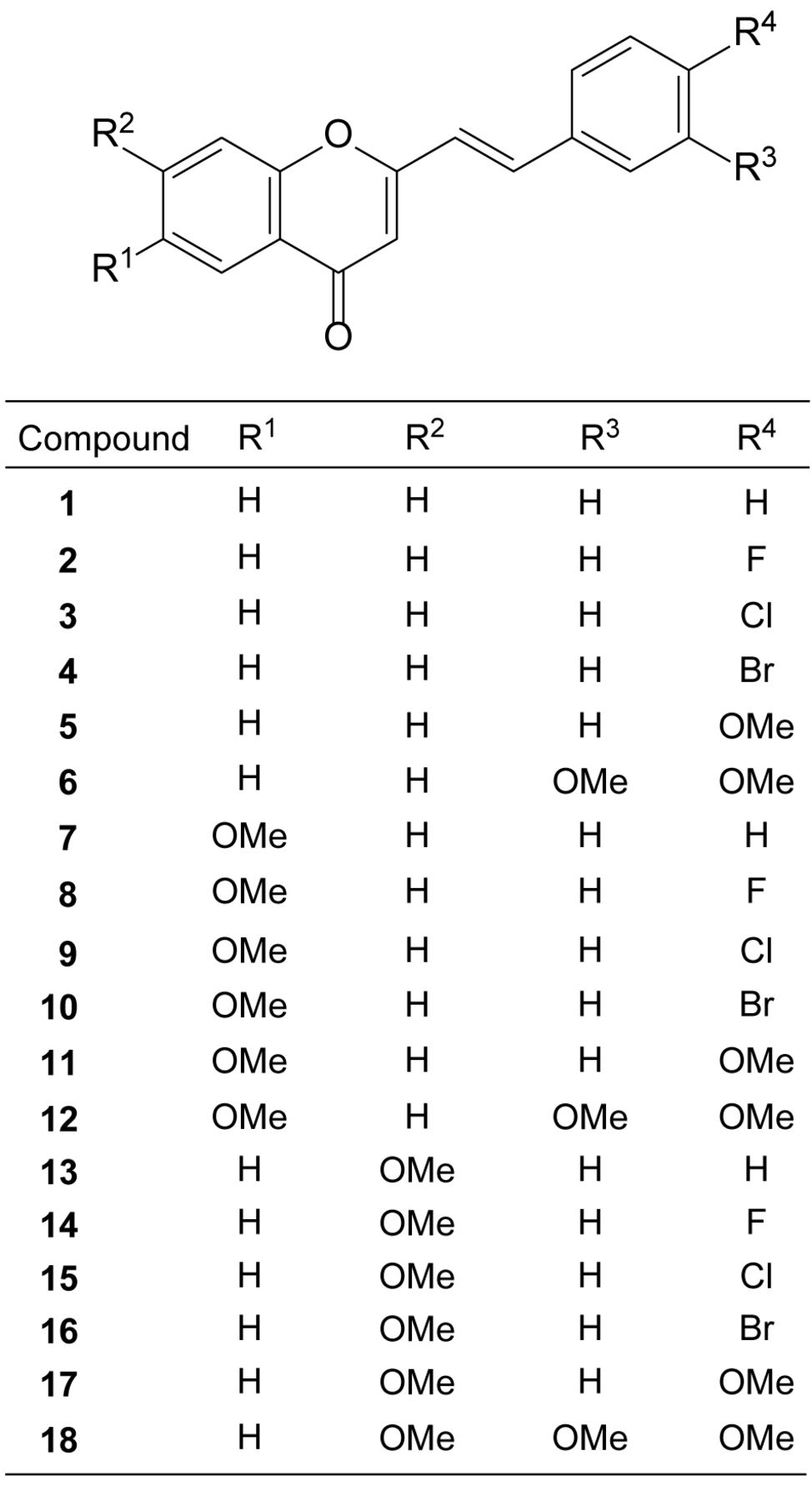
In the present study, we investigated the cytotoxicity of eighteen synthetic 2-styrylchromone derivatives (Figure 1) against four human oral squamous cell carcinoma (OSCC) cells lines (Ca9-22, HSC-2, HSC-3, HSC-4) and three human normal oral mesenchymal cells [human gingival fibroblast (HGF), human periodontal ligamental fibroblast (HPLF) and human pulp cell (HPC)], and performed quantitative structure–activity relationship (QSAR) analysis.
Materials and Methods
Materials. Dulbecco's modified Eagle's medium (DMEM) was purchased from GIBCO BRL (Grand Island, NY, USA); fetal bovine serum (FBS), doxorubicin, 3-(4,5-dimethylthiazol-2-yl)-2,5-diphenyltetrazolium bromide (MTT), ribonuclease (RNase) A from Sigma-Aldrich Inc. (St. Louis, MO, USA); dimethyl sulfoxide (DMSO), actinomycin D (FUJIFILM Wako Chem., Osaka, Japan); 100 mm dishes from True Line (Nippon Genetics Co., Ltd., Tokyo, Japan) and 96-well plates from TPP (Techno Plastic Products AG, Trasadingen, Switzerland).
Synthesis of test compounds. 2-[(1E)-2-Phenylethenyl]-4H-1-benzopyran-4-one [1], 2-[(1E)-2-(4-fluorophenyl)ethenyl]-4H-1-benzopyran-4-one [2], 2-[(1E)-2-(4-chlorophenyl)ethenyl]-4H-1-benzopyran-4-one [3], 2-[(1E)-2-(4-bromophenyl)ethenyl]-4H-1-benzopyran-4-one [4], 2-[(1E)-2-(4-methoxyphenyl)ethenyl]-4H-1-benzopyran-4-one [5], 2-[(1E)-2-(3,4-dimethoxy)ethenyl]-4H-1-benzopyran-4-one [6], 6-methoxy-2-[(1E)-2-phenylethenyl]-4H-1-benzopyran-4-one [7], 2-[(1E)-2-(4-fluorophenyl)ethenyl]-6-methoxy-4H-1-benzopyran-4-one [8], 2-[(1E)-2-(4-chlorophenyl)ethenyl]-6-methoxy-4H-1-benzopyran-4-one [9], 2-[(1E)-2-(4-bromophenyl) ethenyl]-6-methoxy-4H-1-benzopyran-4-one [10], 6-methoxy-2-[(1E)-2-(4-methoxyphenyl)ethenyl]-4H-1-benzopyran-4-one [11], 2-[(1E)-2-(3,4-dimethoxy)ethenyl]-6-methoxy-4H-1-benzopyran-4-one [12], 7-methoxy-2-[(1E)-2-phenylethenyl]-4H-1-benzopyran-4-one [13], 2-[(1E)-2-(4-fluorophenyl)ethenyl]-7-methoxy-4H-1-benzopyran-4-one [14], 2-[(1E)-2-(4-chlorophenyl)ethenyl]-7-methoxy-4H-1-benzopyran-4-one [15], 2-[(1E)-2-(4-bromophenyl)ethenyl]-7-methoxy-4H-1-benzopyran-4-one [16], 7-methoxy-2-[(1E)-2-(4-methoxyphenyl) ethenyl]-4H-1-benzopyran-4-one [17], and 2-[(1E)-2-(3,4-dimethoxy) ethenyl]-7-methoxy-4H-1-benzopyran-4-one [18] were synthesized by the condensation of the corresponding 2-methylchromones with selected benzaldehyde derivatives, according to previous methods (19). All compounds were dissolved in DMSO at 40 mM and stored at −20°C before use.
Cell culture. Human OSCC cell lines (Ca9-22, derived from gingival tissue; HSC-2, HSC-3. HSC-4, derived from tongue) and human normal oral mesenchymal cells (HGF, HPLF, HPC) at 10-18 population doubling levels were cultured at 37°C in DMEM supplemented with 10% heat-inactivated FBS, 100 units/ml, penicillin G and 100 μg/ml streptomycin sulfate under a humidified 5% CO2 atmosphere, as described previously (20, 21).
Assay for cytotoxic activity. Cells were inoculated at 6×10³ cells/cm2 in a 96-microwell plate. After 48 h, the medium was replaced with fresh medium containing (1/2)2 serially diluted test compounds. Cells were incubated for 48 h and the relative viable cell number was then determined in triplicate by MTT method, as described previously (20, 21). Control cells were treated with the same amounts of DMSO and the cell damage induced by DMSO was subtracted. The concentration of compound that reduced the viable cell number by 50% (CC50) was determined from the dose–response curve.
Calculation of tumor-specificity index (TS). TS was calculated by the following equation: TS=Mean CC50 (HGF + HPLF + HPC)/meanCC50 (Ca9-22 + HSC-2 + HSC-3 + HSC-4) (D/B) or CC50 (HGF)/C50 (Ca9-22) [both derived from gingival tissue (22)] (C/A in Table I), as described previously (20, 21).
Calculation of potency-selectivity expression (PSE). PSE, that is the product of tumor-specificity and cytotoxicity against tumor cells, was calculated by the following equation: PSE={Mean CC50 (normal cells)/[mean CC50 (OSCC cell lines)]2}×100 [as shown in (D/B2) ×100 or (C/A2) ×100] (Table I) (20, 21).
Cell-cycle analysis. Treated and untreated cells (approximately 106 cells) were harvested from 100 mm dish, fixed with paraformaldehyde and then treated with ribonuclease A. After staining with propidium iodide in the presence of 0.01% Nonidet-40, filtering through cell strainers and the stained cells were subjected to cell sorting (SH800 Series; SONY Imaging Products and Solutions Inc., Kanagawa, Japan) and cell-cycle analysis with Cell Sorter Software version 2.1.2. (SONY Imaging Products and Solution Inc.), as described previously (21).

Structure of eighteen 2-styrylchromone derivatives [1-18] investigated in this study.
Estimation of CC50 values for computational analysis. The negative log CC50 (pCC50) values were used for the comparison of cytotoxicity between compounds, as described previously (21). The mean pCC50 values for normal cells and tumor cell lines were defined as N and T, respectively. The difference (T–N) was used as a tumor-specificity index in the following analyses (21).
Calculation of chemical descriptors. The 3D structure of each chemical structure (MarvinSketch 18.10.0, ChemAxon, Budapest, Hungary) (23), was optimized by CORINA Classic (Molecular Networks GmbH, Germany) with forcefield calculations (amber-10: EHT) in Molecular Operating Environment (MOE) version 2018.0101 (Chemical Computing Group Inc., Quebec, Canada) (24). The number of structural descriptors calculated from MOE and Dragon (Dragon 7 version 7.0.2 (Kode srl., Pisa, Italy) (25) was 354 and 5,255, respectively. Among them, the number of descriptors used for analysis was 287 and 2,830 (total 3,117), respectively.
Cytotoxic activity of eighteen 2-styrylchromone derivatives [1-18] against oral malignant and non-malignant cells. Each value represents the mean of triplicate determinations. Two sets of tumor-specificity index (TS) and potency-selectivity expression (PSE) values were determined using human oral squamous cell carcinoma (OSCC) cell lines compared to human normal oral mesenchymal cells, and paired cells derived from the same (gingival) tissue.
Statistical analysis. The CC50 values were expressed as mean±S.D. of triplicate assays. The relation among cytotoxicity, tumor-specificity index and chemical descriptors was investigated using simple regression analyses by JMP Pro version 14.1.0 (SAS Institute Inc., Cary, NC, USA). The significance level was set at p<0.05.
Results
Cytotoxicity and tumor-specificity. Among eighteen synthetic 2-styrylchromones, [5] showed the highest cytotoxicity against four OSCC cell lines (mean CC50=1.9 μM) (B in Table I), followed by [11] (3.8) > [3] (10.6) > [14] (12.7) > [9] (14.8) > [10] (15.6) > [2] (19.8) > [4] (24.5) > [7] (37.6) > [1] (37.7) > [6] (45.4) > [17] (49.5) > [18] (56.6) > [16] (65.8) > [8] (67.5) > [15] (68.0) > [13] (115.5) > [12] (256.7 μM). On the other hand, [16] showed the highest cytotoxicity against three normal oral mesenchymal cells (mean CC50=27.3 μM) (D in Table I), followed by [15] (44.8) > [4] (69.5) > [1] (79.2) > [10] (87.0) > [6] (99.8) > [5] (159.1) > [2] (175.6) > [17] (199.3) > [8] (208.6) > [7] (214.2) > [3] (218.1) > [14] (221.0) > [13] (230.9) > [18] (272.2) > [9] (314.9) > [12] (315.9) > [11] (335.7 μM) (Table I). When tumor-specificity (TS) was calculated by the ratio of mean CC50 for non-malignant (normal oral cells) to that of malignant (OSCC cells) (D/B in Table I), [11] showed the highest TS value (89.1), followed by [5] (84.1) > [9] (21.2) > [3] (20.6) > [14] (17.4) > [2] (8.9) > [7] (5.7) > [10] (5.6) > [18] (4.8) > [17] (4.0) > [8] (3.1) > [4] (2.8) > [6] (2.2) > [1] (2.1) > [13] (2.0) > [12] (1.2) > [15] (0.7) > [16] (0.4). [5] also showed the highest PSE value [(D/B2) ×100 in Table I] (4443.4), followed by [11] (2364.8) > [3] (195.3) > [9] (142.8) > [14] (136.3) > [2] (44.9) > [10] (35.7) > [7] (15.2) > [4] (11.6) > [18] (8.5) > [17] (8.1) > [1] (5.6) > [6] (4.8) > [8] (4.6) > [13] (1.7) > [15] (1.0) > [16] (0.6) > [12] (0.5).
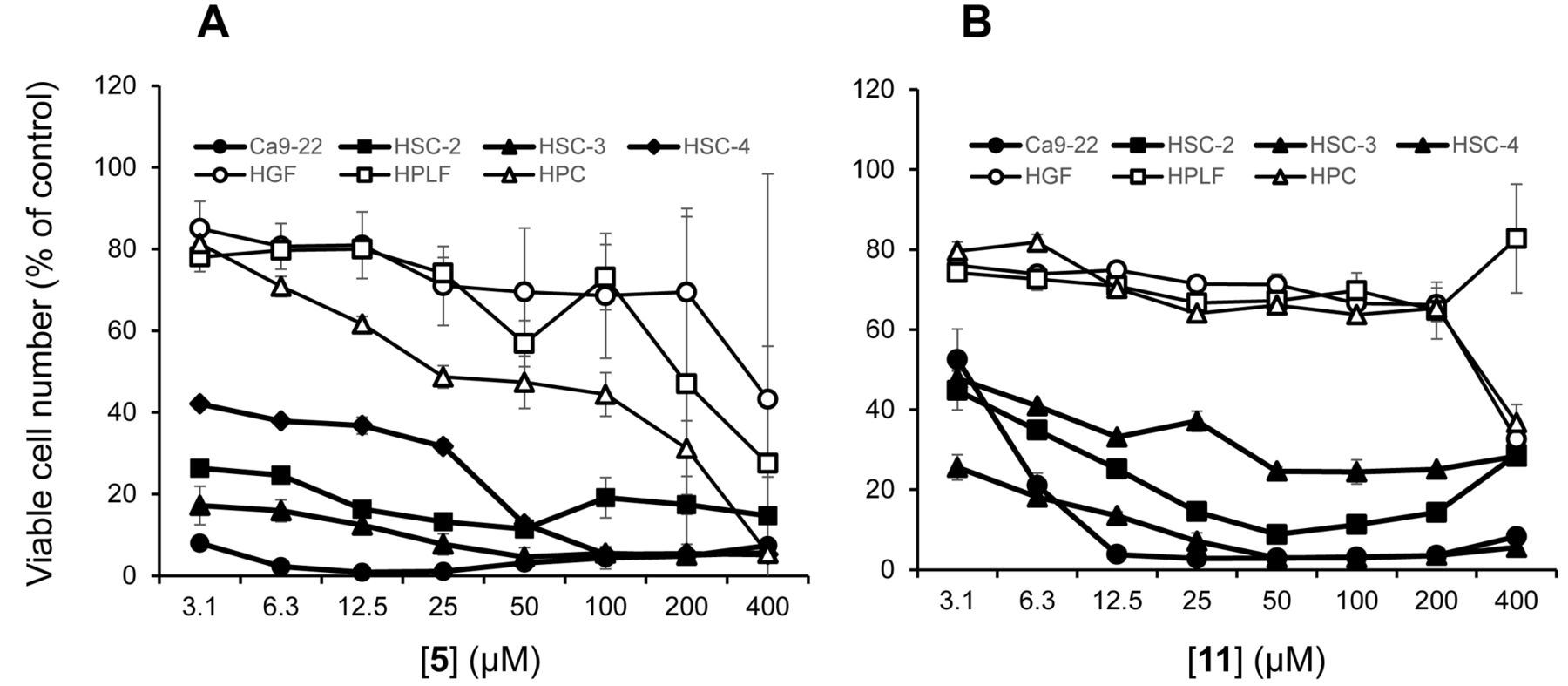
Cytotoxicity of compounds [5, 11] against four human OSCC cell lines, Ca9-22 (●), HSC-2 (▪), HSC-3 (▲) and HSC-4) (◆), and three human normal mesenchymal oral cells, HGF (○), HPLF (□) and HPC (▵). Cells were incubated for 48 h without (control) or with the indicated concentrations of [5] (A) or [11] (B), and cell viability was determined by the MTT method, and expressed as a percentage to that of control. Each value represents the mean±S.D. of triplicate assays.
The prominent TS and PSE values of [5, 11] were not changed when Ca9-22 and HGF, both derived from gingival tissues, were used as target cell: TS=148.6, 102.4 (C/A) and PSE=7592.1, 3531.5 [(C/A2) ×100 in Table I], respectively. Dose-response curves (Figure 2A and 2B) showed clearly that all OSCC cells (Ca9-22, HSC-2, HSC-3, HSC-4) were more sensitive to [5, 11] than normal oral cells (HGF, HPLF, HPC). Cell-cycle analysis demonstrated that both [5, 11] accumulated the subG1 and G2/M phase cells, suggesting the induction of apoptosis (Figure 3).
Computational analysis. QSAR analysis of cytotoxicity and tumor-specificity of eighteen 2-styrylchromone derivatives [1-18] were next performed. Since significant correlation (p<0.05) was found between cytotoxicity against tumor and normal cells, and TS with 139, 472 and 67 chemical descriptors (data not shown), top six chemical descriptors were chosen for QSAR analysis (Figures 4, 5 and 6; Table II).
Cytotoxicity of eighteen 2-styrylchromones against human OSCC cell lines was negatively correlated with J_D (topological shape) (r2=0.555, p=0.0004), balabanJ (topological shape) (r2=0.533, p=0.0006), CATS2D_09_AL (hydrogen-bond acceptor and lipophilicity) (r2=0.486, p=0.0013), RDF085p (3D shape and polarizability) (r2=0.471, p=0.0017), L2s (3D shape, size and electric state) (r2=0.468, p=0.0017) and H2u (3D shape) (r2=0.452, p=0.0022) (Figure 4).
Cytotoxicity of eighteen 2-styrylchromones against normal oral cells was positively correlated with descriptors H2v (3D shape and size) (r2=0.537, p=0.0005), H2p (3D shape and polarizability) (r2=0.502, p=0.0010), TDB09r (3D shape and size) (r2=0.492, p=0.0012), TDB07m (3D shape and size)(r2=0.484, p=0.0013) and Mor08m (3D shape and size) (r2=0.478, p=0.0015), but negatively correlated with Mor11e (3D shape and electric state) (r2=0.470, p=0.0017) (Figure 5).
TS of eighteen 2-styrylchromones was positively correlated with H8i (3D shape and ionization potential) (r2=0.516, p=0.0008), HATS5e (3D shape, size and electric state) (r2=0.462, p=0.0019) and Ks (3D shape, size and electric state) (r2=0.455, p=0.0021), but negatively correlated with J_Dz(m) (topological shape and size) (r2=0.504, p=0.0010), P2s (3D shape, size and electric state) (r2=0.457, p=0.0021) and balabanJ (topological shape) (r2=0.431, p=0.0031) (Figure 6).
Discussion
The present study demonstrated that two compounds: 2-[(1E)-2-(4-methoxyphenyl)ethenyl]-4H-1-benzopyran-4-one [5] and 6-methoxy-2-[(1E)-2-(4-methoxyphenyl)ethenyl]-4H-1-benzopyran-4-one [11], showed comparable tumor-specificity with doxorubicin, an anthracycline anticancer drug (26, 27) (TS=84.1, 89.1 vs. 67.4 in D/B; 148.6, 102.4 vs. 58.8 in C/A, Table I). However, [5, 11] showed much lower PSE values compared to doxorubicin, possibly due to much lower cytotoxicity against OSCC cell lines (Table I). Structure–activity relationship suggests the importance of having OCH3 group in R4 position in [5], and two OCH3 groups in R1 and R4 positions in [11]. However, [12] having three OCH3 groups in R1, R3 and R4 positions, showed significantly reduced tumor-specificity (Figure 1, Table I). This may be explained by the dependence of tumor-specificity on molecular shape, since the cytotoxicity of eighteen 2-styrylchromones against tumor cell lines was significantly (p<0.002) correlated with topological and 3D shape, size, hydrogen-bond acceptor, lipophilicity, polarizability and electric state (Figure 4), and tumor-specificity was also significantly (p<0.004) correlated with topological and 3D shape, size, ionization potential and electric state (Figure 6).

Effect of [5, 11] on cell-cycle distribution in HSC-2 cells. HSC-2 cells were incubated for 24 h with the indicated concentrations of [5], [11] or 1 μM actinomycin D (Act D) as a positive control and then assessed for cell-cycle distribution by a cell sorter.
The present study also demonstrated that both [5, 11] produced subG1 cell population (a marker of apoptosis) and induced mitotic arrest (Figure 3). This is consistent with a previous report that 4’-methoxy-2-styrylchromone stabilized microtubules in a manner similar to paclitaxel, inducing abnormal mitotic spindles characterized by the formation of a monopolar structure, leading to mitotic arrest in human breast adenocarcinoma MCF-7 and lung adenocarcinoma NCI-H460 cell lines (16). It should be noted that both [5, 11] induced apoptosis (subG1 population) of HSC-2 cells more potently than actinomycin D, positive control of apoptosis (28). Induction of both apoptosis and G2+M arrest may further potentiate the antitumor potential.

Top six chemical descriptors that showed higher correlation with cytotoxicity of eighteen 2-styrylchromone derivatives [1-18] against OSCC cells. The mean negative log CC50 values (T) against tumor cells were plotted. CC50: concentration of compound that reduced the viable cell number by 50%. Top six chemical descriptors were: J_D (topological shape), balabanJ (topological shape), CATS2D_09_AL (hydrogen-bond acceptor and lipophilicity), RDF085p (3D shape and polarizability), L2s (3D shape,size and electric state) andH2u (3D shape).
We previously reported that [5] (identical with the compound 3 in (18)) induced internucleosomal DNA fragmentation and activation of caspase-3, 8 and 9 in human promyelocytic leukemia HL-60 cells, but produced large DNA fragment (assessed on agarose gel electrophoresis) in HSC-2 cells (18). Lipophilic property of [5] (logP=2.6) (18) may facilitate its intracellular entry. As far as we know, there are only two papers about the antitumor potential of 4’-methoxy-2-styrylchromone including ours (16, 18). Compound [5, 11] can, thus, be a lead compound for designing a new type of anticancer drug.

Top six chemical descriptors that showed higher correlation with cytotoxicity of eighteen 2-styrylchromone derivatives [1-18] against normal oral cells. The mean negative log CC50 values (N) against normal cells were plotted. Top six chemical descriptors were: H2v (3D shape and size), H2p (3D shape and polarizability), TDB09r (3D shape and size), TDB07m (3D shape and size), Mor08m (3D shape and size) and Mor11e (3D shape and electric state).

Top six chemical descriptors that showed higher correlation with tumor-specificity of eighteen 2-styrylchromone derivatives [1-18]. The mean negative logTS values (T-N) were plotted. Top six chemical descriptors were: H8i (3D shape and ionization potential), J_Dz(m) (topological shape and size), HATS5e (3D shape, size and electric state), P2s (3D shape, size and electric state), Ks (3D shape, size and electric state) and balabanJ (topological shape).
We recently found that the tumor-specificity of 3-(N-cyclicamino)chromone derivatives (29), 2-(N-cyclicamino) chromone (21), pyrano[4,3-b]chromones (30) and furo[2,3-b]chromones (31) was also well correlated with descriptors that reflect the molecular shape. The next step of our research is to estimate the structure that should show higher tumor-specificity based on the accumulated QSAR data base, and then synthesize such compound for the confirmation of tumor-specificity. Repeating this process may make it possible to manufacture clinically applicable compounds. The other direction of approach is to synthesize the 13C-labeled compound to monitor its intracellular distribution for the identification of target molecule.
Properties of descriptors that significantly correlated with cytotoxicity against tumor cells (T) and normal cells (N), and tumor-specificity (T-N).
Acknowledgements
This work was partially supported by KAKENHI from the Japan Society for the Promotion of Science (JSPS) (15K08111, 16K11519).
Footnotes
Authors' Contributions
Y.U. and J.N. performed the QSAR analysis. HX.S. and H.S. performed the cytotoxicity assay. K.B., A.T. and M.T. performed the cell cycle analysis. S.E., K.T. and Y.S. synthesized 18 test compounds. Y.U., H.S. and Y.S. authored or reviewed drafts of the article.
This article is freely accessible online.
Conflicts of Interest
The Authors confirm that there are no known conflicts of interest associated with this publication and there has been no significant financial support for this work that could have influenced its outcome.
- Received November 12, 2019.
- Revision received November 16, 2019.
- Accepted November 20, 2019.
- Copyright© 2019, International Institute of Anticancer Research (Dr. George J. Delinasios), All rights reserved


