


Article Figures & Data
Figures
- Figure 1.
Co-treatment of KBV20C cancer cells with repositioned drugs DON or SID and HAL increased apoptosis. (A-B) KBV20C cells were plated on 96-well plates and grown to 30-40% confluence. The cells were then stimulated for 48 h with 50 ng/ml HAL (HAL), 5 μM donepezil (DON), 5 μM sildenafil citrate (SID), 50 ng/ml HAL plus 5 μM donepezil (HAL+DON), 50 ng/ml HAL plus 5 μM sildenafil citrate (HAL+SID), or 0.1% DMSO (Con). Cell viability assay was performed as described in “Materials and methods”. The data are presented as the mean±S.D. of at least two experiments repeated in triplicate experiments. Statistical analysis was conducted using one-way analysis of variance (ANOVA) followed by multiple-comparison test; *p<0.05 compared to the corresponding control. (C-D) KBV20C cells were grown on 60 mm-diameter dishes and treated with 50 ng/ml HAL (HAL), 5 μM donepezil (DON), 5 μM sildenafil citrate (SID), 50 ng/ml HAL plus 5 μM donepezil (HAL+DON), 50 ng/ml HAL plus 5 μM sildenafil citrate (HAL+SID), or 0.1% DMSO (Con). After 1 day, all cells were observed using an inverted microscope at ×10 magnification (scale bar=100 μm). (E) KBV20C cells were grown on 60 mm-diameter dishes and stimulated with 50 ng/ml HAL (HAL), 5 μM donepezil (DON), 5 μM sildenafil citrate (SID), 50 ng/ml HAL plus 5 μM donepezil (HAL+DON), 50 ng/ml HAL plus 5 μM sildenafil citrate (HAL+SID), or 0.1% DMSO (Con). After 24 h, Annexin V analyses were performed as described in Materials and Methods. (F) KBV20C cells were plated on 60 mm-diameter dishes and treated with 50 ng/ml HAL (HAL), 10 μM donepezil (DON), 10 μM sildenafil citrate (SID), 50 ng/ml HAL plus 10 μM donepezil (HAL+DON), 50 ng/ml HAL plus 10 μM sildenafil citrate (HAL+SID), or 0.1% DMSO (Con). After 24 h, western blot analysis was performed using antibodies against C-PARP, LC3B, Beclin1, Atg5, and β-actin.
- Figure 2.
Co-treatment with SID sensitized KBV20C cells to HAL treatment to a greater extent than DON. KBV20C cells were grown on 60 mm-diameter dishes and treated with 50 ng/ml HAL (HAL), 5 μM donepezil (DON-5), 10 μM donepezil (DON-10), 5 μM sildenafil citrate (SID-5), 10 μM sildenafil citrate (SID-10), 50 ng/ml HAL plus 5 μM donepezil (HAL+DON-5), 50 ng/ml HAL plus 10 μM donepezil (HAL+DON-10), 50 ng/ml HAL plus 5 μM sildenafil citrate (HAL+SID-5), 50 ng/ml HAL plus 10 μM sildenafil citrate (HAL+SID-10), or 0.1% DMSO (Con). A-B) After 24 h, FACS analyses were performed as described in Materials and Methods. (C-D) After 24 h, western blot analysis was performed using antibodies against C-PARP, cylin D1, CDK4, p21, cyclin B1, pRb, cyclin E, pErk, pH2AX, pAkt, pJnk, and β-actin.
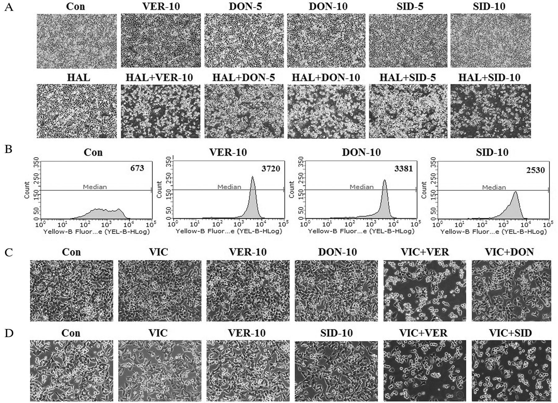
- Figure 3.
SID and verapamil showed similar sensitization of KBV20C cells, but through a different mechanism, to HAL treatment. (A) KBV20C cells were grown on 60 mm-diameter dishes and treated with 50 ng/ml HAL (HAL), 10 μM verapamil (VER-10), 5 μM donepezil (DON-5), 10 μM donepezil (DON-10), 5 μM sildenafil citrate (SID-5), 10 μM sildenafil citrate (SID-10), 50 ng/ml HAL plus 10 μM verapamil (HAL+VER), 50 ng/ml HAL plus 5 μM donepezil (HAL+DON-5), 50 ng/ml HAL plus 10 μM donepezil (HAL+DON-10), 50 ng/ml HAL plus 5 μM sildenafil citrate (HAL+SID-5), 50 ng/ml HAL plus 10 μM sildenafil citrate (HAL+SID-10), or 0.1% DMSO (Con). After 1 day, all cells were observed using an inverted microscope at ×4 magnification. (B) KBV20C cells were grown on 60 mm-diameter dishes and treated with 10 μM verapamil (VER-10), 10 μM donepezil (DON-10), 10 μM sildenafil citrate (SID-10), or 0.1% DMSO (Con). After 24 h, all cells were stained with rhodamine123 and examined by FACS analysis, as described in Materials and Methods. (C-D) KBV20C cells were grown on 60 mm-diameter dishes and treated with 5 nM vincristine (VIC), 10 μM verapamil (VER-10), 10 μM donepezil (DON-10), 10 μM sildenafil citrate (SID-10), 5 nM vincristine plus 10 μM verapamil (VIC+VER), 5 nM vincristine plus 10 μM donepezil (VIC+DON), 5 nM vincristine plus 10 μM sildenafil citrate (VIC+SID), or 0.1% DMSO (Con). After 1 day, all cells were observed using an inverted microscope at ×10 magnification.
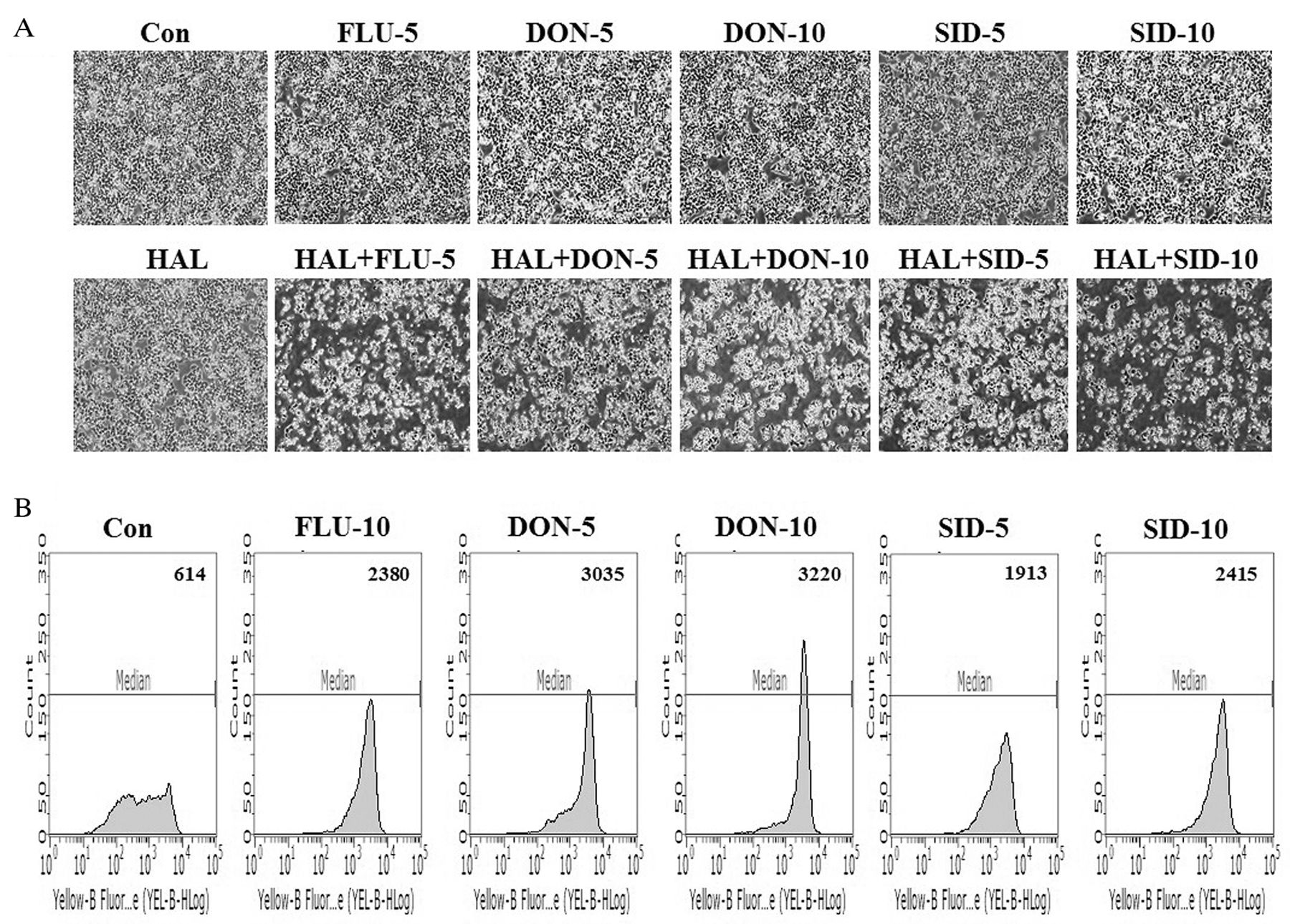
- Figure 4.
SID and FLU similarly sensitize KBV20C cells to HAL treatment by inhibiting P-gp. (A) KBV20C cells were grown on 60 mm-diameter dishes and treated with 50 ng/ml HAL (HAL), 5 μM fluphenazine (FLU-5), 5 μM donepezil (DON-5), 10 μM donepezil (DON-10), 5 μM sildenafil citrate (SID-5), 10 μM sildenafil citrate (SID-10), 50 ng/ml HAL plus 5 μM fluphenazine (HAL+FLU-5), 50 ng/ml HAL plus 5 μM donepezil (HAL+DON-5), 50 ng/ml HAL plus 10 μM donepezil (HAL+DON-10), 50 ng/ml HAL plus 5 μM sildenafil citrate (HAL+SID-5), 50 ng/ml HAL plus 10 μM sildenafil citrate (HAL+SID-10), or 0.1% DMSO (Con). After 1 day, all cells were observed using an inverted microscope at ×4 magnification. (B) KBV20C cells were grown on 60 mm-diameter dishes and treated with 10 μM fluphenazine (FLU-10), 5 μM donepezil (DON-5), 10 μM donepezil (DON-10), 5 μM sildenafil citrate (SID-5), 10 μM sildenafil citrate (SID-10), or 0.1% DMSO (Con). After 24 h, all cells were stained with rhodamine123 and examined by using FACS analysis, as described in Materials and Methods.


In this issue


Jump to section
Related Articles
Cited By...
- Co-treatment With Aripiprazole and Vincristine Sensitizes P-Glycoprotein-Overexpressing Drug-resistant MCF-7/ADR Breast Cancer Cells
- Low-dose Pimecrolimus, an FDA-approved Calcineurin Inhibitor, Sensitizes Drug-resistant Cancer Cells via Strong P-gp Inhibition
- Co-treatment of Low Dose Pacritinib, a Phase III Jak2 Inhibitor, Greatly Increases Apoptosis of P-gp Over-expressing Cancer Cells With Multidrug Resistance
- PKM2 Is Overexpressed in Glioma Tissues, and Its Inhibition Highly Increases Late Apoptosis in U87MG Cells With Low-density Specificity
- Sensitization Effects of Repurposed Blood Pressure-regulating Drugs on Drug-resistant Cancer Cells
- Drug Repositioning With an Anticancer Effect: Contributions to Reduced Cancer Incidence in Susceptible Individuals
- A Low Dose of Aripiprazole Has the Strongest Sensitization Effect Among 19 Repositioned Bipolar Drugs in P-gp-overexpressing Drug-resistant Cancer Cells
- Histamine Receptor Antagonists, Loratadine and Azelastine, Sensitize P-gp-overexpressing Antimitotic Drug-resistant KBV20C Cells Through Different Molecular Mechanisms
- Co-treatment With HIV Protease Inhibitor Nelfinavir Greatly Increases Late-phase Apoptosis of Drug-resistant KBV20C Cancer Cells Independently of P-Glycoprotein Inhibition
- Tyrosine Kinase Inhibitors Imatinib and Erlotinib Increase Apoptosis of Antimitotic Drug-resistant KBV20C Cells Without Inhibiting P-gp