
Main menu

  • Home
  • Current Issue
  • Archive
  • Info for
    • Authors
    • Editorial Policies
    • Subscribers
    • Advertisers
    • Editorial Board
    • Special Issues
  • Journal Metrics
  • Other Publications
    • In Vivo
    • Cancer Genomics & Proteomics
    • Cancer Diagnosis & Prognosis
  • More
    • IIAR
    • Conferences
    • 2008 Nobel Laureates
  • About Us
    • General Policy
    • Contact
  • Other Publications
    • Anticancer Research
    • In Vivo
    • Cancer Genomics & Proteomics

User menu

  • Register
  • Subscribe
  • My alerts
  • Log in
  • My Cart

Search

  • Advanced search
Anticancer Research
  • Other Publications
    • Anticancer Research
    • In Vivo
    • Cancer Genomics & Proteomics
  • Register
  • Subscribe
  • My alerts
  • Log in
  • My Cart
Anticancer Research

Advanced Search

  • Home
  • Current Issue
  • Archive
  • Info for
    • Authors
    • Editorial Policies
    • Subscribers
    • Advertisers
    • Editorial Board
    • Special Issues
  • Journal Metrics
  • Other Publications
    • In Vivo
    • Cancer Genomics & Proteomics
    • Cancer Diagnosis & Prognosis
  • More
    • IIAR
    • Conferences
    • 2008 Nobel Laureates
  • About Us
    • General Policy
    • Contact
  • Visit us on Facebook
  • Follow us on Linkedin
Research ArticleClinical Studies

Treatment of Naïve HCC Combined with Segmental or Subsegmental Portal Vein Tumor Thrombosis: Liver Resection Versus TACE Followed by Radiotherapy

JEONG IL YU, GYU-SEONG CHOI, DO HOON LIM, EONJU LEE, JAE-WON JOH, CHOON HYUCK DAVID KWON, JONG MAN KIM, SEONWOO KIM, SOOK-YOUNG WOO, SEUNG WOON PAIK and HEE CHUL PARK
Anticancer Research August 2018, 38 (8) 4919-4925; DOI: https://doi.org/10.21873/anticanres.12808
JEONG IL YU
1Department of Radiation Oncology, Samsung Medical Center, Sungkyunkwan University School of Medicine, Seoul, Republic of Korea
  • Find this author on Google Scholar
  • Find this author on PubMed
  • Search for this author on this site
GYU-SEONG CHOI
2Department of Surgery, Samsung Medical Center, Sungkyunkwan University School of Medicine, Seoul, Republic of Korea
  • Find this author on Google Scholar
  • Find this author on PubMed
  • Search for this author on this site
DO HOON LIM
1Department of Radiation Oncology, Samsung Medical Center, Sungkyunkwan University School of Medicine, Seoul, Republic of Korea
  • Find this author on Google Scholar
  • Find this author on PubMed
  • Search for this author on this site
EONJU LEE
3Department of Radiation Oncology, Samsung Changwon Hospital, Changwon, Republic of Korea
  • Find this author on Google Scholar
  • Find this author on PubMed
  • Search for this author on this site
JAE-WON JOH
2Department of Surgery, Samsung Medical Center, Sungkyunkwan University School of Medicine, Seoul, Republic of Korea
  • Find this author on Google Scholar
  • Find this author on PubMed
  • Search for this author on this site
CHOON HYUCK DAVID KWON
2Department of Surgery, Samsung Medical Center, Sungkyunkwan University School of Medicine, Seoul, Republic of Korea
  • Find this author on Google Scholar
  • Find this author on PubMed
  • Search for this author on this site
JONG MAN KIM
2Department of Surgery, Samsung Medical Center, Sungkyunkwan University School of Medicine, Seoul, Republic of Korea
  • Find this author on Google Scholar
  • Find this author on PubMed
  • Search for this author on this site
SEONWOO KIM
4Statistics and Data Center, Samsung Medical Center, Seoul, Republic of Korea
  • Find this author on Google Scholar
  • Find this author on PubMed
  • Search for this author on this site
SOOK-YOUNG WOO
4Statistics and Data Center, Samsung Medical Center, Seoul, Republic of Korea
  • Find this author on Google Scholar
  • Find this author on PubMed
  • Search for this author on this site
SEUNG WOON PAIK
5Department of Medicine, Samsung Medical Center, Sungkyunkwan University School of Medicine, Seoul, Republic of Korea
  • Find this author on Google Scholar
  • Find this author on PubMed
  • Search for this author on this site
HEE CHUL PARK
1Department of Radiation Oncology, Samsung Medical Center, Sungkyunkwan University School of Medicine, Seoul, Republic of Korea
6Department of Medical Device Management and Research, Samsung Advanced Institute for Health Sciences and Technology, Sungkyunkwan University, Seoul, Republic of Korea
  • Find this author on Google Scholar
  • Find this author on PubMed
  • Search for this author on this site
  • For correspondence: hee.ro.park{at}skku.edu
  • Article
  • Figures & Data
  • Info & Metrics
  • PDF
Loading

Abstract

Background/Aim: There is no definite consensus regarding management of hepatocellular carcinoma (HCC) with portal vein tumor thrombosis (PVTT). This study aimed to compare clinical outcomes according to initial treatment modality for treatment of naïve HCC combined with subsegmental (Vp1)/ segmental (Vp2) PVTT with liver resection (LR) versus trans-arterial chemoembolization (TACE) followed by radiotherapy (RT). Materials and Methods: From our institutional registry, we enrolled 78 patients diagnosed with HCC combined with Vp1 or Vp2 PVTT and treated with LR or TACE followed by RT (TACE-RT) as a primary treatment. Results: LR was more frequently applied for younger, nodular tumor morphology, or solitary tumor. Overall, LR yielded significantly better progression-free survival (PFS) (p=0.02, 41.9% vs. 15.7% at 2-years), and marginally higher overall survival (OS) (p=0.09, 75.8% vs. 61.5% at 2-years). There was an interaction effect between primary treatment and tumor morphology, and a significantly higher PFS was observed after LR in nodular morphology, in contrast with the lower PFS that was achieved after LR in infiltrative or massive morphology. Conclusion: Although LR yielded higher PFS than TACE-RT in HCC with Vp1 or Vp2 PVTT with similar acute complications, the difference in PFS between the LR and TACE-RT groups was significantly affected by tumor morphology.

  • HCC
  • radiation therapy
  • surgery
  • TACE
  • thrombosis

As the second most common cause of cancer-related death worldwide (1), hepatocellular carcinoma (HCC) is an important unresolved health problem. A considerable portion of patients are still diagnosed as unresectable and treated palliatively (2-4). Portal vein tumor thrombosis (PVTT) is one of the most important obstacles to successful curative local treatments (5).

There is no definite consensus regarding management of HCC with PVTT, especially in patients with minimal vascular invasion on subsegmental (Vp1) and/or segmental (Vp2) portal vein. Although the Barcelona Clinic for Liver Cancer's (BCLC) staging system recommends sorafenib based on two randomized clinical trials (5), superior outcomes have been reported from local modalities like surgery, trans-arterial chemoembolization (TACE), or a combination of TACE and radiotherapy (RT) (6-9). There is also growing evidence that TACE plus RT (TACE-RT) offers superior outcomes for unresectable HCC compared with TACE alone (10). A large cohort study from Japan found that liver resection (LR) yielded excellent survival outcomes in patients with HCC and PVTT (11). The clinical outcomes of these local modalities are affected by PVTT location and superior outcomes usually occur in patients with Vp1-2 PVTT.

Because PVTT can weaken liver function by prohibiting blood flow and can act as a source of intrahepatic and extrahepatic metastasis, early eradication is important for improving clinical outcomes for HCC combined with PVTT (12). At the same time, however, considering the unfavorable survival outcomes and frequent recurrence of HCC combined with PVTT, unnecessary aggressive management should be avoided.

This retrospective registry study was conducted to compare clinical outcomes from local modalities, LR and TACE-RT, for treatment-naïve HCC patients with Vp1-2 PVTT and investigated which subgroup received the most benefit by LR.

Patients and Methods

The HCC registry of the Samsung Medical Center includes newly diagnosed HCC patients who received primary care at Samsung Medical Center; our institution has been prospectively collecting patient data since January 2005. Detailed information about the registry has been described previously (13).

This study comparing clinical outcomes from LR and TACE-RT in HCC patients with Vp1-2 PVTT was approved and received an exemption from written consent by the Samsung Medical Center Institutional Review Board. Our study subjects were patients who received care between January 2010, after the set-up of the current scheduled TACE-RT protocol, and July 2014 (14).

All liver resections were performed by four highly experienced liver surgeons (J.W.J., C.H.K., G.S.C and J.M.K). Among the HCC patients with combined Vp1-2 PVTT, those with the following characteristics were considered for LR rather than other local modalities: 1) Child-Pugh class A, 2) serum total bilirubin <1.5 mg/dl, 3) indocyanine green retention test at 15 min (ICG-R15) <20%, 4) no ascites, or 5) patent portal flow in expected remaining liver lobe. Other operative procedures practiced by our institution have been previously detailed (15).

Patients who did not indicate a preference for LR or whose physicians did not prefer LR for them underwent TACE followed by RT (TACE-RT) at a two-week interval. The detailed TACE-RT protocol was described in a previous study (14). Other details about RT including target definition, dose determination, and verification using image guidance were described in previous reports (16).

Patients who underwent LR were evaluated postoperatively every 2-3 months, and the patients who received TACE-RT were evaluated 1 month after RT completion and at 2-3-month intervals thereafter. Treatment responses and disease progression were assessed by CT and/or MRI using the modified Response Evaluation Criteria in Solid Tumors (mRECIST) (17). Local recurrence was defined as an increase of 20% in diameter of the viable tumor encompassed by a PTV in the follow-up CT or MRI at least. The other recurrence developed in the liver outside of the PTV was defined as intrahepatic elsewhere recurrence.

To compare the difference between the groups, the chi-square test or Fisher's exact test and the Mann-Whitney U-test were used. Overall survival (OS) and progression-free survival (PFS) were calculated as the duration from the starting date of primary treatment to the date when a new event was first detected or, to the date of the last follow-up visit; survival rates were estimated using the Kaplan–Meier method.

For each prognostic factor, main effects and interaction effects of primary treatment were investigated. AFP was log-transformed due to highly-skewness before the analysis. Cox proportional hazards models were used, and assumption from this model was checked with the Kolmogorov supremum test. Multicollinearity was examined using the variance inflation factor (VIF) and lack of multicollinearity was confirmed when VIF was <4. All statistical analyses were executed using SAS version 9.4 (SAS Institute, Cary, NC, USA) and R 3.3.2 (Vienna, Austria; http://www.R-project.org/). A p<0.05 was considered statistically significant and all statistical tests were evaluated as two-sided.

Results

Patients. During the study period of January 2010 to July 2014, 249 patients were enrolled in the Samsung Medical Center's HCC Registry as having HCC with either Vp1 or Vp2 PVTT. Among the 140 treatment-naïve HCC patients with Vp1 or Vp2 PVTT, 78 who were treated with LR (n=31) or TACE-RT (n=47) as their primary treatment were analyzed. The baseline characteristics of enrolled patients according to primary treatment are displayed in Table I.

Adverse events according to primary treatment. There was no treatment related death within 30 days after treatment in both groups. Assessment of other treatment related toxicities proceeded in terms of toxicity-grade elevation according to the Common Terminology Criteria for Adverse Events version 4.0 (CTCAE V4.0), to evaluate treatment complications as objectively and accurately as possible to reduce bias from the initial differences in characteristics. There was one patient (3.2%) who needed to be readmitted for wound repair because of an operation-site infection. Hematologic toxicities developed more frequently in patients who received TACE-RT (6-30% in LR vs. 30-40% in TACE RT), but other laboratory results were not significantly different according to treatment modalities. Aggravation due to anorexia, nausea, and vomiting developed more frequently after TACE-RT than LR, but these toxicities were generally manageable and resolved by the first post-treatment follow-up.

Patterns of recurrence and salvage treatment. Among the 31 patients who received LR, 20 (64.5%) experienced their first recurrence intrahepatically (15, 48.4%) or extrahepatically (5, 16.1%), and seven patients died (22.6%) from recurrence. TACE was mainly used for palliative treatment either alone (9, 29.0%) or in combination with RT (4, 12.9%) after intrahepatic recurrence. One patient received a combination of TACE and radiofrequency ablation (RFA), and the last one received RT. Among the five patients who experienced extrahepatic recurrence, three were treated with systemic therapy (sorafenib in two patients and cytotoxic chemotherapy in one patient). Of two other patients who were managed with supportive care only, one received RT for bone metastasis.

Among the 47 patients treated with TACE-RT as their primary treatment, 41 (87.2%) experienced either an intrahepatic local recurrence (six patients: isolated in two, combined with intrahepatic elsewhere in three, combined with intrahepatic elsewhere and extrahepatic metastasis in one), an intrahepatic non-local recurrence (19 patients; isolated in 15, combined with extrahepatic metastasis in four), or extrahepatic recurrence (16 patients); 21 patients (22.6%) died after recurrence. Additional local treatments were used in 18 out of 20 patients who had recurrences that were local and/or intrahepatic or elsewhere, including LR in one, RFA in one, RT in two, and TACE in 14. Systemic therapy was used for 12 of 21 patients (sorafenib in 10, cytotoxic chemotherapy in one, and clinical trial in one) who had extrahepatic recurrence with or without intrahepatic/local recurrence; local modalities were attempted first in five of these patients. The remaining six patients received supportive care only.

View this table:
  • View inline
  • View popup
  • Download powerpoint
Table I.

Baseline characteristics of patients treated with LR or TACE-RT.

Figure 1.
  • Download figure
  • Open in new tab
  • Download powerpoint
Figure 1.

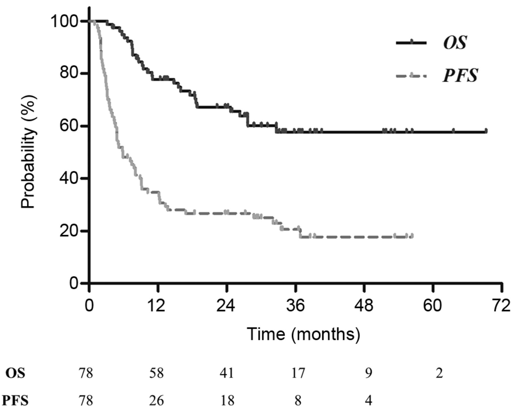
Kaplan–Meier curves for progression-free survival (PFS) and overall survival (OS) for all 78 enrolled patients. The PFS and OS were 26.4% and 67.2% at 2 years, respectively.

Survival outcomes according to primary treatment. Among the 78 patients treated with LR or TACE-RT, 61 experienced disease progression (78.2%) and 28 (35.9%) died during the follow-up period that ranged from 2.5-69.3 months (median, 24.6 months). The PFS and OS for all enrolled patients after 2 years were 26.4% and 67.2%, respectively; the Kaplan–Meier survival curves are as shown in Figure 1. The estimated PFS and OS rates were 41.9% and 75.8% in LR group, and 15.7% and 61.5% in TACE-RT group (Figure 2). Without controlling for differences in characteristics between the two groups, there was a significant difference in PFS (p=0.02, hazard ratio [HR]=0.53, 95% confidence interval [CI]=0.31-0.23), but a non-significant difference in OS (p=0.09, HR=0.44, 95%CI=0.19-1.05).

To determine the optimal primary treatment according to baseline characteristics, we conducted analyses comparing the clinical outcomes from the two groups that considered possible interaction effects among significant prognostic factors for PFS. The results of univariable analyses of probable prognostic factors with consideration of interaction effects of the primary treatment are displayed in Table II.

Tumor size was a significant prognostic factor for PFS after adjusting for primary treatment (p=0.03, HR=1.11, 95%CI=1.01-1.22), while primary treatment was also a significant prognostic factor for PFS after adjusting for tumor morphology (p=0.02, HR=0.45, 95%CI=0.23-0.88). There were also interaction effects between primary treatment and tumor morphology (p=0.002, HR=7.46, 95%CI=2.07-26.88). The significantly higher PFS was observed after LR in the subgroup with nodular morphology (p=0.03, 41.9% in LR vs. 15.7% in TACE-RT at 2-years). In contrast, lower PFS occurred after LR in the subgroup with diffuse or infiltrative morphology (p=0.02, not reached in LR vs. 15.3% in TACE-RT at 2-years). The Kaplan–Meier curves for PFS according to primary treatment and tumor morphology is provided in Figure 3.

Figure 2.
  • Download figure
  • Open in new tab
  • Download powerpoint
Figure 2.

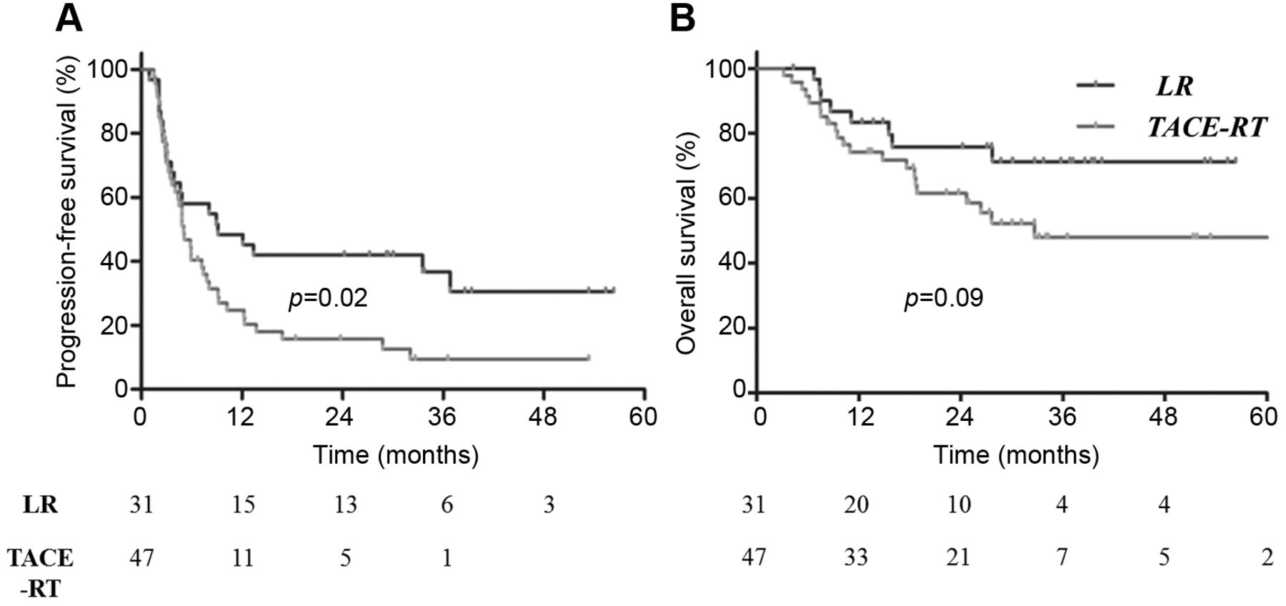
Kaplan–Meier curves for PFS and OS according to primary treatment. The PFS (A) was significantly better and OS (B) was marginally superior in the group treated with LR as a primary treatment when differences in characteristics between the two groups were not taken into consideration.

View this table:
  • View inline
  • View popup
  • Download powerpoint
Table II.

Univariable analysis for PFS and OS with interaction effects with primary treatment.

On the contrary, OS outcomes according to the primary treatment were not significantly affected by other baseline characteristics that were significant prognostic factors of OS without consideration of interaction effects, and there was no significant prognostic factor according to our analyses that included interaction effects.

Figure 3.
  • Download figure
  • Open in new tab
  • Download powerpoint
Figure 3.

Kaplan–Meier curves for PFS according to primary treatment and tumor morphology. A significantly higher PFS was noticed after LR in the subgroup with nodular morphology (A), in contrast to lower PFS that was observed after LR in the subgroup with diffuse or infiltrative morphology (B).

Discussion

In this study, we compared clinical outcomes according to local initial treatment modalities, LR and TACE-RT, in treatment-naïve HCC patients with Vp1-2 PVTT, the clinical outcomes from LR were superior to those from TACE-RT, and the OS of LR was marginally superior to that of TACE-RT. PFS, AFP level, tumor size, morphology, and multiplicity were all significant prognostic factors. Interestingly, the difference in PFS between the LR and TACE-RT groups was significantly affected by tumor morphology.

PVTT is one of the most reliable prognostic factors for HCC patients (18, 19). It causes liver function deterioration, portal pressure increment, interference of successful TACE, intrahepatic metastasis, and extrahepatic metastasis (20-23). Though sorafenib is the only recommended treatment by the BCLC guidelines (5), for patients with minor tumor thrombosis on Vp1 and/or Vp2, the usefulness of LR was consistently reported, mainly in Japan (24, 25). A recent survey study of the Liver Cancer Study Group of Japan reported cumulative survival rates of 48.2% in Vp1 and 29.2% in Vp2 after LR (25).

Based on its proven survival efficacy for unresectable HCC, TACE is one of the most widely used local modalities for HCC cases (26). However, the reported clinical outcomes from treatment with TACE alone were generally inferior to LR in HCC patients with PVTT (7).

With the rapid developments in technology and radiobiology, RT has recently become a frequently administered treatment for HCC (27-30). Because RT is less invasive than LR as a local modality, RT is often performed for cases of locally advanced HCC without distant metastasis, such as cases with combined PVTT. TACE is frequently combined with other treatments, mostly prior to RT, in order to maximize local control and obtain additional information for targeting the tumor using image guidance. According to a recent meta-analysis, adding RT to TACE yielded significantly better complete responses (odds ratio [OR]=1.57, 95%CI=1.26-1.96) and one-year OS rates (OR=1.38, 95%CI=1.16-1.65) (11).

There are no published randomized controlled trials that directly compared LR and TACE-RT in HCC patients with PVTT. Thus, the preference for localized treatment in these patients remains controversial because of reported conflicting results. Tang et al. reported significantly better cumulative survival outcomes from TACE-RT than LR in HCC patients with PVTT (18). Many other studies, including large cohort studies, however, reported outcomes that show that LR outperforms other treatment modalities, including TACE-RT, even after propensity score matching analysis (7, 19, 24, 25). These results, however, should be carefully interpreted because of selection biases that are an inevitable limitation of retrospective studies.

In this study, there was a significant PFS difference between LR and TACE-RT. As reported in other cohort studies, we reaffirmed that LR could provide the most promising oncologic outcome in selected HCC patients with Vp1 orVp2. The difference in PFS, however, is mostly recognized after six months of follow-up. The early progression within 6 months could represent aggressive biological characteristics of disease that are not different between the two groups, as shown in Figure 3A. It might be more appropriate to identify the patients with aggressive disease or an inferior outcome and apply TACE-RT to avoid a more invasive modality.

PFS was paradoxically affected by LR or TACE-RT according to tumor morphology. PFS was significantly worse in patients treated with LR, particularly if they had infiltrative or massive-type HCC combined with Vp1 or Vp2 PVTT. In contrast, significantly better survival outcomes were obtained by LR in patients with nodular-type HCC. LR should be preferentially considered in these patients.

This study has several limitations. First, a small number of cases was analyzed, which presented a serious obstacle to generalizing our findings before being validated by large-scale prospective studies. Second, the study is an inevitable selection bias between the two groups because the primary treatment of LR or TACE-RT was determined according to very strict selection criteria and it is a retrospective, single institution study. The difference in survival outcomes could be largely affected by these differences in characteristics.

Despite the above-mentioned major limitations, this study offers important and valuable findings. As far as we know, this is the only study to compare clinical outcomes between LR and TACE-RT as a primary treatment in treatment-naïve HCC with Vp1/2 PVTT. Our findings highlight important prognostic factors for PFS in treatment-naïve HCC with Vp1/2 PVTT, which include tumor multiplicity, AFP level, and tumor size. Additionally, PFS was significantly affected by choice of primary local modality, LR or TACE-RT. This study provides crucial decision-making information regarding the optimal primary treatment modality in these patients.

In treatment-naïve HCC with Vp1/2 PVTT, LR yielded significantly better PFS and marginally superior OS than TACE-RT. The superiority of LR for PFS, however, was inverted in the subgroup that had diffuse or infiltrative tumor morphology. Tumor morphology could be a valuable criterium for choosing a primary treatment in patients. Further large-scale studies on this topic are required.

Acknowledgements

This research was supported by a Basic Science Research Program through the National Research Foundation of Korea (NRF), which was funded by the Ministry of Education (NRF-2017R1D1A1B03031275), and a grant from the Marine Biotechnology Program (20150220), which was funded by the Ministry of Oceans and Fisheries, Korea.

Footnotes

  • ↵* These Authors contributed equally to this study.

  • Conflicts of Interest

    The Authors declare that they have no conflicts of interest to this work.

  • Received May 23, 2018.
  • Revision received June 14, 2018.
  • Accepted June 15, 2018.
  • Copyright© 2018, International Institute of Anticancer Research (Dr. George J. Delinasios), All rights reserved

References

  1. ↵
    1. Global Burden of Disease Cancer Collaboration,
    2. Fitzmaurice C,
    3. Allen C,
    4. Barber RM,
    5. Barregard L,
    6. Bhutta ZA,
    7. Brenner H,
    8. Dicker DJ,
    9. Chimed-Orchir O,
    10. Dandona R,
    11. Dandona L,
    12. Fleming T,
    13. Forouzanfar MH,
    14. Hancock J,
    15. Hay RJ,
    16. Hunter-Merrill R,
    17. Huynh C,
    18. Hosgood HD,
    19. Johnson CO,
    20. Jonas JB,
    21. Khubchandani J,
    22. Kumar GA,
    23. Kutz M,
    24. Lan Q,
    25. Larson HJ,
    26. Liang X,
    27. Lim SS,
    28. Lopez AD,
    29. MacIntyre MF,
    30. Marczak L,
    31. Marquez N,
    32. Mokdad AH,
    33. Pinho C,
    34. Pourmalek F,
    35. Salomon JA,
    36. Sanabria JR,
    37. Sandar L,
    38. Sartorius B,
    39. Schwartz SM,
    40. Shackelford KA,
    41. Shibuya K,
    42. Stanaway J,
    43. Steiner C,
    44. Sun J,
    45. Takahashi K,
    46. Vollset SE,
    47. Vos T,
    48. Wagner JA,
    49. Wang H,
    50. Westerman R,
    51. Zeeb H,
    52. Zoeckler L,
    53. Abd-Allah F,
    54. Ahmed MB,
    55. Alabed S,
    56. Alam NK,
    57. Aldhahri SF,
    58. Alem G,
    59. Alemayohu MA,
    60. Ali R,
    61. Al-Raddadi R,
    62. Amare A,
    63. Amoako Y,
    64. Artaman A,
    65. Asayesh H,
    66. Atnafu N,
    67. Awasthi A,
    68. Saleem HB,
    69. Barac A,
    70. Bedi N,
    71. Bensenor I,
    72. Berhane A,
    73. Bernabe E,
    74. Betsu B,
    75. Binagwaho A,
    76. Boneya D,
    77. Campos-Nonato I,
    78. Castaneda-Orjuela C,
    79. Catala-Lopez F,
    80. Chiang P,
    81. Chibueze C,
    82. Chitheer A,
    83. Choi JY,
    84. Cowie B,
    85. Damtew S,
    86. das Neves J,
    87. Dey S,
    88. Dharmaratne S,
    89. Dhillon P,
    90. Ding E,
    91. Driscoll T,
    92. Ekwueme D,
    93. Endries AY,
    94. Farvid M,
    95. Farzadfar F,
    96. Fernandes J,
    97. Fischer F,
    98. TT GH,
    99. Gebru A,
    100. Gopalani S,
    101. Hailu A,
    102. Horino M,
    103. Horita N,
    104. Husseini A,
    105. Huybrechts I,
    106. Inoue M,
    107. Islami F,
    108. Jakovljevic M,
    109. James S,
    110. Javanbakht M,
    111. Jee SH,
    112. Kasaeian A,
    113. Kedir MS,
    114. Khader YS,
    115. Khang YH,
    116. Kim D,
    117. Leigh J,
    118. Linn S,
    119. Lunevicius R,
    120. El Razek HMA,
    121. Malekzadeh R,
    122. Malta DC,
    123. Marcenes W,
    124. Markos D,
    125. Melaku YA,
    126. Meles KG,
    127. Mendoza W,
    128. Mengiste DT,
    129. Meretoja TJ,
    130. Miller TR,
    131. Mohammad KA,
    132. Mohammadi A,
    133. Mohammed S,
    134. Moradi-Lakeh M,
    135. Nagel G,
    136. Nand D,
    137. Le Nguyen Q,
    138. Nolte S,
    139. Ogbo FA,
    140. Oladimeji KE,
    141. Oren E,
    142. Pa M,
    143. Park EK,
    144. Pereira DM,
    145. Plass D,
    146. Qorbani M,
    147. Radfar A,
    148. Rafay A,
    149. Rahman M,
    150. Rana SM,
    151. Soreide K,
    152. Satpathy M,
    153. Sawhney M,
    154. Sepanlou SG,
    155. Shaikh MA,
    156. She J,
    157. Shiue I,
    158. Shore HR,
    159. Shrime MG,
    160. So S,
    161. Soneji S,
    162. Stathopoulou V,
    163. Stroumpoulis K,
    164. Sufiyan MB,
    165. Sykes BL,
    166. Tabares-Seisdedos R,
    167. Tadese F,
    168. Tedla BA,
    169. Tessema GA,
    170. Thakur JS,
    171. Tran BX,
    172. Ukwaja KN,
    173. Uzochukwu BSC,
    174. Vlassov VV,
    175. Weiderpass E,
    176. Wubshet Terefe M,
    177. Yebyo HG,
    178. Yimam HH,
    179. Yonemoto N,
    180. Younis MZ,
    181. Yu C,
    182. Zaidi Z,
    183. Zaki MES,
    184. Zenebe ZM,
    185. Murray CJL,
    186. Naghavi M
    : Global, Regional, and National Cancer Incidence, Mortality, Years of Life Lost, Years Lived With Disability, and Disability-Adjusted Life-years for 32 Cancer Groups, 1990 to 2015: A Systematic Analysis for the Global Burden of Disease Study. JAMA Oncol 3: 524-548, 2017.
    OpenUrlPubMed
  2. ↵
    1. Borzio M,
    2. Dionigi E,
    3. Rossini A,
    4. Toldi A,
    5. Francica G,
    6. Fornari F,
    7. Salmi A,
    8. Farinati F,
    9. Vicari S,
    10. Marignani M,
    11. Terracciano F,
    12. Ginanni B,
    13. Sacco R
    : Trend of improving prognosis of hepatocellular carcinoma in clinical practice: an Italian in-field experience. Dig Dis Sci 60: 1465-1473, 2015.
    OpenUrlPubMed
    1. Jepsen P,
    2. Andersen MW,
    3. Villadsen GE,
    4. Ott P,
    5. Vilstrup H
    : Time-trends in incidence and prognosis of hepatocellular carcinoma in Denmark: A nationwide register-based cohort study. Liver Int 37: 871-878, 2017.
    OpenUrlPubMed
  3. ↵
    1. Lim D,
    2. Ha M,
    3. Song I
    : Trends in major cancer mortality in Korea, 1983-2012, with a joinpoint analysis. Cancer Epidemiol 39: 939-946, 2015.
    OpenUrl
  4. ↵
    1. Forner A,
    2. Llovet JM,
    3. Bruix J
    : Hepatocellular carcinoma. Lancet 379: 1245-1255, 2012.
    OpenUrlCrossRefPubMed
  5. ↵
    1. Cho JY,
    2. Paik YH,
    3. Park HC,
    4. Yu JI,
    5. Sohn W,
    6. Gwak GY,
    7. Choi MS,
    8. Lee JH,
    9. Koh KC,
    10. Paik SW,
    11. Yoo BC
    : The feasibility of combined transcatheter arterial chemoembolization and radiotherapy for advanced hepatocellular carcinoma. Liver Int 34: 795-801, 2014.
    OpenUrlCrossRefPubMed
  6. ↵
    1. Kokudo T,
    2. Hasegawa K,
    3. Matsuyama Y,
    4. Takayama T,
    5. Izumi N,
    6. Kadoya M,
    7. Kudo M,
    8. Ku Y,
    9. Sakamoto M,
    10. Nakashima O,
    11. Kaneko S,
    12. Kokudo N
    : Survival benefit of liver resection for hepatocellular carcinoma associated with portal vein invasion. J Hepatol 65: 938-943, 2016.
    OpenUrlCrossRefPubMed
    1. Lee JM,
    2. Jang BK,
    3. Lee YJ,
    4. Choi WY,
    5. Choi SM,
    6. Chung WJ,
    7. Hwang JS,
    8. Kang KJ,
    9. Kim YH,
    10. Chauhan AK,
    11. Park SY,
    12. Tak WY,
    13. Kweon YO,
    14. Kim BS,
    15. Lee CH
    : Survival outcomes of hepatic resection compared with transarterial chemoembolization or sorafenib for hepatocellular carcinoma with portal vein tumor thrombosis. Clin Mol Hepatol 22: 160-167, 2016.
    OpenUrlPubMed
  7. ↵
    1. Rim CH,
    2. Seong J
    : Application of radiotherapy for hepatocellular carcinoma in current clinical practice guidelines. Radiat Oncol J 34: 160-167, 2016.
    OpenUrlPubMed
  8. ↵
    1. Yu JI,
    2. Park HC
    : Radiotherapy as valid modality for hepatocellular carcinoma with portal vein tumor thrombosis. World J Gastroenterol 22: 6851-6863, 2016.
    OpenUrlPubMed
  9. ↵
    1. Huo YR,
    2. Eslick GD
    : Transcatheter arterial chemoembolization plus radiotherapy compared with chemoembolization alone for hepatocellular carcinoma: A Systematic Review and Meta-analysis. JAMA Oncol 1: 756-765, 2015.
    OpenUrlPubMed
  10. ↵
    1. Yu JI,
    2. Park JW,
    3. Park HC,
    4. Yoon SM,
    5. Lim do H,
    6. Lee JH,
    7. Lee HC,
    8. Kim SW,
    9. Kim JH
    : Clinical impact of combined transarterial chemoembolization and radiotherapy for advanced hepatocellular carcinoma with portal vein tumor thrombosis: An external validation study. Radiother Oncol 118: 408-415, 2016.
    OpenUrlPubMed
  11. ↵
    1. Kim KM,
    2. Sinn DH,
    3. Jung SH,
    4. Gwak GY,
    5. Paik YH,
    6. Choi MS,
    7. Lee JH,
    8. Koh KC,
    9. Paik SW
    : The recommended treatment algorithms of the BCLC and HKLC staging systems: does following these always improve survival rates for HCC patients? Liver Int 36: 1490-1497, 2016.
    OpenUrlPubMed
  12. ↵
    1. Yu JI,
    2. Park HC,
    3. Lim DH,
    4. Kim CJ,
    5. Oh D,
    6. Yoo BC,
    7. Paik SW,
    8. Kho KC,
    9. Lee JH
    : Scheduled interval trans-catheter arterial chemoembolization followed by radiation therapy in patients with unresectable hepatocellular carcinoma. J Korean Med Sci 27: 736-743, 2012.
    OpenUrlPubMed
  13. ↵
    1. Lee JS,
    2. Kim JM,
    3. Lee S,
    4. Choi JY,
    5. Cho W,
    6. Choi GS,
    7. Park JB,
    8. Kwon CH,
    9. Kim SJ,
    10. Joh JW
    : The prognosis in cases of hepatocellular carcinoma after hepatectomy: young patients versus older patients. Korean J Hepatobiliary Pancreat Surg 19: 154-160, 2015.
    OpenUrlPubMed
  14. ↵
    1. Yu JI,
    2. Park HC,
    3. Lim DH,
    4. Choi Y,
    5. Jung SH,
    6. Paik SW,
    7. Kim SH,
    8. Jeong WK,
    9. Kim YK
    : The role of diffusion-weighted magnetic resonance imaging in the treatment response evaluation of hepatocellular carcinoma patients treated with radiation therapy. Int J Radiat Oncol Biol Phys 89: 814-821, 2014.
    OpenUrlPubMed
  15. ↵
    1. Lencioni R,
    2. Llovet JM
    : Modified RECIST (mRECIST) assessment for hepatocellular carcinoma. Semin Liver Dis 30: 52-60, 2010.
    OpenUrlCrossRefPubMed
  16. ↵
    1. Tang QH,
    2. Li AJ,
    3. Yang GM,
    4. Lai EC,
    5. Zhou WP,
    6. Jiang ZH,
    7. Lau WY,
    8. Wu MC
    : Surgical resection versus conformal radiotherapy combined with TACE for resectable hepatocellular carcinoma with portal vein tumor thrombus: a comparative study. World J Surg 37: 1362-1370, 2013.
    OpenUrlCrossRefPubMed
  17. ↵
    1. Zhang YF,
    2. Le Y,
    3. Wei W,
    4. Zou RH,
    5. Wang JH,
    6. OuYang HY,
    7. Xiao CZ,
    8. Zhong XP,
    9. Shi M,
    10. Guo RP
    : Optimal surgical strategy for hepatocellular carcinoma with portal vein tumor thrombus: a propensity score analysis. Oncotarget 7: 38845-38856, 2016.
    OpenUrlPubMed
  18. ↵
    1. Kadouchi K,
    2. Higuchi K,
    3. Shiba M,
    4. Okazaki H,
    5. Yamamori K,
    6. Sasaki E,
    7. Tominaga K,
    8. Watanabe T,
    9. Fujiwara Y,
    10. Oshitani N,
    11. Arakawa T
    : What are the risk factors for aggravation of esophageal varices in patients with hepatocellular carcinoma? J Gastroenterol Hepatol 22: 240-246, 2007.
    OpenUrlCrossRefPubMed
    1. Llovet JM,
    2. Bustamante J,
    3. Castells A,
    4. Vilana R,
    5. Ayuso Mdel C,
    6. Sala M,
    7. Bru C,
    8. Rodes J,
    9. Bruix J
    : Natural history of untreated nonsurgical hepatocellular carcinoma: rationale for the design and evaluation of therapeutic trials. Hepatology 29: 62-67, 1999.
    OpenUrlCrossRefPubMed
    1. Oh D,
    2. Shin SW,
    3. Park HC,
    4. Cho SK,
    5. Lim DH,
    6. Paik SW
    : Changes in arterioportal shunts in hepatocellular carcinoma patients with portal vein thrombosis who were treated with chemoembolization followed by radiotherapy. Cancer Res Treat 47: 251-258, 2015.
    OpenUrlPubMed
  19. ↵
    1. Toyosaka A,
    2. Okamoto E,
    3. Mitsunobu M,
    4. Oriyama T,
    5. Nakao N,
    6. Miura K
    : Pathologic and radiographic studies of intrahepatic metastasis in hepatocellular carcinoma; the role of efferent vessels. HPB Surg 10: 97-103, 1996.
    OpenUrlCrossRefPubMed
  20. ↵
    1. Ikai I,
    2. Arii S,
    3. Okazaki M,
    4. Okita K,
    5. Omata M,
    6. Kojiro M,
    7. Takayasu K,
    8. Nakanuma Y,
    9. Makuuchi M,
    10. Matsuyama Y,
    11. Monden M,
    12. Kudo M
    : Report of the 17th Nationwide Follow-up Survey of Primary Liver Cancer in Japan. Hepatol Res 37: 676-691, 2007.
    OpenUrlCrossRefPubMed
  21. ↵
    1. Kudo M,
    2. Izumi N,
    3. Ichida T,
    4. Ku Y,
    5. Kokudo N,
    6. Sakamoto M,
    7. Takayama T,
    8. Nakashima O,
    9. Matsui O,
    10. Matsuyama Y
    : Report of the 19th follow-up survey of primary liver cancer in Japan. Hepatol Res 46: 372-390, 2016.
    OpenUrlCrossRefPubMed
  22. ↵
    1. Llovet JM,
    2. Bruix J
    : Systematic review of randomized trials for unresectable hepatocellular carcinoma: Chemoembolization improves survival. Hepatology 37: 429-442, 2003.
    OpenUrlCrossRefPubMed
  23. ↵
    1. Cho B
    : Intensity-modulated radiation therapy: a review with a physics perspective. Radiat Oncol J 36: 1-10, 2018.
    OpenUrlPubMed
    1. Shiba S,
    2. Shibuya K,
    3. Katoh H,
    4. Koyama Y,
    5. Okamoto M,
    6. Abe T,
    7. Ohno T,
    8. Nakano T
    : No Deterioration in Clinical Outcomes of Carbon Ion Radiotherapy for Sarcopenia Patients with Hepatocellular Carcinoma. Anticancer Res 38: 3579-3586, 2018.
    OpenUrlAbstract/FREE Full Text
    1. Uemoto K,
    2. Doi H,
    3. Shiomi H,
    4. Yamada K,
    5. Tatsumi D,
    6. Yasumoto T,
    7. Takashina M,
    8. Koizumi M,
    9. Oh RJ
    : Clinical assessment of micro-residual tumors during stereotactic body radiation therapy for hepatocellular carcinoma. Anticancer Res 38: 945-954, 2018.
    OpenUrlAbstract/FREE Full Text
  24. ↵
    1. Yu JI,
    2. Yoo GS,
    3. Cho S,
    4. Jung SH,
    5. Han Y,
    6. Park S,
    7. Lee B,
    8. Kang W,
    9. Sinn DH,
    10. Paik YH,
    11. Gwak GY,
    12. Choi MS,
    13. Lee JH,
    14. Koh KC,
    15. Paik SW,
    16. Park HC
    : Initial clinical outcomes of proton beam radiotherapy for hepatocellular carcinoma. Radiat Oncol J 36: 25-34, 2018.
    OpenUrlPubMed
PreviousNext
Back to top

In this issue

Anticancer Research
Vol. 38, Issue 8
August 2018
  • Table of Contents
  • Table of Contents (PDF)
  • Index by author
  • Back Matter (PDF)
  • Ed Board (PDF)
  • Front Matter (PDF)
Print
Download PDF
Article Alerts
Sign In to Email Alerts with your Email Address
Email Article

Thank you for your interest in spreading the word on Anticancer Research.

NOTE: We only request your email address so that the person you are recommending the page to knows that you wanted them to see it, and that it is not junk mail. We do not capture any email address.

Enter multiple addresses on separate lines or separate them with commas.
Treatment of Naïve HCC Combined with Segmental or Subsegmental Portal Vein Tumor Thrombosis: Liver Resection Versus TACE Followed by Radiotherapy
(Your Name) has sent you a message from Anticancer Research
(Your Name) thought you would like to see the Anticancer Research web site.
CAPTCHA
This question is for testing whether or not you are a human visitor and to prevent automated spam submissions.
10 + 5 =
Solve this simple math problem and enter the result. E.g. for 1+3, enter 4.
Citation Tools
Treatment of Naïve HCC Combined with Segmental or Subsegmental Portal Vein Tumor Thrombosis: Liver Resection Versus TACE Followed by Radiotherapy
JEONG IL YU, GYU-SEONG CHOI, DO HOON LIM, EONJU LEE, JAE-WON JOH, CHOON HYUCK DAVID KWON, JONG MAN KIM, SEONWOO KIM, SOOK-YOUNG WOO, SEUNG WOON PAIK, HEE CHUL PARK
Anticancer Research Aug 2018, 38 (8) 4919-4925; DOI: 10.21873/anticanres.12808

Citation Manager Formats

  • BibTeX
  • Bookends
  • EasyBib
  • EndNote (tagged)
  • EndNote 8 (xml)
  • Medlars
  • Mendeley
  • Papers
  • RefWorks Tagged
  • Ref Manager
  • RIS
  • Zotero
Reprints and Permissions
Share
Treatment of Naïve HCC Combined with Segmental or Subsegmental Portal Vein Tumor Thrombosis: Liver Resection Versus TACE Followed by Radiotherapy
JEONG IL YU, GYU-SEONG CHOI, DO HOON LIM, EONJU LEE, JAE-WON JOH, CHOON HYUCK DAVID KWON, JONG MAN KIM, SEONWOO KIM, SOOK-YOUNG WOO, SEUNG WOON PAIK, HEE CHUL PARK
Anticancer Research Aug 2018, 38 (8) 4919-4925; DOI: 10.21873/anticanres.12808
Twitter logo Facebook logo Mendeley logo
  • Tweet Widget
  • Facebook Like
  • Google Plus One

Jump to section

  • Article
    • Abstract
    • Patients and Methods
    • Results
    • Discussion
    • Acknowledgements
    • Footnotes
    • References
  • Figures & Data
  • Info & Metrics
  • PDF

Related Articles

Cited By...

  • Expanding Sorafenib Treatment for Hepatocellular Carcinoma Beyond Barcelona Clinic Liver Cancer Stage C Patients: A National Study
  • Google Scholar

More in this TOC Section

  • Optimizing Biopsy Decisions in PI-RADS 3-4 Lesions: Integrating PSA-derived Biomarkers to Reduce Unnecessary Procedures
  • Surgical Outcomes and Postoperative Changes in Nutritional Indexes and Sarcopenia Markers in Oldest-old Patients With Resected Biliary Tract Cancer: A Retrospective Cohort Study
  • Effect of Tissue Sample Type on The Evaluation of PD-L1 (SP142) Expression in Breast Cancer
Show more Clinical Studies

Keywords

  • HCC
  • radiation therapy
  • surgery
  • TACE
  • Thrombosis
Anticancer Research

© 2026 Anticancer Research

Powered by HighWire