


Abstract
Background/Aim: Recombinant antibodies have been investigated and used in applications such as targeting modules of drug-delivery systems (DDS) against cancers. This study aimed to prepare recombinant antibodies against HER2, containing sortase A (SrtA) recognition sequence, that are applicable as targeting modules in DDS after linkage with the drug-carrier containing oligoglycine-acceptor peptide by SrtA transpeptidation. Materials and Methods: The recombinant trastuzumab fragment antibodies (scFvs and Fab) with the SrtA-recognition motif (LPXTG) at their C-terminal were constructed and expressed in Escherichia coli and Chinese hamster ovary (CHO) cells, respectively. The reactivity of the purified recombinant antibodies towards HER2-expressing cells was also evaluated via immunofluorescent staining. Results: Fab demonstrated higher yield and purity and better reactivity towards HER2-expressing cells (HCT-15 and HeLa) when compared to scFvs. Conclusion: The CHO expression system possesses superior yield and purity when compared to the E. coli expression system with respect to the preparation of recombinant antibodies applicable in targeting modules for DDS (DDS-TM). Moreover, a Fab variant prepared in this study demonstrated the potential to be a DDS-TM against HER2-expressing cancer cells.
Drug-delivery systems (DDS) are considered to be effective treatment approaches for various diseases, particularly in the treatment of cancer so as to relieve the side-effects caused by anticancer drugs on normal (non-target) cells, owing to their potential in targeting and controlling drug release and minimizing dose. In order to establish an effective DDS for cancer therapeutics, the development of novel DDS tools against cancers has been attempted in our laboratory using functional proteins produced from bacteria. For instance, the bacterial pore-forming protein toxin called cholesterol-dependent cytolysin (CDC) was investigated in order to be used for the construction of a DDS tool. This investigation led to the development of several DDS tools such as chimera CDCs carrying a lung tumor-binding peptide (LTBP) (1) or IgG-binding domain of protein A (Z-domain) fused CDCs (2) as targeting modules. A bacterial transpeptidase, sortase A (SrtA), derived from Gram-positive bacteria was also characterized in order to apply the bacterial functional protein in the direct modification of DDS drug carriers (3).
SrtA is considered a promising protein engineering tool in the development of DDS tools, because SrtA recognizes the LPXTG motif in proteins, cleaves it between T and G, and joins the C-terminal of the generated LPXT motif to the N-terminal of oligoglycine in mild condition (4-6). In those studies, ligations between two proteins/molecules with individual functions were successfully performed using SrtA reaction, e.g., the molecules such as GFP and various antibodies holding LPXTG motif and the molecules with oligoglycine such as peptide/protein, toxin and fluorophore. The investigations proved that SrtA transpeptidation provides brilliant flexibility in protein modification without interference on protein activity. Therefore, we intended to apply the SrtA-mediated reaction in preparation of anticancer-DDS by ligation of cancer-targeting molecules that can effectively recognize cancer cells and oligoglycine-containing drug-carrier/effecter such as liposome and toxin. In such a background, we previously investigated SrtA with respect to the construction of a tool applicable for DDS by modifying liposomes with a cancer-cell targeting module (LTBP) (7).
Oligonucleotides used in this study.
Antibodies and their derivatives are useful as DDS-targeting modules against cancer cells. The antibodies that are applied in therapeutics, such as drug conjugates and DDS tools, against various tumors have been investigated for several years (8, 9). Recently, recombinant fragment antibodies such as single-chain variable fragments (scFvs) and antigen-binding fragments (Fabs), also known as smaller derivatives of IgG that possess only one antigen-binding region (10, 11), are receiving more attention than whole IgGs.
In the present study, we focused on trastuzumab, a recombinant humanized monoclonal antibody against human epidermal growth factor receptor 2 (HER2) that is overexpressed in breast and cervical cancers, as it is used for the clinical treatment of breast cancer (12-14). In order to develop a novel DDS against cancer cells overexpressing HER2, the trastuzumab-derived recombinant scFvs and Fab were designed as SrtA donor substrates containing a SrtA-recognition motif (LPXTG) at their C-terminal to directly modify the DDS carrier surfaces and expressed in E. coli and Chinese hamster ovary (CHO) cells, respectively. Both recombinant fragment antibodies were purified and evaluated for yield, purity, and targeting ability towards HER2-positive cells.
Materials and Methods
Cell lines and culture conditions. A human colorectal adenocarcinoma cell line, HCT-15 (TKG0504; Cell Resource Center for Biomedical Research, Tohoku University, Sendai, Japan), and a human cervix epithelioid carcinoma cell line, HeLa (RCB0007; RIKEN BioResource Center, Tsukuba, Japan), were cultured in RPMI-1640 and EMEM respectively, supplemented with 10% (v/v) FBS and antibiotics (benzylpenicillin potassium and streptomycin sulfate salt) at 37°C in 5% CO2. Moreover, two CHO cell lines, an EB-B2 cell line derived from CHO-K1 (Onitsuka M. et al., manuscript in preparation) and a CHO-HcD6 cell line (15), were used for the transient expression of Trastuzumab Fab fragment and Trastuzumab IgG1, respectively.
Trastuzumab-derived scFvs expression system in E. coli. The template plasmid, pFscFv(LH)-Fc (Onitsuka M. et al., manuscript in preparation), was constructed using a bacterial codon-optimized scFv(LH)-Fc fragment, which was synthesized according to the amino acid sequence of Trastuzumab (Drug bank ID: DB00072) and pFLAG-CTS™ Expression Vector (Merck KGaA, Darmstadt, Germany). In order to construct the scFv expression system in E. coli, the sequences coding VH and VL of Trastuzumab were first amplified via PCR using PrimeSTAR® HS DNA Polymerase (TaKaRa Bio, Shiga, Japan) and pFscFv(LH)-Fc as the template along with the primer sets described in Table I (No.1 and No.2 for VH, and No.3 and No.4 for VL). Subsequently, a linker fragment encoding (G4S)3 was amplified using PrimeSTAR® HS DNA Polymerase, an annealed oligonucleotide (No.5 and No.6 in Table I) as a template, and the No.7 and No.8 primer set (Table I). The purified VH, VL, and (G4S)3 fragments were joined via fusion PCR to produce inserts encoding VH-(G4S)3-VL (scFv_HL) and VL-(G4S)3-VH (scFv_LH). Each fragment was inserted into the BamHI/SalI-digested cloning site of a pQE-9 variant vector (Qiagen, Hilden, Germany) with deleted N-terminal His-tag (6 residues of His) encoding sequences and added SrtA-recognition motif (LPXTG) and His-tag encoding sequences between the SalI and HindIII restriction sites. Transformation of the SHuffle® T7 Express Competent E. coli (C3029H; New England Biolabs, MA, USA) containing each expression plasmid, pscFv_HL or pscFv_LH (Figure 1A), as well as transformant selection was performed according to standard methods. After confirming the sequence of the expression vector from the selected clone, the recombinant protein was expressed overnight in the presence of 0.1 mM IPTG at 16°C according to the C3029H manual. Both recombinants were purified using nickel-affinity chromatography as previously reported (1).

Recombinant targeting modules (scFvs) prepared using a bacterial expression system. A. Lower, Map of expression vector for scFvs (pscFv_HL/ pscFv_LH). Upper, Schematic diagram of recombinant scFvs possessing SrtA recognition sequence LPETGG followed by His-tag for Ni-affinity purification. B: SDS-PAGE image (CBB staining) of the purified recombinant scFvs. Both scFv_HL and scFv_LH (indicated by arrowhead) were prepared with high purity and the calculated molecular weight of each antibody was observed to be 27.9 kDa.
Trastuzumab-derived Fab expression system in CHO cells. The pOTC vector, prepared by inserting the PCR-amplified oriP region from pEBMulti-Neo (Wako Pure Chemical, Osaka, Japan) into the SspI site of pCAG-Hyg TARGET tag-C (Wako Pure Chemical), was employed to prepare the recombinant antibodies. First, to prepare a Trastuzumab IgG, two vectors denominated as pOTC-tHC and pOTC-tLC were constructed by inserting the synthesized Trastuzumab heavy-chain or light-chain coding sequences into pOTC between the KpnI and BamHI restriction sites, respectively (Onitsuka M. et al., manuscript in preparation). Next, in order to prepare the Trastuzumab Fab carrying LPETGG sequence which changes the Fab to a substrate of SrtA transpeptidation reaction, pOTC-tLC (light-chain vector) was directly used, while heavy-chain vector was re-constructed as follows. A cassette for LPETGG, prepared by annealing the No.15 and No.16 oligonucleotides (Table I), was inserted between the BamHI and NotI restriction sites of pOTC. Subsequently, the Trastuzumab VHCH1 coding sequence with the nonapeptide hinge area in pOTC-tHC was amplified using PrimeSTAR® HS DNA Polymerase and the primer set No.13 and No.14 (Table I) followed by KpnI and BamHI digestion. The purified fragment was then inserted between the KpnI and BamHI restriction sites of pOTC with LPETGG encoding sequence to construct pOTC-tHChg9. The transformation of E. coli DH5αZ1 with pOTC-tHChg9 and transformant selection on LB agar plate containing 100 μg/ml of hygromycin B were performed according to standard methods. After confirming the pOTC-tLC and pOTC-tHChg9 sequences from the selected clones, the plasmids (Figure 2A) were used to transform EB-B2 cells.
The constructed EB-B2 cells were cultured in duplicates in 500 ml Erlenmeyer flasks containing 100 ml of BalanCD Transfectory CHO medium (Irvine Scientific, Santa Ana, CA, USA) with 2 mM L-glutamine. When viable cell density reached 4×10⁶ cells/ml, the culture medium was replaced with fresh BalanCD Transfectory CHO medium and the cells were transfected with 320 μg of expression plasmid mixture (3:1, pOTC-tHChg9:pOTC-tLC) using Polyethylenimine Max (Polysciences Inc, Warrington, PA, USA). The transfected cells were then incubated at 32°C and 80% humidity with shaking at 120 rpm, and the culture was maintained for 12 days. At day 2 of incubation, 10 ml of the nutrient medium transfectory supplement (Irvine Scientific) was added. Recombinant Fab was purified from the culture supernatant via HiTrap™ Protein L affinity chromatography using an ÄKTAprime™ Plus system (GE Healthcare, Buckingham, UK) with a one-step pH gradient from pH 7.0 to pH 2.5 ~ pH 2.7, and the eluate was neutralized with equilibration buffer (1 M Tris-HCl, pH 8.0). After dialysis against PBS containing 1 mM EDTA, the purity of the recombinant was confirmed using SDS-PAGE.
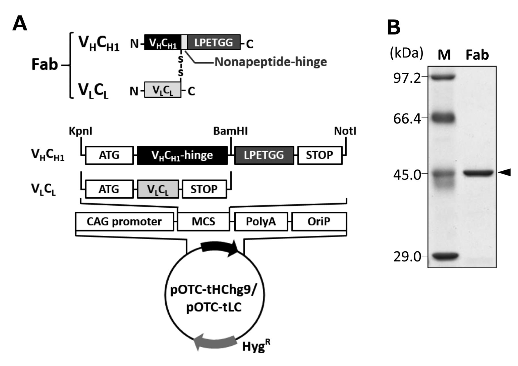
Recombinant targeting module (Fab) prepared using a mammalian cell expression system. A. Lower, Map of expression vector for Fab (pOTC-tHChg9/pOTC-tLC). Only pOTC-tHChg9 has the sequence for LPETGG at the downstream of the sequence for hinge region. Upper, Schematic diagram of recombinant Fab possessing SrtA recognition sequence LPETGG at the C-terminal of heavy chain fragment, which is connected with light chain by disulfide bond. B: SDS-PAGE image (CBB staining) of the purified recombinant Fab. Fab (indicated by arrowhead) was prepared with high purity and the calculated molecular weight was observed to be about 45.0 kDa, which is lower than its calculated molecular weight (53.8 kDa).
Semi-quantitative real-time PCR analysis. Total RNA from HCT-15 and HeLa cells was prepared using the NucleoSpin Plus extraction kit (Macherey-Nagel, Düren, Germany) and cDNA was synthesized using the PrimeScript™ RT reagent kit with gDNA Eraser (TaKaRa Bio). Real-time PCR for HER2 cDNA was performed on the Thermal Cycler Dice Real Time System Lite (TaKaRa Bio) using a standard 2-step PCR protocol along with the No.17 and No.18 primer set shown in Table I (16). The results were analyzed using the in-built TP700 software (TaKaRa Bio) against the gene encoding human GAPDH as the standard.
Immunofluorescence imaging. HCT-15 (1.0×10⁶ cells/well) and HeLa (0.4×10⁶ cells/well) cells were seeded onto sterile cover glasses in 6 well-plates and incubated for 2 days until 80% or higher confluency. The cells were then washed with FBS- and antibiotic-free culture medium, fixed with 4% (w/v) paraformaldehyde at room temperature for 15 min, and washed with PBS. Next, the cells were treated with blocking solution, PBS containing 1% (w/v) BSA, for 1 h at 4°C. Subsequently, the fixed cells were incubated for 1 h at 4°C with 1 μM of recombinant antibody (scFv_HL, scFv_LH, or Fab) as the primary antibody, purified Trastuzumab IgG1 as the positive control, and blank PBS as the background control. After washing with PBS, the cells were probed with Alexa Fluor 488-labeled goat anti-human IgG (H+L) (Thermo Fisher Scientific, Rockford, IL, USA) or Alexa Fluor 488-conjugated anti-His-tag (Qiagen) secondary antibodies at 4°C for 1 h. After washing with PBS again, each sample was mounted on a glass slide using Ultramount Aqueous Permanent Mounting Medium (Dako North America, Carpinteria, CA, USA). The cells were observed using the IX71 inverted microscope (Olympus, Tokyo, Japan) and the images were acquired using the cellSens software (Olympus).
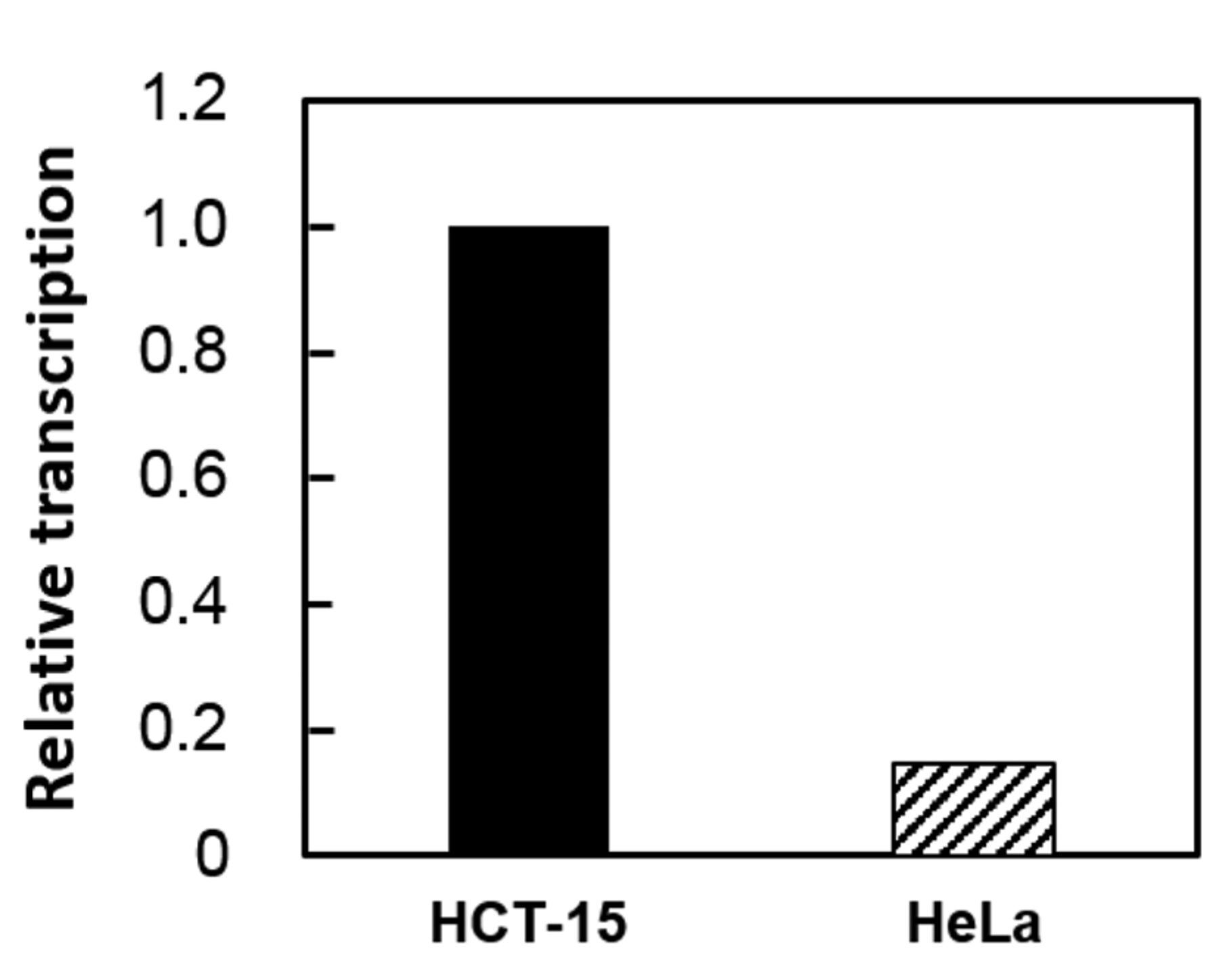
Transcription of the HER2 gene in human cell lines. Transcription of the gene encoding HER2 was investigated in HCT-15 and HeLa cell lines. The gene encoding GAPDH was used as an internal standard for the assay. This experiment was conducted twice, and one of the results is shown.

Reactivity of the anti-HER2 recombinant fragment antibodies against HER2-positive cells. HCT-15 and HeLa cells were treated with anti-HER2 recombinant fragment antibodies as primary antibodies and Alexa Fluor 488-labeled secondary antibodies. The fluorescent image of Alexa Fluor 488 was observed using the IX71 fluorescence microscope (Olympus, Tokyo, Japan). BG: Samples prepared without anti-HER2 recombinant treatment. Scale bars indicate 10 μm.
Results
Production and purification of Trastuzumab-derived scFvs with SrtA-recognition motifs. To apply Trastuzumab-derived scFvs as targeting modules for DDS (DDS-TM) against HER2-positive cancer cells, a bacterial expression system for scFvs was designed that contained LPETGG consisting of a SrtA-recognition motif (consensus motif: LPXTG) necessary for transpeptidation to the liposome with oligoglycine motif and His-tag for purification at the C-terminal side (Figure 1A). In this study, two scFv expression systems were constructed i.e., “VH-linker-VL” type (scFv_HL) and “VL-linker-VH” type (scFv_LH). These scFvs (calculated molecular weight: 27.9 kDa) were produced in E. coli and purified via nickel-affinity chromatography with sufficient purity (Figure 1B). The scFv_HL and scFv_LH yields were observed to be 9.5 mg/l and 11.3 mg/l, respectively.
Production and purification of Trastuzumab-derived Fab with SrtA-recognition motif. A mammalian CHO cell expression system for Fab was also designed, which contained the LPETGG motif at the C-terminal of heavy-chain fragment (VHCH1) that followed the nonapeptide hinge (Figure 2A). Fab present in the culture supernatant demonstrated high yield (80.0 mg/l) and superior purity according to CBB staining after SDS-PAGE under non-reducing conditions. The apparent molecular weight was smaller than the calculated value (53.8 kDa; Figure 2B). Gel filtration chromatography showed that Fab was highly purified and contained only a small amount of light-chain aggregates (data not shown).
Comparison of HER2 gene transcription in HCT-15 and HeLa cells. Semi-quantitative real-time PCR was performed to compare HER2 gene transcription in HCT-15 and HeLa cells. Consequently, this gene was observed to be transcribed in both cell lines. However, the transcriptional level of the HER2 gene in HeLa was about 1/7 to 1/6 of that in HCT-15 (Figure 3).
Reactivity of Trastuzumab-derived recombinant antibodies towards HER2-positive cells. Immunofluorescence imaging was performed to evaluate the reactivity of the three recombinant antibodies towards the tested cell lines. The results showed that Fab reactivity towards HCT-15 expressing high level HER2 was superior to scFv reactivity. However, the reactivity of the recombinant antibodies towards HeLa expressing low level HER2 was either faintly detectable or almost undetectable (Figure 4).
Discussion
DDS are thought to be highly promising in anticancer therapeutics owing to their ability of selectively delivering drugs to target cancer cells. Using bacterial functional proteins, we have been investigating the construction of DDS tools composed of drug-carrier liposomes and targeting modules on their surface. Previously, recombinant proteins prepared using bacterial expression systems were used as DDS-TM (1, 2). However, the selection of targeting modules is limited with these systems. In order to achieve better versatility of the DDS-TM, SrtA (a bacterial transpeptidase) -mediated transpeptidation was adopted to prepare novel DDS tools as a wide range of proteins or peptides with LPXTG (a SrtA-recognition motif) could be joined to the N-terminal of oligoglycine introduced as lipopeptides in drug-carrier liposome (3, 7).
In the present study, trastuzumab-derived recombinant fragment antibodies (scFv and Fab) containing the SrtA-recognition motif (LPXTG) at their C-terminal were prepared (Figures 1 and 2). The CHO cell expression system provided an 8-fold higher yield per liter of culture media and superior purity when compared to the E. coli expression system, although much longer culture periods were necessary. This is because the expression system in mammalian cells such as CHO undergo appropriate post-translational modifications, particularly with respect to mammal protein expression. Thus, CHO cells are ideal in producing commercial therapeutic antibodies (17, 18). On the other hand, bacterial expression systems such as those in E. coli are frequently adopted because they are effective in reducing costs and time with respect to target recombinant preparation. However, the target recombinants produced in E. coli expression systems often form inclusion bodies that are insoluble and difficult to purify as active proteins (19). Moreover, our experience also shows purified scFvs to be less stable and Fab to be more stable against freeze-thawing (data not shown). This may be because the constant domain provides structural stability to Fab against temperature changes (20). Cumulatively, our results suggest that the CHO cell expression system exhibits superiority in the production of recombinant proteins, such as fragment antibodies, which can be applied to construct DDS tools.
In conclusion, the results of the present study demonstrate a Fab variant of trastuzumab with LPETGG on its C-terminal prepared as a substrate for SrtA in a CHO cell expression system, that can be used to construct novel DDS tools that target HER2-positive cancer cells. Currently, the development of a liposome-based DDS tool using LPETGG-tagged Fabs to modify the surface of liposomes is being attempted.
Acknowledgements
This research was partially supported by the Program for the Strategic Research Foundation at Private Universities (2012–2016) initiated by the Japanese Ministry of Education, Culture, Sports, Science, and Technology, and by the Japan Agency for Medical Research and Development (grant number: JP15km0908001). The authors are also grateful to Editage (www.editage.jp) for providing English language editing.
- Received April 11, 2018.
- Revision received May 16, 2018.
- Accepted May 17, 2018.
- Copyright© 2018, International Institute of Anticancer Research (Dr. George J. Delinasios), All rights reserved


