


Article Figures & Data
Figures
- Figure 1.
Combination of ritonavir and delanzomib inhibited renal cancer cell growth in vitro and in vivo. A: Inhibition of cell viability by ritonavir–delanzomib combination (MTS assay; mean±SD, n=6). Cells were treated for 48 h with 25-50 μM ritonavir with/without 10-50 nM delanzomib. B: Photomicrographs of renal cancer cells treated with ritonavir and delanzomib for 48 h (original magnification ×100). Note that many of the cells treated with the combination were floating. C: Colony-formation assay. The combination treatment was effective in inhibiting renal cancer cell growth (mean±SD, n=3). Significantly different at: *p=0.0463, **p=0.0431, and ***p=0.0495. Cells were treated with 50 μM ritonavir with/without 25 nM delanzomib for 48 h and then incubated for 10 days. D: In vivo efficacy of ritonavir–delanzomib combination. A subcutaneous tumor model was made using Renca cells. The control group received dimethyl sulfoxide intraperitoneally, and the other three groups received ritonavir (15 mg/kg), delanzomib (30 μg/kg) or both (mean±SD, n=5). Significantly different at: *p=0.0472, **p=0.0463, and ***p=0.009.
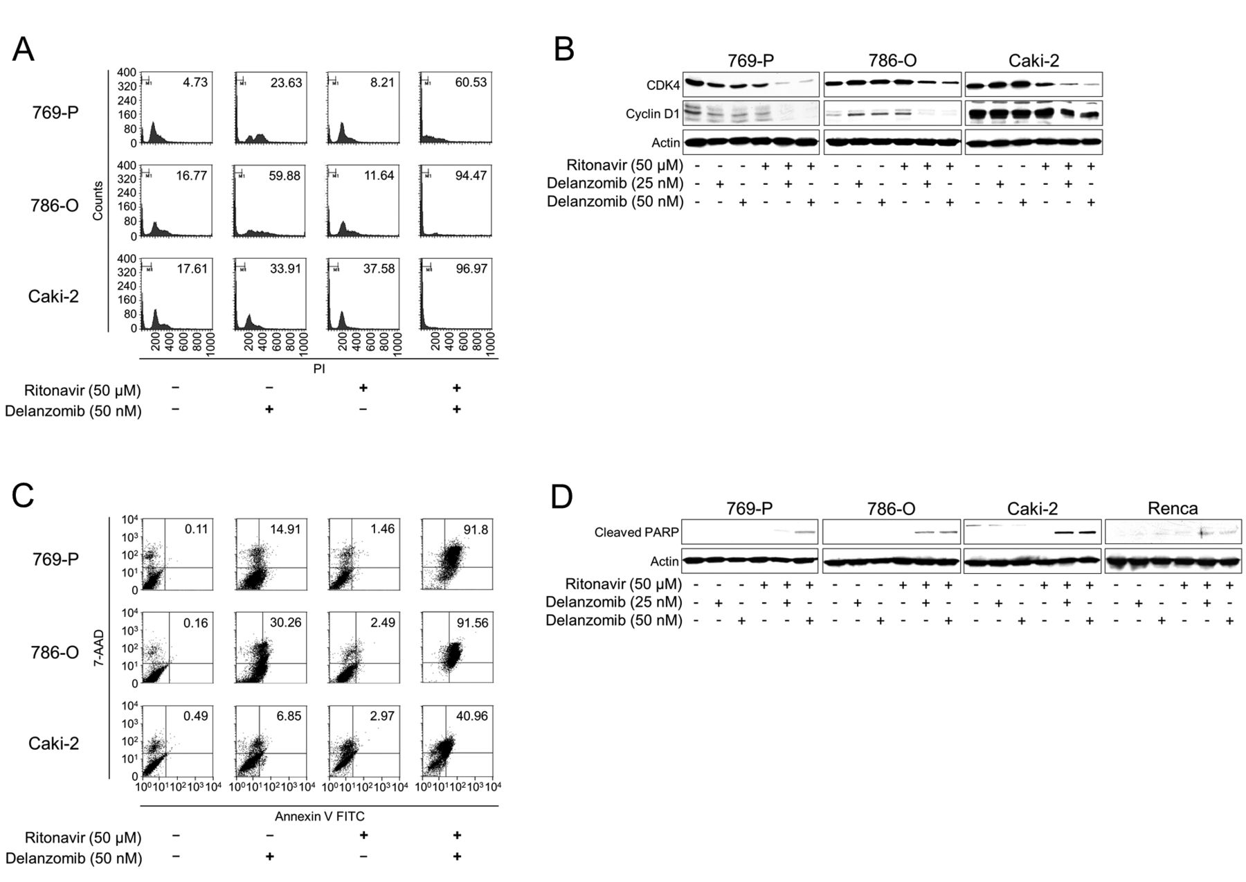
- Figure 2.
Combination of ritonavir and delanzomib perturbed the cell cycle and induced apoptosis of renal cancer cells. A: Cell-cycle analysis. Cells were treated for 48 h with 50 μM ritonavir with/without 50 nM delanzomib. Ten thousand cells were counted and changes in the cell cycle were evaluated using flow cytometry. The inset number in each graph shows the percentage of cells in the sub-G1 fraction. Representative results of flow cytometry are shown. B: Western blot analysis for cyclin D1 and cyclin-dependent kinase 4 (CDK4). Cells were treated for 48 h with 25 or 50 nM delanzomib with or without 50 μM ritonavir. Actin was used as a loading control. Representative blots are shown. C: Annexin V assay. Cells were treated for 48 h with 50 μM ritonavir with/without 50 nM delanzomib. Ten thousand cells were counted and apoptotic cells were detected by annexin V assay using flow cytometry. The inset number in each graph is the percentage of annexin-V-positive cells. Representative results of flow cytometry are shown. D: Western blot analysis for cleaved poly(ADP-ribose) polymerase (PARP). Cells were treated for 48 h with 25 or 50 nM delanzomib with or without 50 μM ritonavir. Actin was used as a loading control. Representative blots are shown.
- Figure 3.
Combination of ritonavir and delanzomib induced endoplasmic reticulum (ER) stress and autophagy, inhibited mammalian target of rapamycin (mTOR) pathway, and caused histone acetylation in renal cancer cells. A: Western blot analysis for the ER stress markers glucose-regulated protein 78 (GRP78), endoplasmic reticulum resident protein 44 (ERp44), and endoplasmic oxidoreductin-1-like protein (ERO1-L), and for ubiquitinated proteins. Cells were treated for 48 h with 25 or 50 nM delanzomib with or without 50 μM ritonavir. Actin was used as a loading control. Representative blots are shown. B: Western blot analysis for ubiquitinated proteins. Cells were treated for 12, 24, and 48 h with 50 μM ritonavir and 50 nM delanzomib. Actin was used for the loading control. Representative blots are shown. C: Western blot analysis for the autophagy marker light chain 3 (LC3). Cells were treated for 48 h with 25 or 50 nM delanzomib with or without 50 μM ritonavir. Actin was used for the loading control. Representative blots are shown. D: Western blot analysis for sestrin 2, AMP-activated protein kinase (AMPK), mTOR, and S6 ribosomal protein (S6) after 48-h treatment with 25 or 50 nM delanzomib with or without 50 μM ritonavir. Actin was used as a loading control. Representative blots are shown. E: Western blot analysis for acetylated histone after 48-h treatment with 25 or 50 nM delanzomib with or without 50 μM ritonavir. Actin was used as a loading control. Representative blots are shown. F: Western blotting for histone deacetylase (HDAC) 1,-2,-3, and-6 after 48-h treatment with 25 or 50 nM delanzomib with or without 50 μM ritonavir. Actin was used as a loading control. Representative blots are shown.


Tables

In this issue


Jump to section
Related Articles
Cited By...
- The Dual Histone Deacetylase-Proteasome Inhibitor RTS-V5 Acts Synergistically With Ritonavir to Induce Endoplasmic Reticulum Stress in Bladder Cancer Cells
- Elucidation of the molecular interactions that enable stable interaction between HIV protease inhibitor ritonavir and human DNA repair enzyme ALKBH2: a molecular dynamics simulation study
- Panobinostat and Nelfinavir Inhibit Renal Cancer Growth by Inducing Endoplasmic Reticulum Stress
- Thiasyrbactins Induce Cell Death via Proteasome Inhibition in Multiple Myeloma Cells