


Abstract
Background/Aim: Contrast nanocarriers as drug-delivery systems, capable of selective delivery to cancer cells and solid tumors, are essential for the development of new diagnostic and therapeutic (theranostic) strategies. The present study aimed to investigate the loading efficiency of chitosan-based polymersomes with fluorescent contrast substances [quantum dots (QDs) and conventional organic dyes] and the possibility to control their release from the polymer matrix into cells by chemical modifications and electroporation. Materials and Methods: All investigated fluorophores were retained within the polymer globule via electrostatic and hydrophilic–hydrophobic interactions, without conjugation with the polymer. The fluorophore-loaded polymersomes were characterized by dynamic light scattering, zeta-potential titration, and fluorescence spectroscopy. The release of fluorophore from the polymersomes, passively or after electroporation, was detected by 5-step spin-ultrafiltration, combined with fluorescence spectroscopy of the upper phase (supernatant) of the filter unit. Passive intracellular delivery of the nanoparticles to HeLa cells was detected by fluorescence confocal microscopy. Results: The QDs were retained tightly and continuously in the polymer matrix, while the organic fluorophores [fluorescein isothiocyanate (FITC), FITC-dextran10,000 and FITC-dextran70,000] were released rapidly from the polymersomes. The detergent Brij significantly increased the retention of FITC-dextran10,000 in the polymer globule. Electroporation up to 1000 V/cm did not induce release of QDs from the polymersomes, but accelerated the release of Brij-treated FITC-dextran10,000 B from the polymer matrix. High-voltage pulses (over 750 V/cm) induced also fragmentation or aggregation of the nanoparticles. QD_labeled polymersomes penetrated passively in cancer cells after 24-hour incubation. Conclusion: The results suggest that QD-labeled polymersomes are appropriate fluorescent probes and a nano-drug delivery system with high tracing opportunities for in vitro and in vivo applications. Furthermore, loading polymersomes with organic dyes with different molecular weights (such as FITC-dextrans) is a simple model for visualizing and predicting the rate of release of small organic molecules (e.g. conventional drugs, other contrasts, stabilizers, and supplements) from the polymer matrix.
In the last 15 years, fluorescence-based detection has achieved prominent progress in the synthesis of new highly luminescent fluorophores, based on nanotechnologies (1). This has enabled the development of a new generation of fluorescent equipment. A wide range of fluorescent nanoprobes have been designed and tested in preclinical studies (2, 3). Currently, it is widely accepted that contrast nanocarriers as drug-delivery systems, which are capable of selective delivery to cancer cells and solid tumors, are essential for the development of new diagnostic and therapeutic (theranostic) strategies. The theranostic idea has received great recognition in pharmacy and medicine. The application of multimodal nanoparticles (loaded with different contrast or therapeutic agents) as target-specific molecules increases the precision of cancer treatment, which is guaranteed to reduce side-effects (4, 5).
Size-controlled long-circulating polymersomes are promising carriers for drug delivery (6). Polymersomes are constructed from different biodegradable copolymers, mainly based on their low (to zero) cytotoxicity and biocompatibility (3, 7). To be suitable carriers for therapeutic and diagnostic agents, the polymersome needs to be characterized by: stability in high-salt physiological fluids, high retention capability for contrast/drug agents and options for their controlled/sustained release (4).
Polymersomes are labeled with contrast agents for tracking their pharmacodynamics and accumulation in tissues and organs using different imaging techniques (e.g. optical imaging, magnetic resonance imaging, and positron-emission tomography). In the case of optical imaging, the most commonly used organic fluorophores are: fluoresceins, porphyrins, cyanines, rhodamines, etc. Most are poorly water-soluble and consequents poorly bioavailable, have a relatively low fluorescence quantum yield and are not attractive for in vivo application (1). They have a small-size and easily leave the polymer matrix if they are in free form.
There are two options for retaining small organic fluorophores in polymersomes. The first approach is to conjugate the fluorophore with polymer (8-10). The advantage of this method is due to the strong covalent bond between both substances, which is difficult to break. This bond allows a precise visualization of the location of the nanoparticles in biological subjects (cells and living organisms). However, the conjugation of the polymer with fluorophore can change the surface charge and behavior of the polymersomes, which can restrict their penetration into cells and tissues.
The second approach is the conjugation of fluorophore with dextran (of different molecular weights) and subsequent incorporation of fluorophore-dextran conjugates into the polymersomes (8-10). This approach allows evaluation of the rate and extent of release of the fluorophore from polymersomes. This can be used as a model for prediction of the rate and extent of release of drugs from nanoparticles, depending on their molecular weight and the structure of the polymer matrix. A disadvantage of this approach is associated with leakage of fluorophore-dextran from the nanoparticles, which restricts the accurate assessment of their penetration into and localization in living biological objects. Quantum dots (QDs) are one option to overcome these limitations. They are also preferred fluorescent markers for imaging of drug delivery in vitro and in vivo due to their unique physicochemical properties (6, 11).
The present study was directed to estimate the loading efficiency of chitosan-based polymersomes with QDs and conventional organic dyes, and the possibility to control their release from the polymer matrix by chemical modification and electroporation. Electroporation is a biophysical technique which increases membrane permeability due to creation of reversible pores after application of external electrical pulses (12). Electro-assisted delivery of nanoparticles in cancer cells or tissues by application of electrical pulses is reported to be a promising biophysical approach for local (targeted) internalization. Recently, we reported an increased voltage-dependent delivery of polymersomes into cancer cells and their accelerated internalization in solid tumors (6, 13, 14).
Materials and Methods
Chemicals and nanoparticles. QDs (Qdots®605 and Qdots®655 ITK™ non-tagged) were purchased from Invitrogen (Carlsbad, CA, USA). Fluorescein isothiocyanate (FITC) was purchased from Thermo Fisher Scientific. FITC-dextran (Mr 10,000), FITC-dextran (Mr 70,000), Brij, and Triton-X100 were purchased from Sigma-Aldrich (Steinheim, Germany).
All chemicals used in this study were analytical or high-performance liquid chromatography grade.
Water-soluble polymersomes were prepared from chemically modified chitosan as described by Lee et al. (15). Labeling of polymersomes with FITC, FITC-dextran or QDs was carried out by passive internalization of fluorophores in the polymer matrix after intensive vortexing for 2 min at room temperature. The size and charge of the nanoparticles were characterized by dynamic light scattering (DLS) and zeta-potential analyzer (Zetasizer Nano ZS™; Malvern Instruments, Malvern, UK). The size and approximate shape of the nanoparticles were also analyzed indirectly, using multistep ultrafiltration (30, 50, 100, and 300 k) on a Vivaspin 6 filter unit (Sartorius, Gottingen, Germany). Nanoparticle fluorescence was analyzed by fluorescence spectroscopy (Shimadzu RF-6000; Shimadzu, Kyoto, Japan).
Spin-ultrafiltration (multistep). To determine the loading efficiency of polymersomes with fluorophore, we conducted a series of experiments using multistep spin-ultrafiltration and spectrofluorimetry (Figure 1). In these experiments, we used different Vivaspin 6 ultrafiltration centrifuge tubes with polyethylene-sulphonyl membrane (Sartorius, Gottingen, Germany), depending on the molecular weight of fluorophore, to concentrate and purify samples with a volume of 2-6 ml.
Fifty microliters of the prepared nanoparticles were dispersed in 450 μl 10 mM phosphate-buffered saline (PBS, pH 7.4), then subjected to ultra-filtration five times with washing by the same buffer using Vivaspin-6 tubes at 4150 × g (4°C). The upper phase, containing nanoparticles, was re-dissolved to 500 μl with PBS and subjected to the next step of ultra-filtration. At each ultra-filtration step, the upper phase (supernatant) and lower phase (filtrate) were subjected to spectrofluorimetric analysis at appropriate spectral parameters for the particular fluorophores: (i) in the case of FITC: λex=492 nm, λem=525 nm; (ii) in the case of QD655: λex=420 nm, λem=655 nm.
Electroporation. The effect of electroporation on the incorporation of fluorophores and its release from polymer matrix was also investigated. An electroporator Chemopulse IV (Institute of Biophysics and Biomedical Engineering, Bulgarian Academy of Sciences, Sofia, Bulgaria), generating bipolar pulses, was used in the experiments (6). The instrument was equipped with a large voltage control in the limits of 100-2200 V, simplified operations, locking against illegal manipulations and enhanced protection against electrical hazards. The electrotreatment consisted of 16 biphasic pulses (burst of pulses), each of them 50+50 μs duration with 20 μs pause between phases and pause between bipolar pulses of 880 μs. Stainless steel electrodes with intra-electrode distance of 10 mm were used. In this study, electrical pulses with intensity of 100-2000 V/cm were applied.
Fluorescence confocal microscopy. The intracellular delivery of QD605-labeled polymersomes to HeLa cells (Sigma-Aldrich, Steinheim, Germany) was detected using fluorescence confocal microscopy (Olympus FV-1000; Olympus, Tokyo, Japan; excitation: 420 nm; emission filter: 575-630 nm).
Cells were grown in Dulbecco's modified Eagle's medium (Sigma-Aldrich), supplemented with 10% fetal bovine serum (Sigma-Aldrich), in a humidified atmosphere (37°C, 5% CO2). Cells were incubated with QD605- or QD655-labeled polymersomes for 24 h in a cell incubator. After incubation, cells were washed three times with PBS prior to imaging.
Results and Discussion
Loading efficiency and passive release of fluorophores from polymersomes. All investigated fluorophores were retained within polymer globules via electrostatic and hydrophilic–hydrophobic interactions, without chemical (covalent) conjugation with the polymer. The polymersomes were hydrophilic/amphiphilic and their surface was positively charged (ζ-potential was in the range 17-22 mV, depending on the type of incorporated fluorophore). Their average size was ~110 nm, which was determined by DLS. The nanoparticles were stable in high-salt physiological fluids such as 10 mM PBS and serum. Aggregation was not detected even after 24 h incubation with serum at 37°C (14).
The first step of the study aimed to investigate the loading capacity of polymersomes with small molecules (FITC) and to increase their retention in the polymer matrix by conjugating with dextran (FITC-dextran10,000 and FITC-dextran70,000) or by forming micelles with detergents. The data shown in Figure 2 demonstrate the dynamics of FITC fluorescence in the supernatant during 5-step ultrafiltration, which is representative of retention of an organic dye in the polymer matrix. FITC left the polymersomes very quickly – over 80% were out of polymer matrix after the first filtration (Figure 2). Better retention was detected for FITC-dextran10,000 and FITC-dextran70,000, at about 45% and 62%, respectively, after the first filtration (Figure 2). However, the rate of passive leakage of FITC-dextran from the polymersomes was also relatively high. The level of FITC fluorescence in the polymersomes was less than 20% of the initial level after the fifth filtration.
The use of detergent in the buffer (in which FITC-dextran10,000 was dissolved) significantly increased the retention of fluorophore in the polymer matrix and reduced its leakage (Figure 2). Two detergents were applied, Triton-X100 and Brij. In the presence of Triton-X100, ~82% of FITC-dextran10,000 was detected in the polymersomes after the first filtration, but only ~7% after the fifth filtration. In the presence of Brij, ~80% of FITC-dextran10,000 was detected in the polymersomes after the first filtration, and ~60% after the fifth filtration. Both detergents formed micelles with the fluorophores and suppressed leakage from the polymer globules. The retention effect of Brij was much better than that of Triton-X100. Neither detergent significantly affected the size of polymersomes as analyzed by DLS.
The data suggest that FITC-dextran is not an appropriate fluorescent marker for tracking polymersomes in vitro and in vivo due to the release of the fluorophore from the polymer matrix. However, FITC-dextran is an appropriate and cheap model for investigating the rate of release of different substances from the polymer matrix, depending on their size.
The second step of the study aimed to investigate the loading capacity of polymersomes with QDs. The fluorescence signal in the supernatant was very high even after five filtrations of QD-labeled polymersomes (Figure 2). Fluorescence was not detected in the filtrate.
The data indicate that QDs are appropriate fluorescence markers for performing quantitative monitoring of pharmacodynamics of polymersomes in vitro and in vivo. In this case, we can be sure that the origin of the fluorescence signal will be entirely due to the QDs within the nanoparticles rather than free QDs. Figure 3 demonstrates the penetration of QD605_labeled polymersomes in cancer cells after 24-h incubation. The fluorescence signal was detected predominantly in the cytoplasm. QD605, which were not coated by polymersomes, did not penetrate into the cells – no fluorescence was detected in this case. Moreover QD605_labeled polymersomes were successfully delivered passively in solid tumors in vivo after their intravenous injection, which was demonstrated on colon cancer-grafted mice (16, 17).
Electro-induced release of fluorophore from polymersomes. The next step of the study aimed to verify whether the application of electrical pulses affects the ability of the polymersomes to retain or release the fluorescence marker, as well as the effect of electroporation on the nanoparticle size. The experiments were performed on QD655_labeled polymersomes and FITC-dextran10,000_labeled Brij-treated polymersomes (Figure 4). The intensity of the electrical pulses varied in the range 200-1000 V/cm. The electroporation was applied after the second ultrafiltration, when the retention level was constant for FITC-dextran10,000_labeled Brij-treated polymersomes. The fluorescence intensity was registered (recorded) simultaneously in the supernatant and filtrate after one additional ultrafiltration step.
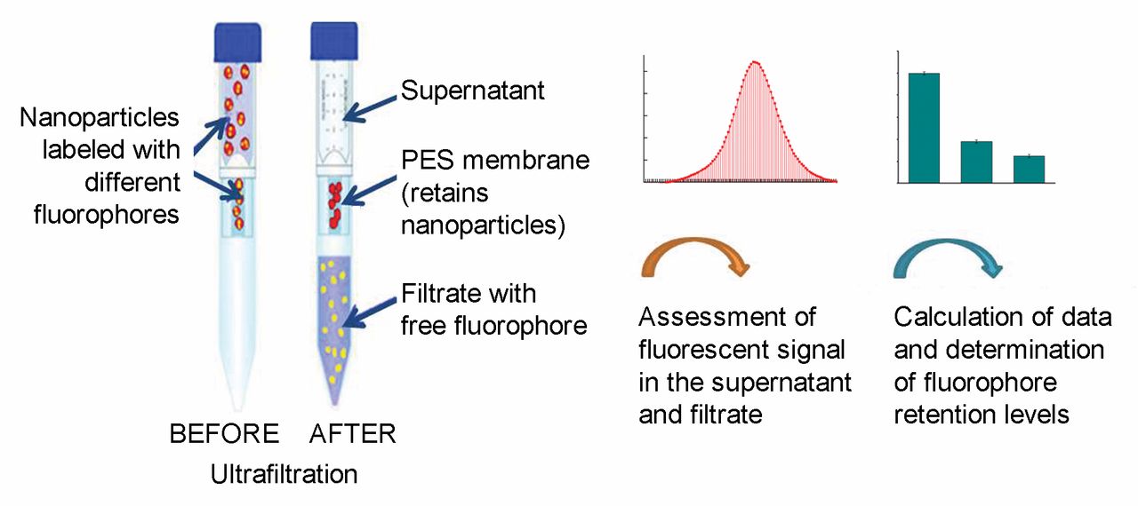
Schematic representation of the experimental design. PES: Polyethylene-sulphonyl membrane.
In the case of QD_labeled polymersomes, the fluorescence signal in the supernatant remained constant after electroporation (Figure 4A, left). The fluorescence intensity in the supernatant was same as before electroporation and no fluorescence was detected in the filtrate (Figure 4B, left). The data suggest that electroporation with pulse intensity up to 1000 V/cm did not induce a release of QDs from the polymer matrix. We also established that increase of electroporation intensity (2000 V/cm) resulted in the appearance of QD fluorescence in the filtrate, which indicates a disruption of the integrity of polymersomes and leakage of QDs from the matrix.
The effect of electroporation on the size of QD655_labeled polymersomes is shown in Table I. The size was not altered significantly up to 1000 V/cm, which indicates that the applied pulses did not affect the structure of the nanoparticles. However, two peaks were recorded in the DLS histograms at 1500 V/cm, at 88 nm and 313 nm. The fraction with small size could be explained by fragmentation and the fraction with large size by aggregation of the nanoparticles.
The fluorescence signal of FITC-dextran10,000_labeled Brij-treated polymersomes in the supernatant decreased after electroporation (Figure 4B, right) and FITC fluorescence appeared in the filtrate (Figure 4A, right). The electroporation accelerated the leakage of fluorophore from the polymer matrix.
The size of FITC-dextran10,000_labeled Brij-treated polymersomes did not change significantly up to 750 V/cm (Table II). However, the nanoparticle size decreased significantly after electroporation with 1000 V/cm and higher, which indicates fragmentation of the polymersomes.

Fluorescence intensity, obtained from the upper phase (supernatant) of Vivaspin filter, after 5-step ultrafiltration of fluorophore-loaded polymersomes. The data are means±SD from three independent experiments for fluorescein isothiocyanate (FITC); FITC-dextran10,000; FITC-dextran70,000; FITC-dextran10,000 plus Triton-X100 (1:32, mol:mol); FITC-dextran10,000 plus Brij (1:128, mol:mol); QD655. In the case of FITC: λex=492 nm, λem=525 nm. In the case of QD655: λex=420 nm, λem=655 nm.
In conclusion, the data demonstrate that the loading efficiency of FITC in the polymersomes was below 20%, accompanied by slow or faster release of fluorophores, depending on the size of conjugated dextran. Electroporation increased the release of FITC-dextran from the nanoparticles. In contrast, the loading efficiency of non-tagged QDs in the polymersomes was 100%. QDs were retained in the polymer globule even after 5-step ultrafiltration. Electroporation (at the selected conditions) did not induce a release of QD from the polymersomes. The results suggest that QD_labeled polymersomes are appropriate fluorescent probes and nano drug-delivery system with high tracing opportunities for in vitro and in vivo applications. Moreover, loading polymersomes with organic dyes of different molecular weights (such as FITC-dextrans) is a simple model for visualizing and predicting the rate of release of small organic molecules (e.g. conventional drugs, other contrasts, stabilizers and supplements) from the polymer matrix.\
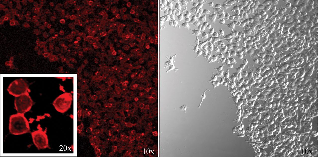
Intracellular delivery of QD605_labeled polymersomes to HeLa cells after 24-h incubation in a humidified atmosphere, as detected by fluorescence confocal microcopy (excitation – 420 nm; emission filter – 575-630 nm). QD605 concentration, 2 nmol/105 cells.
Size of quantum dot605_labeled polymersomes as determined before and after electroporation.
Size of fluorescein isothiocyanate-dextran10.000_labeled Brij-treated polymersomes as determined before and after electroporation.

A: Fluorescence intensity (upper panel), obtained from the upper phase (supernatant) of a Vivaspin filter, from fluorophore-loaded polymersomes: QD605_labeled (left) and fluorescein isothiocyanate (FITC)-dextran10,000_labeled Brij-treated polymersomes (right) after electroporation and subsequent ultrafiltration. The data are means±SD from three independent experiments. B: Representative fluorescence spectra of the supernatant and filtrate, obtained after electroporation of fluorophore-loaded polymersomes and subsequent ultrafiltration: left: QD605_labeled polymersomes, treated with 1000 V/cm (λex=420 nm); right: FITC-dextran10,000_labeled Brij-treated polymersomes, treated with 500 V/cm (λex=492 nm). In the case of FITC: λex=492 nm, λem=525 nm. In the case of QD655: λex=420 nm, λem=655 nm.
Acknowledgements
The study was supported by the Project № DFNP-17-137/01.08.2017 of the Program for Career Development of Young Scientists and Ph.D. students, Bulgarian Academy of Sciences (granted to S.S.), as well as by the Center of Innovation Stream from the Japanese Science and Technology Agency (JST, Japan).
- Received November 20, 2017.
- Revision received December 1, 2017.
- Accepted December 7, 2017.
- Copyright© 2018, International Institute of Anticancer Research (Dr. George J. Delinasios), All rights reserved
In this issue


Jump to section
Related Articles
Cited By...
- No citing articles found.