


Abstract
Background/Aim: We evaluated the association between positron emission tomography (PET) textural features and glucose transporter 1 (GLUT1) expression level and further investigated the prognostic significance of textural features in lung adenocarcinoma. Patients and Methods: We evaluated 105 adenocarcinoma patients. We extracted texture-based PET parameters of primary tumors. Conventional PET parameters were also measured. The relationships between PET parameters and GLUT1 expression levels were evaluated. The association between PET parameters and overall survival (OS) was assessed using Cox's proportional hazard regression models. Results: In terms of PET textural features, tumors expressing high levels of GLUT1 exhibited significantly lower coarseness, contrast, complexity, and strength, but significantly higher busyness. On univariate analysis, the metabolic tumor volume, total lesion glycolysis, contrast, busyness, complexity, and strength were significant predictors of OS. Multivariate analysis showed that lower complexity (HR=2.017, 95%CI=1.032-3.942, p=0.040) was independently associated with poorer survival. Conclusion: PET textural features may aid risk stratification in lung adenocarcinoma patients.
- Non-small cell lung cancer
- adenocarcinoma
- positron emission tomography
- texture analysis
- glucose transporter 1
Non-small cell lung cancer (NSCLC) accounts for 85% of all lung cancers and is the leading cause of cancer-related death (1). NSCLC is a heterogeneous disease, including both adenocarcinomas and squamous cell carcinomas. The biological behaviors and treatment-related outcomes of adenocarcinomas and squamous cell carcinomas differ (2, 3). Adenocarcinoma is the most common subtype of NSCLC and patients with early-stage lung adenocarcinomas exhibit a broad prognostic spectrum. Although recent advances in targeted therapy are impressive, survival rates remain unsatisfactory.
18F-fluorodeoxyglucose (FDG) positron emission tomography (PET) is widely used for staging NSCLC, to assess treatment response, and to predict prognosis (4-6). The glucose transporter 1 (GLUT1) plays an important role in FDG transport; FDG-PET assesses tumor metabolism in a noninvasive manner. A previous study suggested that the significance to be attributed to FDG accumulation varied by NSCLC histology in terms of tumor glucose metabolism and estimation of prognosis (7). Elevated GLUT1 expression correlated with high-level FDG uptake by lung adenocarcinomas but not squamous cell carcinomas (8, 9). High-level FDG uptake by the primary tumor was associated with poor prognoses of early-stage lung adenocarcinoma patients (10, 11). Therefore, the utility of PET may differ depending on NSCLC histology.
Recently, textural analysis of PET data has emerged as a new tool for assessing intratumoral heterogeneity. Increasing evidence suggests that heterogeneity evidenced by FDG-PET is associated with diagnosis of malignancy and/or poor prognosis (12, 13). Several studies showed that quantitative imaging features may predict the prognosis of NSCLC patients (14-16). One study found that adenocarcinomas and squamous cell carcinomas differed in terms of metabolic heterogeneity (17). Thus, we hypothesized that the GLUT1 expression level might affect the PET textural features of lung adenocarcinoma. We investigated the association between GLUT1 expression levels and PET textural features in lung adenocarcinoma patients, and the prognostic utility of textural features.
Patients and Methods
Patients. We reviewed the medical records of 139 consecutive lung cancer patients with adenocarcinomas who underwent FDG-PET/CT prior to surgical resection between January 2009 and December 2013 at Ajou University Hospital, Suwon, South Korea. We excluded 34 patients with tumor diameters <2 cm because their spatial resolutions were limited, creating partial-volume effects when PET imaging was used to explore tracer distribution. Ultimately, we included a total of 105 adenocarcinoma patients. For comparative purposes, we also reviewed the PET images of 70 stage-matched squamous cell carcinoma patients. This retrospective study was approved by the Ethics Committee of our institution and, given the nature of the work, the requirement for informed consent was waived.
FDG PET/CT protocol. FDG-PET/CT was performed using a Discovery STE scanner (GE Healthcare, Milwaukee, WI, USA). All patients fasted for at least 6 h prior to scanning; all serum glucose levels at the time of FDG injection were <150 mg/dl. Unenhanced CT was performed 60 min after the injection of 5 MBq/kg FDG, using 16-slice helical CT (120 keV; 30-100 mA in the AutomA mode; section width 3.75 mm). Emission PET data were acquired from the thigh to the head at 3.0 min/frame in the three-dimensional mode. Attenuation-corrected PET images derived from the CT data were reconstructed with the aid of an ordered-subset, expectation maximization algorithm (20 subsets, 2 iterations).
FDG PET image analysis. PET textural analysis has been described in detail in a previous report (18). To delineate tumors, we used the gradient-based segmentation method (‘PET Edge’) of MIM version 6.4 software (MIM Software Inc., Cleveland, OH, USA). Textural analysis quantifying PET-revealed intratumoral heterogeneity was performed with the aid of the Chang-Gung Image Texture Analysis toolbox (CGITA, http://code.google.com/p/cigita); this is an open-source software package implemented in MATLAB (version 2012a; MathWorks Inc., Natick, MA, USA) (19). Of the many textural matrices available in CGITA, we employed principally the neighborhood gray-tone difference matrices (NGTDMs); coarseness, contrast, busyness, complexity and strength. Conventional PET parameters of primary tumors, including the maximum SUV, the metabolic tumor volume (MTV), and total lesion glycolysis (TLG), were also measured.
Histopathological analysis and immunohistochemistry. Histological subclassification was performed by a single pathologist (YWK) using the IASLC/ATS/ERS classifications (20). A representative tumor-section paraffin block (a donor block) was selected for each case, and two 2-mm-diameter tumor cores were obtained using a trephine apparatus. The trephinated paraffin cores were placed in plastic molds, which were then filled with liquid paraffin and cooled. An automatic immunohistochemical staining device (Benchmark XT; Ventana Medical Systems, Tucson, AZ, USA) was used to stain formalin-fixed, paraffin-embedded tissue sections. Briefly, 5-μm-thick sections were transferred to poly-L-lysine-coated adhesive slides and dried at 62°C for 30 min. After standard epitope retrieval for 30 min in ethylenediaminetetraacetic acid (pH 8.0) in the autostainer, the samples were incubated with a rabbit polyclonal antibody against cleaved GLUT1 (dilution 1:100; Cell Marque, Rocklin, CA, USA); subsequently incubated with biotinylated anti-mouse immunoglobulin, peroxidase-conjugated streptavidin (LSAB kit, DAKO, Glostrup, Denmark), and 3,3’-diaminobenzidine; and then counterstained with Harris' hematoxylin. GLUT1 expression levels (percentages) were measured.
Clinical characteristics of 105 patients.
Difference of PET parameters according to GLUT1 expression.
Statistical analysis. Differences between the two groups were compared using the unpaired t-test for continuous variables. The variables included in the survival analysis were age, sex, pathological tumor stage, pathological node stage, American Joint Committee on Cancer (AJCC) stage, treatment modality, conventional PET parameters, and NGTDM parameters derived from FDG-PET. In statistical analyses, continuous variables were grouped into two categories by reference to the median values, with the exception of age. Overall survival (OS) was used as a measure of patient outcome and was defined as the period from initial diagnosis to death from any cause. Survival curves were estimated using the Kaplan-Meier method, and differences between the two groups were compared using the log-rank test.
Univariate and multivariate Cox regression analysis for overall survival using PET parameters.
Results
Patient characteristics. The demographic data of the 105 adenocarcinoma patients are listed in Table I. Sixty-three patients (60.0%) were male and 42 (40.0%) were female, with a median age of 62 years. Most primary tumors (92.4%) were of stage T2. The mean tumor diameter was 3.9±1.7 cm. Of all patients, 43 (40.9%), 25 (23.8%), and 37 (34.3%) were of stages I, II, and III, respectively. Sixty patients (57.1%) underwent adjuvant chemotherapy or radiotherapy after surgical treatment. The median follow-up time was 39.4 months.
Association between GLUT1 expression and PET parameters. Significant correlations were evident between the GLUT1 expression level and the MTV (r=0.273, p=0.005), coarseness (r=−0.266, p=0.006), and busyness (r=0.262, p=0.007). No significant association was apparent between GLUT1 expression level and any of SUVmax, TLG, contrast, complexity, or strength.
We divided the tumors into two groups (exhibiting low- and high-level GLUT1 expression) using the median GLUT1 expression level (50%). Table II summarizes differences in the PET parameters of the two groups. Some NGTDMs and conventional PET parameters differed significantly between the groups. Tumors expressing high levels of GLUT1 exhibited significantly higher SUVmax, MTV, and TLG values than tumors expressing low levels of GLUT1. Of the NGTDM parameters, tumors expressing high levels of GLUT1 exhibited significantly lower coarseness, contrast, complexity, and strength, but significantly higher busyness, than tumors expressing low levels of GLUT1.
Survival analysis. At the time of study completion, 41 patients (39.0%) had deceased. On univariate analysis, MTV, TLG, contrast, busyness, complexity, and strength were significant predictors of OS (Table III). After adjusting for age, AJCC stage, and prescription of adjuvant therapy, multivariate analysis showed that lower complexity (HR=2.017, 95%CI=1.032-3.942, p=0.040) was independently associated with poorer survival. The Kaplan-Meier curve showed that survival was significantly poorer in patients with tumors of lower complexity (<44; Figure 1).
Patients with tumors expressing high levels of GLUT1 had a lower OS than those with tumors expressing low levels of GLUT1 (p=0.004). When compared with overall stage-matched squamous cell carcinoma patients (n=70), patients with adenocarcinomas of high complexity showed a better prognosis than squamous cell carcinoma patients; adenocarcinomas of low complexity were associated with the poorest prognosis (Figure 2).
Discussion
Several studies evaluated the clinical significance of PET textural features in NSCLC patients (14-17); however, few studies evaluated lung adenocarcinoma as distinct from all histological types of NSCLC. Our study is important because accumulating evidence suggests that glucose metabolism differs between adenocarcinomas and squamous cell carcinomas in NSCLC patients (7, 9, 21). We included 105 adenocarcinoma patients who underwent surgery, and evaluated the prognostic significance of local textural features derived from NGTDMs, which evaluate differences between each voxel and neighboring voxels on adjacent image planes, thus functioning in a manner very similar to human perception of the image (22). We hypothesized that GLUT1 expression levels might affect local textural features, and found that the expression level was weakly associated with the coarseness and busyness of lung adenocarcinomas. In addition, tumors expressing high levels of GLUT1 differed significantly in terms of textural features and metabolic parameters from tumors expressing low levels of GLUT1. Thus, spatial heterogeneity of FDG uptake, directly associated with GLUT1 expression within a tumor, may contribute to the development of local textural features.

Kaplan-Meir survival curve for overall survival according to complexity. Median was used to the determine cut-off.
We found that the complexity of local textural features was independently associated with the OS of lung adenocarcinoma patients although no PET textural features have been widely accepted for lung cancer yet. Our results support the suggestion that intratumoral heterogeneity measured via PET textural analysis is associated with survival. Interestingly, patients with adenocarcinomas of high complexity survived better than did squamous cell carcinoma patients, and those with adenocarcinomas of low complexity had the poorest prognosis. Thus, evaluation of PET textural features may improve risk stratification of NSCLC patients.
Our study had several limitations. The work was retrospective in nature and the patients had cancers of various stages, although all underwent surgical resection with or without adjuvant treatment. We did not include the status of epidermal growth factor receptor mutations to the analysis due to the unavailability of the data for the whole cohort of patients.
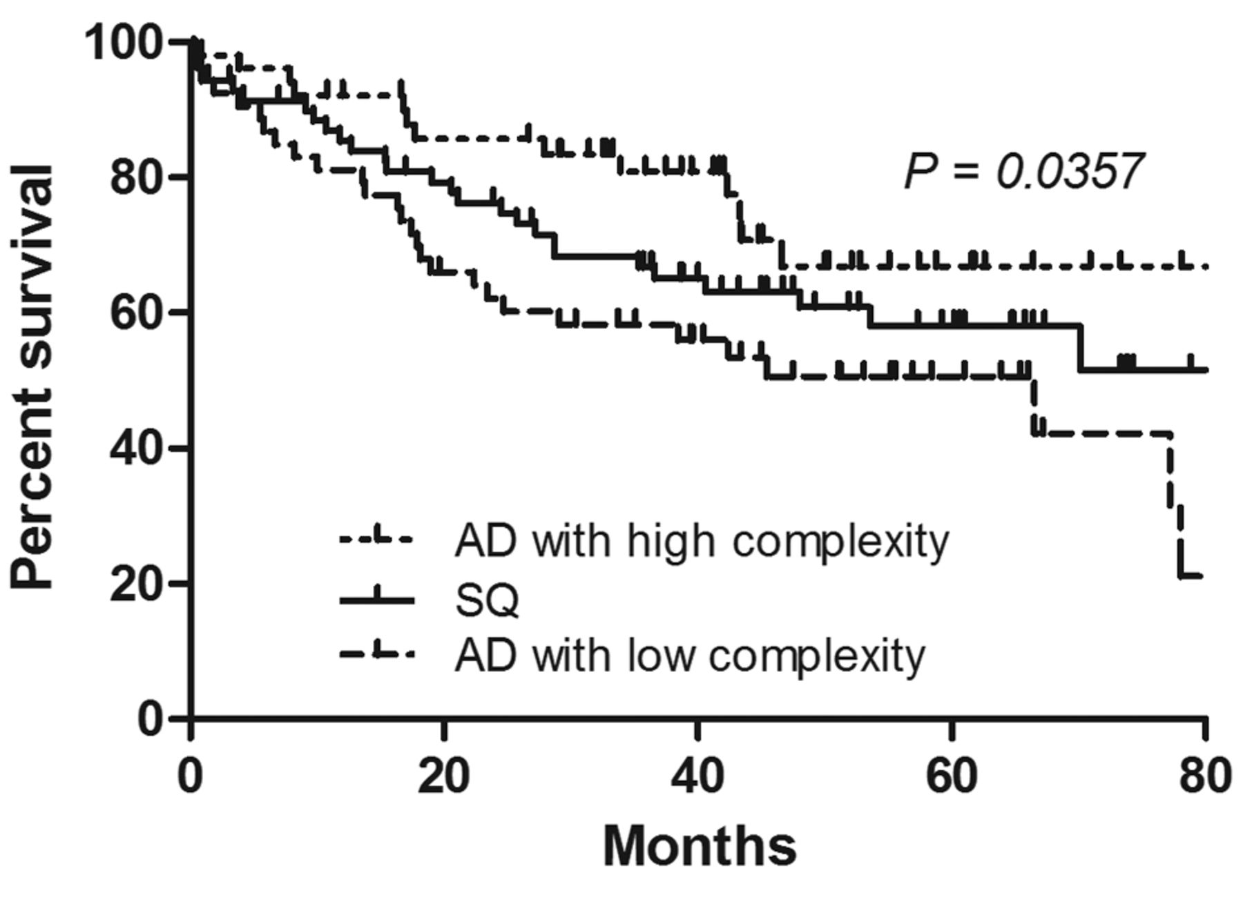
Overall survival according to histological subtype and complexity in adenocarcinoma. AD: Adenocarcinoma; SQ: squamous cell carcinoma.
Conclusion
GLUT1 expression level may influence local PET textural features. Intratumoral heterogeneity as assessed via PET textural analysis may improve risk stratification of patients with lung adenocarcinomas.
Acknowledgements
This study was supported by Basic Science Research Program through the National Research Foundations of Korea (NRF) funded by the Ministry of Education, Science and Technology (NRF-2016R1C1B2011583).
- Received November 20, 2017.
- Revision received November 27, 2017.
- Accepted November 28, 2017.
- Copyright© 2018, International Institute of Anticancer Research (Dr. George J. Delinasios), All rights reserved


