


Abstract
Aim: The aim of the present study was to investigate the efficacy of the traditional Chinese medicine (TCM), astragaloside IV (AS-IV) and curcumin on tumor growth and angiogenesis in an orthotopic nude-mouse model of human hepatocellular carcinoma (HCC). We have previously shown the usefulness of orthotopic models of human cancer for evaluation of the efficacy of TCM. Materials and Methods: Nude mice with orthotopic HepG2 HCC were treated with vehicle control (0.01 ml/g normal saline), cisplatinum (2 mg/kg), AS-IV (20 mg/kg), curcumin (100 mg/kg) or AS-IV plus curcumin (20 mg/kg + 100 mg/kg). Tumor inhibition in each group was evaluated by tumor weight at autopsy. The effect of AS-IV and curcumin on tumor angiogenesis was assessed by CD34 staining and expression of fibroblast growth factor-2 (FGF2), matrix metalloproteinase 2 (MMP2), vascular endothelial growth factor (VEGF), hepatocyte growth factor (HGF), thrombosis-related factor tissue factor (TF) and coagulation factor VII (FVII), as well as microRNAs miR-122 and miR-221. Results: AS-IV and curcumin alone and in combination significantly reduced mean tumor weight compared to vehicle control (p<0.05). Tumor microvessel count was reduced by AS-IV and curcumin alone. Expression of FGF2, MMP2, VEGF, HGF, TF and FVII was reduced by AS-IV and curcumin alone. AS-IV and curcumin alone up-regulated expression of miR-122 and down-regulated that of miR-221. The combination of AS-IV and curcumin demonstrated significant synergistic effects on microvessel count as well as on expression of angiogenic and thrombosis-related factors and microRNAs. Conclusion: The present study indicates future clinical potential of combination therapy with AS-IV and curcumin for HCC.
Hepatocellular carcinoma (HCC) is a highly aggressive and hypervascular tumor, associated with high morbidity (1), and is recalcitrant to therapy (2). Targeting angiogenesis could be an effective therapeutic strategy for controlling the progression of HCC.
Traditional Chinese Medicine (TCM) has been widely used for anticancer treatment in China with a long history. Astragaloside IV (AS-IV) is a representative constituent of Astragalus membranaceus (Fisch.) Bge. Var. mongholicus (Bge.) Hsiao and has various pharmacological activities, including anti-tumor, anti-inflammatory and anti-diabetic (3, 4). Curcumin (diferuloylmethane) is the chief component of the spice turmeric and is derived from the rhizome of the East Indian plant Curcuma longa. Curcumin exhibits anti inflammatory, antioxidant and anticancer efficacy (5). AS-IV was reported to reduce invasiveness and angiogenesis of gastric cancer cells and down-regulate the expression of fibroblast growth factor-2 (FGF-2) and suppress angiogenesis of HCT-116 colon cancer cells (6, 7). Curcumin showed anti-metastatic and anti-angiogenesis efficacy against small-cell lung cancer through inhibition of matrix metalloproteinase-2 and -9 (MMP-2/-9) activation and down-regulating signal transduction and activator of transcription 3 (STAT3) expression (8, 9). However, anti-angiogenic efficacy of AS-IV and curcumin on HCC has not been investigated to our knowledge.
Our laboratories have demonstrated the usefulness of orthotopic nude-mouse models of human cancer for the evaluation of efficacy of TCM herbal mixtures and compounds (10-14).
In the present study, we used a nude-mouse orthotopic liver-cancer model to evaluate the anti-angiogenic and anti-tumor efficacy of AS-IV and curcumin, alone and in combination. Expression of angiogenesis-related factors and genes in the treated tumors was examined to explore the underlying anti-angiogenic mechanism of these herbal agents.
Materials and Methods
Cell culture. The human hepatocarcinoma cell line HepG2 was obtained from Yangzhou College, Yangzhou, China and cultured in RPMI-1640 medium (GIBCO, Grand Island, NY, USA) with 10% calf serum (Tianhang Biotechnology Company, Zhejiang, China) plus 1% penicillin and streptomycin at 37°C in a 5% CO2 atmosphere. Logarithmic-growth-phase HepG2 cells (5×10⁶) were treated with 0.25% trypsin (GIBCO) and suspended in 200 μl serum-free RPMI-1640 for subsequent injection into nude mice
HCC orthotopic mouse model. Male BALB/c nude mice (5-6 weeks old) were purchased from Slack Laboratory Animal Company (Shanghai, China). Animal care was in accordance with institutional animal care guidelines. All mice were maintained in a HEPA-filtered environment at 24-25°C, and humidity was maintained at 50-60%. All animals were fed with autoclaved laboratory rodent diet. Animal experiments were approved by the Animal Committee of Nanjing Origin Biosciences, China (OR1507).
Stocks of HepG2 tumors were established by subcutaneously injecting HepG2 cells (5×10⁶) into the flank of nude mice. The subcutaneous tumors were harvested at the exponential growth phase and resected under aseptic conditions. Necrotic tissues were removed, and viable tissues were cut with scissors and minced into 1 mm3 pieces. For orthotopic tumor implantation, animals were anesthetized by injection of 0.02 ml of solution of 50% ketamine, 38% xylazine, and 12% acepromazine maleate. The surgical area was sterilized with povidone iodine. A transverse incision was made below the right costal margin. A tumor fragment was transplanted into an incision within the liver parenchyma. To prevent bleeding, the implanted tumor was dabbed with sterile cotton. The abdominal wound was closed with 5-0 silk sutures. All surgical procedures and animal manipulations were conducted under HEPA-filtered laminar-flow hoods with a ×8 surgical microscope (Shanghai Precision Instruments, Shanghai, China).
Treatment. Astragaloside IV (AS-IV) (98.3% purity by high-performance liquid chromatography [HPLC]) and curcumin (98.2% purity by HPLC) were obtained from Zelang Medical Technologicy (Nanjing, China). Cisplatinum was purchased from Qilu Pharmaceutical (Shandong, China). The mice were randomly divided into five groups of eight after the third day of orthotopic tumor implantation. The vehicle-control group was given saline via oral gavage at 0.01 ml/g. The cisplatinum group was treated by intraperitoneal injection at a dose of 2 mg/kg. The AS-IV group was treated via oral gavage at a dose of 20 mg/kg. The curcumin group was treated via oral gavage at a dose of 100 mg/kg. The combination group (AS-IV plus curcumin) received AS-IV and curcumin via oral gavage at doses of 20 mg/kg and 100 mg/kg, respectively. The cisplatinum group was treated once every 3 days for 21 days. The control, AS-IV, curcumin and AS-IV plus curcumin groups were treated daily for 21 days. Mouse body weight and clinical signs were recorded over the course of the experiments. All mice were sacrificed 2 days after the last treatment. At autopsy the tumor was removed and weighed. The inhibition of tumor in each group was calculated with the formula: inhibition (%)=100 × (mean tumor weight of the control group – mean tumor weight of the treated group)/mean tumor weight of the control.
Immunohistochemistry. Tumor tissues collected at autopsy were fixed in 10% buffered formalin, paraffin-embedded and sliced into 4 μm sections for immunohistochemical staining of hematopoietic-progenitor-cell antigen CD34 and vascular-endothelial growth factor (VEGF). Slides were deparaffinized in xylene, followed by rehydration in ethanol. Endogenous peroxidase was blocked by incubation with 3% hydrogen peroxide. The slides were incubated with primary antibody to CD34 (ab81289, 1:100; Abcam, Cambridge, UK) and to VEGF (ab46154, 1:200; Abcam) at 37°C for 2 h. After washing with phosphate-buffered saline, the slides were incubated with the secondary antibody labeled by horseradish peroxidase (HRP) at room temperature for 30 min. The sections were counterstained with Mayer's hematoxylin. The whole slide was first viewed at ×100 magnification in order to identify a ‘hot spot’ representing the area of the highest vessel density. The field was then switched to ×400 magnification for analysis. The microvessel count (MVC) represented by CD34 staining was calculated as the average number of CD34+ vessels in four fields of view. The positively-stained areas and the integral optical density (IOD) of VEGF in each field were analyzed by Image-Pro Plus 6.0 software (Media Cybernetics, Silver Spring, MD, USA). The final VEGF value was calculated from five randomly-selected fields of each section using the following formula: mean OD value= (IOD/positive area).
Enzyme-linked immunosorbent assay (ELISA). Blood was collected and serum was prepared for all mice at the end of the study. The serum FGF-2 and MMP-2 levels were determined with ELISA kits (Xinbosheng biotechnology, Nanjing, China) according to their instructions. Briefly, the flat-bottom 96-well microtiter plates were coated with anti- FGF-2 and anti-MMP-2 and incubated overnight at 4°C then blocked with bovine serum albumin buffer at 37°C for 3 h. The wells were then loaded with diluted serum samples or standard (100 μl/well) and incubated at 37°C for 2 h. The plates were washed five times and incubated with biotinylated anti-FGF-2 and anti-MMP-2 at 37°C for 1 h. After washing five times, the plates were incubated with horseradish-peroxidase-labeled avidin at 37°C for 30 min. After washing five times, the plates were incubated with tetramethylbenzidine substrate (100 μl/well) at 37°C for 30 min. The color reaction was stopped by the addition of H2SO4 (2 mol/l, 50 μl/well). The optical density (OD) was then measured at 450 nm using a microplate reader (ELx800uv; Bio-Tec Instruments, Houston, TX, USA). The concentrations of FGF-2 and MMP-2 were calculated according to calibration curve.
Primer sequences for real-time polymerase chain reaction (PCR).
Real-time polymerase chain reaction (RT-PCR). Total RNA was isolated from tumor tissues collected at autopsy in 1 ml TRIzol reagent (Invitrogen Life Tech, Carlsbad, CA, USA). Real-Time PCR was performed in a final volume of 20 μl containing 1 μl cDNA template, 2 μl primer, 10 μl of SYBR Green Master Mix (TOYOBO, Japan), and 7 μl diethy pyrocarbonate water (KeyGen BioTech, Nanjing, China). Conditions used for PCR consisted of 40 cycles at 95°C for 5 min, 95°C for 15 s, 60°C for 20 s, and 72°C for 40 s. The melting curve was analyzed at the temperature range of 65-95°C. Primers were designed as shown in Table I. Glyceraldehyde 3-phosphate dehydrogenase (GAPDH) served as an internal reference for thrombosis-related factor tissue factor (TF), coagulation factor VII (FVII) and hepatocyte growth factor (HGF). U6 snRNA served as an internal references for hsa-mir-122a and hsa-miR-221. Gene expression was calculated using the 2−ΔΔCt method.
Western blotting. Protein from tumor tissues collected at autopsy was extracted with RIPA buffer (Beyotime BioTech, Shanghai, China). Protein concentration was determined with the Bradford protein assay kit (KeyGen BioTech). Total proteins were separated by sodium salt-polyacrylamide gel electrophoresis and electrophoretically transferred to a polyvinylidene fluoride membrane (Millipore, Bedford, MA, USA). The membranes were incubated with 5% nonfat milk solution for blocking non-specific binding and with primary antibodies to TF ((ab104513; 1:400), anti-FVII (ab97614; 1:600), anti-HGF (ab178395; 1:800) (Abcam). After washing twice with tris-buffered saline, the membranes were incubated with appropriate secondary antibodies conjugated with HRP for 2 h at room temperature. Analysis of electro-chemi-luminescence was performed according to the manufacturer's instructions using a Bio-Rad imaging system and Quantity One version software (Bio-Rad, Hercules, CA, USA) was used to quantify the density of bands.
Statistical analysis. All data are presented as the mean values±standard deviation and were analyzed using SPSS 17.0 software (Chicago, IL, USA). Comparisons between different groups were conducted using a one-way ANOVA, with least significant difference used for inter-group comparison. A value of p<0.05 was considered to indicate a statistically-significant difference.
Results
AS-IV and curcumin inhibited tumor growth. The effect of AS-IV and curcumin on tumor growth was evaluated in the HepG2 HCC orthotopic mouse model. Tumor weight was significantly reduced by cisplatinum, AS-IV, curcumin, and AS-IV plus curcumin as compared to the vehicle control (p<0.05) (Figure 1). A greater inhibitory efficacy on tumor growth was observed with the AS-IV plus curcumin combination than AS-IV or curcumin alone, although statistical significance was not achieved (p>0.05) (Figure 1).
AS-IV and curcumin reduced tumor vascularization. As shown in Figure 2A, CD34-positive staining was indicated by the presence of brown particles. In the vehicle-control group, there was a notable increase in CD34-positive staining in tumor tissues and the capillary networks were misaligned, tortuous and irregular in the tumor tissues of all groups. Decreased MVC was found in AS-IV, curcumin and AS-IV plus curcumin combination groups compared with the vehicle control and cisplatinum groups (p<0.01). The combination-treated group showed a significantly greater decrease in MCV than did the groups treated with AS-IV, or curcumin alone (p<0.05), indicating synergistic anti-angiogenic efficacy of the AS-IV and curcumin combination (Figure 2B).
AS-IV and curcumin down-regulated expression of angiogenic factors. The expression of VEGF in tumors of all treatment groups was significantly down-regulated compared with those of the vehicle control (p<0.01). The lowest VEGF expression was found in the AS-IV–curcumin combination group compared to the cisplatinum-, AS-IV-, and curcumin-treated groups of mice (p<0.01) (Figure 3A and B).

Efficacy of astragaloside IV (AS-IV) and curcumin on tumor growth in the HepG2 HCC orthotopic nude-mouse model. A: Representative orthotopic liver tumors of each treated group at autopsy. B: Representative excised liver tumor of each treated group at autopsy. C: Mean tumor weight of each treated group. Results are presented as the mean±SD (n=8). aSignificantly different at p<0.05 vs. vehicle control. D: Percentage of tumor growth of each treated group relative to control.
The serum levels of FGF-2 and MMP-2 were significantly reduced in all treatment groups compared with the vehicle control (p<0.01).The serum FGF-2 and MMP2 levels in groups treated with AS-IV, curcumin, and AS-IV plus curcumin groups were lower than in the cisplatinum-treated group (p<0.01). The AS-IV–curcumin combination had the most effective inhibition of serum FGF-2 and MMP-2 among all treated groups (p<0.01) (Figure 3C and 3D).
HGF mRNA and protein expression was down-regulated by all treatments as compared with the vehicle control (p<0.01). The AS-IV plus curcumin combination-treated mice had significantly lower HGF mRNA expression than the other treated groups (p<0.01) (Figure 3E-G).
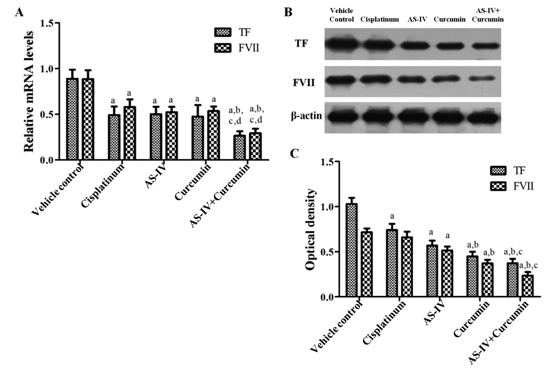
AS-IV and curcumin inhibited the expression of thrombosis-related factors. TF and FVII mRNA expression were down-regulated by cisplatinum, AS-IV, curcumin, and the combination of AS-IV with curcumin compared with the vehicle control (p<0.01) (Figure 4A).
The combination-treated group had significantly lower TF and FVII mRNA expression levels than the cisplatinum, AS-IV, and curcumin groups (p<0.01) (Figure 4A). AS-IV, curcumin, and the combination AS-IV+ curcumin decreased TF and FVII protein expression compared with the vehicle control (p<0.01. Significantly decreased TF and FVII protein expression was found in the AS-IV plus curcumin combination group compared with the cisplatinum- and AS-IV-treated groups (p<0.05) (Figure 4B and C).
The combination of AS-IV and curcumin up-regulated miR-122 expression and down-regulated miR-221 expression. The cisplatinum-, AS-IV-, and curcumin-treated groups did not show significant differences in miR-122 expression as compared with the vehicle control group (p>0.05). However, treatment with the AS-IV–curcumin combination significantly up-regulated the expression of miR-122 (p<0.01) (Figure 5A). miR-221 expression was significantly down-regulated in all treatment groups compared with the vehicle control (p<0.01). The AS-IV–curcumin combination group showed a greater decrease in miR-221 expression than did the other treatment groups (p<0.01) (Figure 5B).
Discussion
The aim of the present study was to investigate the efficacy of AS-IV and curcumin on the inhibition of tumor growth and angiogenesis in an orthotopic nude-mouse model of HCC. A new finding of our study is that AS-IV and curcumin in combination had a synergistic effect for treatment of HCC, leading to enhanced inhibition of tumor growth and angiogenesis in a mouse model of HCC. When examined for the mechanism, we found that this synergistic efficacy of AS-IV and curcumin was associated with regulation of some angiogenesis-related factors and microRNAs.
Angiogenesis is a dynamic process involving several key factors possessing pro-angiogenic activity. VEGF stimulates endothelial-cell proliferation and induces budding in neovascularization (15). FGF-2 binds to heparin sulfate proteoglycans initially, and then the complex binds to the FGF receptor, leading to proliferation and angiogenesis (16, 17). MMP-2 not only degrades type IV collagen, but also induces cellular proliferation and angiogenesis (18). HGF is a stromal-cell factor able to promote tumor angiogenesis through stimulation of vascular endothelial cell proliferation, migration and activation of the protein kinase B and extracellular signal-regulated kinases. Auyeung et al. (19) found that Astragalus saponins modulated angiogenesis in human gastric adenocarcinoma cells through inhibition of VEGF and MMP-2 protein. Di et al. (20) demonstrated that curcumin inhibited the transcript levels of two major angiogenesis factors, VEGF and FGF-2, in human breast cancer cells. Our results were consistent with these reports but demonstrated a greater inhibitory effect of AS-IV and curcumin in combination on these angiogenic factors than AS-IV or curcumin treatment alone.

Efficacy of astragaloside IV (AS-IV) and curcumin on tumor vascularization in the HepG2 HCC orthotopic nude-mouse model. A: Representative immunohistochemical staining for CD34 in the tumor at ×400 magnification. B: Microvessel count (MVC) of each treated group. Results are presented as the mean±SD. Significantly different at p<0.01 vs. avehicle control, bcisplatinum, cAS-IV and dcurcumin groups.
TF and FVII have been reported to contribute to tumor angiogenesis and metastasis progression by initiating the exogenous coagulation pathway in some cancer types (21-23). In this study, we show for the first time that suppression of TF and FVII may be involved in the anti-angiogenesis efficacy of AS-IV and curcumin.
MicroRNAs belong to an abundant family of endogenous, short and non-coding RNAs. MicroRNAs that have oncogenic or tumor-suppressive properties play an important role in cancer onset and progression. miR-122, as a tumor-suppressor gene, accounts for approximately 70% of the total miRNA in the liver (24) and inhibits metastasis of HCC through anti-angiogenic activity (25). miR-221 acts as an oncogene in HCC and is essential for angiogenesis by NOTCH signaling and induces proliferation and migration of endothelial cells (26). Our result suggests that up-regulation of miR-122 and down-regulation of miR-221 might be responsible for the anti-angiogenesis mechanisms of AS-IV and curcumin in HCC.

Efficacy of astragaloside IV (AS-IV) and curcumin on the expression of angiogenic factors. A: Representative immunohistochemical staining for vascular endothelial growth factor (VEGF) in the tumor at ×400 magnification. B: Optical density of immunohistochemistry staining for VEGF. C, D: Serum fibroblast growth factor-2 (FGF-2) (C) and matrix metalloproteinase-2 (MMP-2) (D) level in each treated group. E-G: hepatocyte growth factor (HGF) mRNA (E) and protein (F, G) expression in each treated group. Results are presented as mean±SD. Significantly different at p<0.01 vs. avehicle control, bcisplatinum, cAS-IV and dcurcumin groups.

Efficacy of astragaloside IV (AS-IV) and curcumin against the expression of thrombosis-related tissue factor (TF) and coagulation factor VII (FVII). mRNA (A) and protein expression (B,C) of TF and FVII in each treated group. Results are presented as the mean±SD. Significantly different at p<0.01 vs. avehicle control, bcisplatinum and cAS-IV groups.

Efficacy of astragaloside IV (AS-IV) and curcumin on the expression of miR-122 and miR-221. A: Tumor miR-122 expression in each treated group. Results are presented as the mean±SD (n=5). Significantly different at p<0.01 vs. avehicle control, bcisplatinum, cAS-IV and dcurcumin groups. B: Tumor miR-221 expression in each treated group. Results are presented as the mean±SD (n=5). Significantly different at p<0.01 vs. avehicle control, bAS-IV and ccurcumin groups.
We previously demonstrated the inhibitory efficacy of Celastrus orbiculatus Thunb on the growth of HCC tumor through VEGF inhibition (10). The present report indicates that AS-IV and curcumin are active against HCC in orthotopic models, further indicating the potential of TCM for HCC. We also previously showed that AS-IV and curcumin were active in an orthotopic model of ovarian cancer (14).
In conclusion, this study shows that AS-IV and curcumin alone and in combination inhibit tumor growth and angiogenesis in an orthotopic nude-mouse model of human hepatocellular carcinoma. AS-IV and curcumin demonstrated synergistic suppressive efficacy against angiogenic factors VEGF, FGF-2, MMP-2 and HGF, as well as thrombosis-related factors TF and FVII. Up-regulation of microRNAs miR-122 and down-regulation of miR-221 may mediate anti-angiogenesis mechanisms of AS-IV and curcumin in HCC. This study indicates the potential for AS-IV and curcumin TCM for treatment of HCC.
Acknowledgements
This research was supported by the National Natural Science Foundation of China (no. 81373990). We also thank all our colleagues in our research group for their generous support. In addition, special thanks go to Prof. Jiangang Shen (School of Chinese Medicine, University of Hong Kong) for revision of this article.
Footnotes
↵* These Authors contributed equally to this work.
This article is freely accessible online.
Conflicts of Interest
The Authors declare no conflict of interest for this study.
- Received November 21, 2016.
- Revision received December 16, 2016.
- Accepted December 20, 2016.
- Copyright© 2017, International Institute of Anticancer Research (Dr. George J. Delinasios), All rights reserved


