
Main menu

  • Home
  • Current Issue
  • Archive
  • Info for
    • Authors
    • Editorial Policies
    • Subscribers
    • Advertisers
    • Editorial Board
    • Special Issues
  • Journal Metrics
  • Other Publications
    • In Vivo
    • Cancer Genomics & Proteomics
    • Cancer Diagnosis & Prognosis
  • More
    • IIAR
    • Conferences
    • 2008 Nobel Laureates
  • About Us
    • General Policy
    • Contact
  • Other Publications
    • Anticancer Research
    • In Vivo
    • Cancer Genomics & Proteomics

User menu

  • Register
  • Subscribe
  • My alerts
  • Log in
  • My Cart

Search

  • Advanced search
Anticancer Research
  • Other Publications
    • Anticancer Research
    • In Vivo
    • Cancer Genomics & Proteomics
  • Register
  • Subscribe
  • My alerts
  • Log in
  • My Cart
Anticancer Research

Advanced Search

  • Home
  • Current Issue
  • Archive
  • Info for
    • Authors
    • Editorial Policies
    • Subscribers
    • Advertisers
    • Editorial Board
    • Special Issues
  • Journal Metrics
  • Other Publications
    • In Vivo
    • Cancer Genomics & Proteomics
    • Cancer Diagnosis & Prognosis
  • More
    • IIAR
    • Conferences
    • 2008 Nobel Laureates
  • About Us
    • General Policy
    • Contact
  • Visit us on Facebook
  • Follow us on Linkedin
Research ArticleExperimental Studies

Paclitaxel, Carboplatin and 1,25-D3 Inhibit Proliferation of Endometrial Cancer Cells In Vitro

TEA KUITTINEN, PÄIVI ROVIO, SYNNÖVE STAFF, TIINA LUUKKAALA, ANNE KALLIONIEMI, SEIJA GRÉNMAN, MARITA LAURILA and JOHANNA MÄENPÄÄ
Anticancer Research December 2017, 37 (12) 6575-6581;
TEA KUITTINEN
1Department of Obstetrics and Gynaecology, Tampere University Hospital, Tampere, Finland
  • Find this author on Google Scholar
  • Find this author on PubMed
  • Search for this author on this site
  • For correspondence: tea.kuittinen{at}xfimnet.fi
PÄIVI ROVIO
1Department of Obstetrics and Gynaecology, Tampere University Hospital, Tampere, Finland
  • Find this author on Google Scholar
  • Find this author on PubMed
  • Search for this author on this site
SYNNÖVE STAFF
1Department of Obstetrics and Gynaecology, Tampere University Hospital, Tampere, Finland
2Laboratory of Cancer Biology, BioMediTech Institute, Faculty of Medicine and Life Sciences, University of Tampere, Tampere, Finland
  • Find this author on Google Scholar
  • Find this author on PubMed
  • Search for this author on this site
TIINA LUUKKAALA
3Research and Innovation Centre, Tampere University Hospital, Tampere, Finland
4Health Sciences, Faculty of Social Sciences, University of Tampere, Tampere, Finland
  • Find this author on Google Scholar
  • Find this author on PubMed
  • Search for this author on this site
ANNE KALLIONIEMI
5BioMediTech Institute and Faculty of Medicine and Life Sciences, University of Tampere, Tampere, Finland
6Fimlab Ltd, Tampere University Hospital, Tampere, Finland
  • Find this author on Google Scholar
  • Find this author on PubMed
  • Search for this author on this site
SEIJA GRÉNMAN
7Department of Obstetrics and Gynaecology, Turku University Hospital, Turku, Finland
8University of Turku, Turku, Finland
  • Find this author on Google Scholar
  • Find this author on PubMed
  • Search for this author on this site
MARITA LAURILA
9Department of Pathology, Fimlab Ltd, Tampere University Hospital, Tampere, Finland
  • Find this author on Google Scholar
  • Find this author on PubMed
  • Search for this author on this site
JOHANNA MÄENPÄÄ
1Department of Obstetrics and Gynaecology, Tampere University Hospital, Tampere, Finland
10Faculty of Medicine and Life Sciences, University of Tampere, Tampere, Finland
  • Find this author on Google Scholar
  • Find this author on PubMed
  • Search for this author on this site
  • Article
  • Figures & Data
  • Info & Metrics
  • PDF
Loading

Abstract

Background/Aim: Endometrial cancer cells are known to be sensitive to carboplatin and paclitaxel. Furthermore, vitamin D (1,25-D3) has been reported to inhibit endometrial cancer cell growth both as a single agent and combined with carboplatin. However, there are no studies comparing the effect of paclitaxel and carboplatin as single agents vs. in combination in endometrial cancer cell lines. Neither has the effect of 1,25-D3 been studied with paclitaxel. The present study investigated the effect of paclitaxel, carboplatin and 1,25-D3 on the growth of endometrial cancer cells in vitro. Materials and Methods: Two endometrial adenocarcinoma cell lines (UT-EC-1 and UT-EC-3) were cultured with different doses of paclitaxel, carboplatin and 1,25-D3. The cellular VDR (vitamin D receptor) mRNA levels were measured and the expression of estrogen (ER) and progesterone (PR) receptors by the cells was determined. Results: In the UT-EC-1 cell line the growth inhibition was 72% with paclitaxel, 54% with carboplatin and 73% with the combination of these compounds. The corresponding numbers in UT-EC-3 were 70%, 33% and 65%, respectively. 1,25-D3 suppressed cell growth 88% with paclitaxel, 63% with carboplatin and 87% with their combination in the UT-EC-1 cell line. Conclusion: In both cell lines, single-agent paclitaxel was as effective as the combination of the compounds and more effective than single carboplatin. 1,25-D3 may further contribute to the cytotoxic effect of these agents.

  • Vitamin D
  • 1,25-D3
  • endometrial cancer in vitro
  • paclitaxel
  • carboplatin
  • growth inhibition
  • VDR

Endometrial cancer is the most common gynecological malignancy in developed countries. In Finland incidence is 13.9 per 100,000 persons a year (1). Most women are diagnosed with early-stage disease and have a high cure rate with surgery alone with a reported 5-year survival rate of 90% (2). Still up to 25% of women will have advanced, high grade or recurrent disease and require additional treatment. In these cases, the therapeutic response to cytotoxic and hormonal agents has been typically only partial and of short duration. There is no generally accepted standard chemotherapy in treatment of advanced and recurrent endometrial carcinoma but simultaneous administration of carboplatin and paclitaxel or carboplatin and pegylated liposomal doxorubicin are widely used (3-8).

Vitamin D is an antiproliferative and immunomodulatory secosteroid hormone with a well-established role in maintenance of calcium synthesis. Active metabolite of vitamin D, 1alpha,25-dihydroxyvitamin D3 (1,25-D3) binds to the vitamin D receptor (VDR) in the cell nucleus. The function and/or expression of the VDR is needed for the growth regulatory effects of 1,25-D3 in vitro and disabled VDR activity leads to 1,25-D3 insensitivity and accelerated tumorigenesis (9). Many different cell types, including normal and malignant endometrial cells, contain VDR and 1alpha hydroxylases to synthesize active 1,25-D3 locally (10, 11). This active metabolite does not enter the circulation and has no effect on calcium homeostasis, but is rather degraded by 24-hydroxylases.

1,25-D3 is well known for its antiproliferative roles including the induction of G0-G1 cell-cycle arrest with associated down-regulation of oncogenic cyclins D1 and D3 and p27/p21 induction (12-15). 1,25-D3 is responsible for VDR-mediated apoptosis and inhibits angiogenesis and differentiation in a variety of cancer types (13, 16-20). 1,25-D3 is a potential antitumor agent because of its ability to regulate cancer cell growth. 1,25-D3 has been found to inhibit endometrial cancer cell growth both as a single agent (21) and combined with carboplatin (22).

Endometrial cancer cells are sensitive to carboplatin, cisplatin and paclitaxel (23, 24). Still there are no studies comparing paclitaxel and carboplatin as single agents vs. in combination in endometrial cancer in vitro or in vivo. In addition, there are no studies examining the effect of vitamin D with paclitaxel or the combination of paclitaxel and carboplatin on endometrial cancer cells. The aim of the present study was to investigate the potential effects of paclitaxel, carboplatin and 1,25-D3 on the growth of endometrial cancer cells both as single agents and in different combinations with each other in vitro.

Materials and Methods

Cell lines. Two endometrial adenocarcinoma cell lines were used (UT-EC-1 and UT-EC-3) in this study. The cell lines were originally established from primary tumors before any treatment using an explant technique (25, 26). UT-EC-1 was established from a curettage specimen of a clinical FIGO Stage II Grade 2 endometrial papillary adenocarcinoma and UT-EC-3 from the hysterectomy specimen of a surgical FIGO Stage Ia Grade 3 endometrial adenocarcinoma (23). The biological properties of these cell lines have been described in detail earlier (23).

Cell cultures and treatments. Prior to the experiments the cells were cultured as described previously (23). In brief, the cells were plated weekly and maintained in logarithmic phase in 75 cm2 culture flasks in Dulbecco's modified Eagle minimal essential medium (DMEM), supplemented with 10% fetal bovine serum (FBS), 1% penicillin/streptomycin, 1% glutamine and 1% non-essential amino acids at 37°C in a humidified atmosphere containing 5% CO2. Both cell lines were tested negative for mycoplasma contamination.

Paclitaxel (Hospira® 6 mg/ml, Maidenhead, UK) and carboplatin (Accord® 10 mg/ml, Middlesex, UK) were provided by the Pharmacy of the Tampere University Hospital. 1,25-D3 was purchased from Sigma-Aldrich (St. Louis, MO, USA). Paclitaxel was diluted in 0.9% sodium chloride to get a 0.1 mM solution. Stock solutions of 100 nM were made in DMEM. Final concentrations used were 1-100 nM and fresh stock solutions were made for each experiment. Carboplatin was diluted in sterile water to get a solution of 100 μg/ml. The final dilutions used were 0.1-20 μg/ml. The dilutions were made immediately before use. 1,25-D3 was diluted in DMEM to get final 0.1-100 nM solutions. The concentrations of drugs were based on previous reports as well as physiological relevance (23, 24, 27, 28).

VDR, ER and PR expression analyses. The VDR mRNA levels in UT-EC-1 and UT-EC-3 cells were measured by qRT–PCR using the LightCycler equipment (Roche, Mannheim, Germany). Total RNA was extracted using the RNeasy Mini Kit (Qiagen, Valencia, CA, USA) and was reverse transcribed with SuperScript™ First-Strand Synthesis System for RT–PCR (Invitrogen, Carlsbad, CA, USA) as described (29). qRT-PCR was performed using 20 μM gene specific primers (Sigma-Aldrich, sense 5’ ATCGGCATGATGAAGGAGTT 3’, antisense 5’ TGCTCCTCAGACAGCTTGG 3’) and 10 μM UPL probe (probe number # 12). The PCR program included the following steps: 10 min denaturation at 95°C followed by 45 cycles of 10 s denaturation at 95°C, 30 s annealing at 60°C and 1 s elongation at 72°C. The expression levels were normalized using glyceraldehyde-3-phosphate dehydrogenase (GAPD) housekeeping gene.

The expression of estrogen (ER) and progesterone (PR) receptors in both cell lines was determined with immunohistochemical staining. ER and PR immunostainings were performed using Ventana Benchmark Ultra automated IHC/ISH Slide staining system (Ventana Medical Systems, AZ, USA). Deparaffinization and antigen retrieval were performed using Roche Ventana reagents (EZ Prep 10X concentrate and Cell Conditioning Solution CC1). ER staining was performed using a Roche Ventana RTU Confirm anti-Estrogen Receptor Rabbit monoclonal antibody (Clone SP1) with 32 min incubation time. PR staining was performed using a Roche Ventana RTU Confirm anti-Progesterone Receptor Rabbit monoclonal antibody (Clone 1E2) with 24 min incubation time. Both immunostainings were detected and visualized using Roche Ventana UltraView DAB IHC Detection Kit.

Cell growth assays. For each experiment, the cells were plated on 24-well plates at 30,000 cells/well. Cells were then treated with indicated doses of paclitaxel (1-100 nM), carboplatin (0.1-20 μg/ml) and vitamin D3 (0.1-100 nM) alone or in combination. After a 3-day incubation period, the cells were trypsinized and counted using the Z2 Coulter Counter (Beckman Coulter, Fullerton, CA). All experiments were done in six replicates and were repeated at least twice.

Statistical analyses. Non-parametrical Mann-Whitney test was used to assess differences between treated and control groups. Exact p-values under 0.05 were considered as statistically significant. All statistical analyses were performed using IBM SPSS Statistics version 23 (IBM SPSS Statistics, IBM Corporation, Chicago, IL).

Results

The cells were first cultured with different concentrations of paclitaxel (1, 3, 5, 10, 50, and 100 nM) and carboplatin (0.1 μg/ml, 0.5 μg/ml, 5 μg/ml, 10 μg/ml, and 20 μg/ml) to obtain the estimated drug concentration causing at least 50% inhibition of cancer cell growth (IC50). In UT-EC-1 cell line the concentration of carboplatin was set in between 0.5-1 μg/ml and the one of paclitaxel was 5 nM. In UT-EC-3 the IC50 was 0.1-1 μg/ml for carboplatin and 1-5 nM for paclitaxel, respectively. A decision was made to use the same concentrations for the both cell lines because the estimated values were so close to each other, 3 nM for paclitaxel and 0.5 μg/ml for carboplatin. Thus, both cell lines were treated either with 3 nM paclitaxel, 0.5 μg/ml carboplatin or with a combination of the two agents and cell growth was compared to untreated controls. In UT-EC-1 cell line growth inhibition with paclitaxel was 72%, with carboplatin 54% and with combination 73%. In UT-EC-3 cell line growth inhibition was 70% with paclitaxel, 33% with carboplatin and 65% with paclitaxel and carboplatin together (Figure 1). In both cell lines, single agent paclitaxel was as effective as the combination of the drugs and more effective than single agent carboplatin. In other words, no additive or synergistic effect was achieved by using them combined with each other. The difference was significant when comparing the effect of single paclitaxel to single carboplatin in both cell lines (p=0.002). There was no statistical significance in either one of the cell lines when comparing single paclitaxel to the combination of the paclitaxel and carboplatin (p=0.240).

Figure 1.
  • Download figure
  • Open in new tab
  • Download powerpoint
Figure 1.

Effect of cytotoxic agents on UT-EC-1 and UT-EC-3 endometrial adenocarcinoma cells growth. The cells were treated for three days with indicated drugs and cell numbers were determined. Mean and SD of six replicates are shown.

In the second part of the study UT-EC-1 and UT-EC-3 cells were treated with the different concentrations of 1,25-D3 (0.1, 1, 10, 50, and 100 nM) to determine its effect on the growth of the cell lines. There was no growth inhibition with 1,25-D3 in UT-EC-3 cells (Figure 2). In UT-EC-1 cells there was a 24% (p=0.002) and 32% (p=0.009) growth inhibition with 50 nM and 100 nM doses of 1,25-D3 as compared to the control, respectively (Figure 2). The vitamin D receptor (VDR) expression status was further examined in both cell lines. The UT-EC-1 cells expressed VDR whereas UT-EC-3 did not (Figure 3). Regarding the ER/PR expression, both cell lines were found to be ER- and PR-negative.

The VDR-positive UT-EC-1 cell line was selected for the subsequent experiment. The 50 nM dose of 1,25-D3 was chosen because it is closer to the physiological and clinically attainable concentrations. The cells were cultured with i) 3 nM paclitaxel and 50 nM 1,25-D3, ii) 0.5 μg/ml carboplatin and 50 nM 1,25-D3 and iii) 3 nM paclitaxel, 0.5 μg/ml carboplatin and 50 nM 1,25-D3. Cells were counted as described previously and growth inhibition was analyzed relative to untreated control cells. Paclitaxel and 1,25-D3 suppressed cell growth 88%, carboplatin and 1,25-D3 63% and paclitaxel, carboplatin and 1,25-D3 87% in comparison to the control (Figure 4). Single-agent paclitaxel combined with 1,25-D3 were as effective as the combination of all three (p=0.002) and more effective than single agent carboplatin combined with 1,25-D3 (p=0.002).

Figure 2.
  • Download figure
  • Open in new tab
  • Download powerpoint
Figure 2.

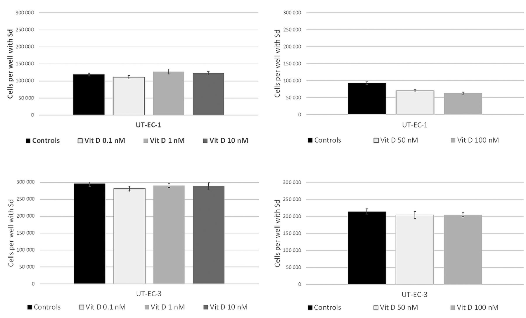
Effect of various concentrations of 1,25-D3 on UT-EC-1 and UT-EC-3 endometrial adenocarcinoma cells growth. The cells were treated for three days with indicated concentrations of 1,25-D3 and cell numbers were determined. Mean and SD of six replicates are shown.

Discussion

In the present study we tested the effects of paclitaxel and carboplatin as single agents and in combination with and without 1,25-D3 on the growth of endometrial cancer in vitro. Our results suggest that paclitaxel alone is as effective as the combination of paclitaxel and carboplatin and more effective than carboplatin alone in inhibiting the growth of endometrial cancer cells. Our data also imply that combining 1,25-D3 with paclitaxel and carboplatin, may further potentiate the growth inhibition of the cancer cells. The additive effect of 1,25-D3 was 17%, 15% and 16% when compared to single-agent paclitaxel, single agent carboplatin and paclitaxel and carboplatin in combination, respectively. On the other hand, adding carboplatin did not provide any additive or synergistic effect, as compared to the combination of paclitaxel and 1,25-D3.

The authors are not aware of any studies comparing clinically single-agent carboplatin and paclitaxel with their combination in endometrial cancer. In phase II studies, the response rate of advanced or recurrent chemo-naïve endometrial cancer to both drugs has been in the order of 35% (30, 31). Hoekstra et al. (32) compared cytoreductive ability of carboplatin and paclitaxel on endometrial carcinoma cells. A concentration of 10 nM paclitaxel induced over 90% cell death in 24 h whereas that induced by carboplatin was only 30-40%. This is in line with our results, the cytoreduction rate of paclitaxel was 25% higher in UT-EC-1 cell line and 53% higher in UT-EC-3 cell line than that of carboplatin.

Figure 3.
  • Download figure
  • Open in new tab
  • Download powerpoint
Figure 3.

VDR expression in UT-EC-1 and UT-EC-3 endometrial adenocarcinoma cell lines.

Figure 4.
  • Download figure
  • Open in new tab
  • Download powerpoint
Figure 4.

Cell growth inhibition in UT-EC-1 endometrial adenocarcinoma cells with different combinations of cytotoxic agents and 1,25-D3. The cells were treated for three days with indicated drugs and cell numbers were determined. Mean and SD of six replicates are shown.

Ballard et al. (33) evaluated micro-culture kinetic (MiCK) apoptosis assay to predict response of endometrial cancer cells in vitro to single or combination chemotherapy. The results were compared to previous GOG trials. They collected 15 endometrial cancer specimens from total abdominal hysterectomies. All grades, stages and histological types were included. The study demonstrated that 25% of patients might be treated with single-agent chemotherapy. Paclitaxel, ifosfamide and cisplatin had high activity as single agents of which paclitaxel had the highest activity. The correlation of the mean kinetic units with the overall response rates of GOG trials was also high. Single agent paclitaxel appeared to show almost the same activity as the combination of paclitaxel and carboplatin (3.1 vs. 3.4). This is in concordance with our results. The present and previous results imply that it may be reasonable to design at least a randomized Phase II study comparing paclitaxel alone to the combination of paclitaxel and carboplatin in advanced/recurrent endometrial cancer.

Our preliminary results show that 1,25-D3 may have an additive inhibitory effect with cytotoxic agents in endometrial cancer cells expressing VDR. Paclitaxel and 1,25-D3 had a growth inhibitory effect of 88% compared to 72% of single paclitaxel. A potential bias of the present study is the fact that the experiments were done partly separately. However, we used non-commercial well-established cell lines, and a synergistic anti-tumor effect was seen in UT-EC-1 cell line also with carboplatin and 1,25-D3 (63% vs. 54%), as well as with the combination of all three (87% vs. 73%). Of note is that the 1,25-D3 sensitive UT-EC-1 cell line expressed strongly VDR, while UT-EC-3 did not. Taking the low toxicity of vitamin D into account, it would be interesting to test this phenomenon also clinically.

Earlier studies support the present findings. Saunders et al. (22) found additive inhibition of endometrial carcinoma cell growth by carboplatin and calcitriol. Lee et al. (17) studied anti-tumor effects of progesterone and 1,25-D3 in endometrial cancer cell lines and immortalized human endometrial epithelial cells. They found that both agents enhanced vitamin D receptor expression and inhibited cell proliferation through caspase-3 activation. Yabushita et al. (21) examined the localization of vitamin D receptor immunohistochemically in 21 endometrial adenocarcinoma specimens, and the effect of 1,25(OH)2D3 on cell growth. The VDR was detected in 14 of the 21 specimens. They cultured one cell line expressing VDR with 50 nM 1,25(OH)2D3, resulting in 44% growth inhibition. In contrast, the cells not expressing VDR were completely uninhibited when cultured with 100 nm 1,25-D3 which is in concordance with our results. According to these findings, endometrial adenocarcinoma is a potential target of 1,25-D3.

VDR-negative breast cancer has been found to relapse earlier than VDR-positive one (34). LaPorta et al. (35) reported that 1,25-D3 down-regulates four genes that are known to promote tumorigenesis in breast cancer cells. It has been also demonstrated that the highest expression of VDR, ER and androgen receptors was related to the best prognosis (36). Further, VDR-agonists have been shown to have growth inhibitory effect in established human breast cancer xenografts in animal models (37). 1,25-D3 has been found to be a potential antiproliferative agent but dose-limiting calcemic effects have been noticed as a major obstacle (37).

Peng et al. (38) reported that estrogen receptor-positive breast cancer cells were sensitive whereas estrogen receptor-negative ones were resistant to the antiproliferative effects of vitamin D and its analogs. Furthermore they found that intact VDR-signaling machinery in the cell nucleus is essential for this action. In our study both cell lines were estrogen and progesterone receptor-negative. However, the VDR-positive UT-EC-1 cell line responded to the growth inhibitory effect with 1,25-D3. The different VDR status in the cell lines might be due to differences in VDR-signaling machinery and local production and deactivation of the 1,25-D3. On the other hand, it seems that in ovarian cancer, expression of VDR is independently regulated from the expression of estrogen and progesterone receptors (39). Whether this also takes place in endometrial cancer cells remains to be elucidated.

In conclusion, we demonstrated with two endometrial carcinoma cell lines that paclitaxel alone seems to be as effective as the combination of carboplatin and paclitaxel in vitro. In addition, 1,25-D3 had a growth-inhibitory effect on the VDR expressing cell line both alone and combined with paclitaxel and carboplatin.

Footnotes

  • This Article is freely accessible online.

  • Received September 16, 2017.
  • Revision received October 15, 2017.
  • Accepted October 17, 2017.
  • Copyright© 2017, International Institute of Anticancer Research (Dr. George J. Delinasios), All rights reserved

References

  1. ↵
    Finnish Cancer Registry, Cancer Statistics at www.cancerregistry.fi. 2013.
  2. ↵
    1. Lewin SN,
    2. Herzog TJ,
    3. Barrena Medel NI,
    4. Deutsch I,
    5. Burke WM,
    6. Sun X,
    7. Wright JD
    : Comparative performance of the 2009 international Federation of gynecology and obstetrics' staging system for uterine corpus cancer. Obstet Gynecol 116: 1141-1149, 2010.
    OpenUrlCrossRefPubMed
  3. ↵
    1. Price FV,
    2. Edwards RP,
    3. Kelley JL,
    4. Kunschner AJ,
    5. Hart LA
    : A trial of outpatient paclitaxel and carboplatin for advanced, recurrent, and histologic high-risk endometrial carcinoma: preliminary report. Semin Oncol 24: 78-82, 1997.
    OpenUrl
    1. Fleming GF,
    2. Brunetto VL,
    3. Cella D,
    4. Look KY,
    5. Reid GC,
    6. Munkarah AR,
    7. Kline R,
    8. Burger RA,
    9. Goodman A,
    10. Burks RT
    : Phase III trial of doxorubicin plus cisplatin with or without paclitaxel plus filgrastim in advanced endometrial carcinoma: A Gynecologic Oncology Group Study. J Clin Oncol 22: 2159-2166, 2004.
    OpenUrlAbstract/FREE Full Text
    1. Hoskins PJ,
    2. Swenerton KD,
    3. Pike JA,
    4. Wong F,
    5. Lim P,
    6. Acquino-Parsons C,
    7. Lee N
    : Paclitaxel and carboplatin, alone or with irradiation, in advanced or recurrent endometrial cancer: a phase II study. J Clin Oncol 19: 4048-4053, 2001.
    OpenUrlAbstract/FREE Full Text
    1. Sovak MA,
    2. Dupont J,
    3. Hensley ML,
    4. Ishill N,
    5. Gerst S,
    6. Abu-Rustum N,
    7. Anderson S,
    8. Barakat R,
    9. Konner J,
    10. Poyner E,
    11. Sabbatini P,
    12. Spriggs DR,
    13. Aghajanian C
    : Paclitaxel and carboplatin in the treatment of advanced or recurrent endometrial cancer: a large retrospective study. Int J Gynecol Cancer 17: 197-203, 2007.
    OpenUrlAbstract/FREE Full Text
    1. Nagao S,
    2. Nishio S,
    3. Okada S,
    4. Otsuki T,
    5. Fujiwara K,
    6. Tanabe H,
    7. Takano M,
    8. Hasumi Y,
    9. Takei Y,
    10. Hasegawa T,
    11. Matsumoto T,
    12. Fujiwara K,
    13. Takekuma M,
    14. Nakamura K,
    15. Shimada M,
    16. Suzuki M,
    17. Kigawa J
    : What is an appropriate second-line regimen for recurrent endometrial cancer? Ancillary analysis of the SGSG012/GOTIC004/Intergroup study. Cancer Chemother Pharmacol 76: 335-342, 2015.
    OpenUrlPubMed
  4. ↵
    1. Vergote I,
    2. Debruyne P,
    3. Kridelka F,
    4. Berteloot P,
    5. Amant F,
    6. Honhon B,
    7. Lybaert W,
    8. Leunen K,
    9. Geldhof K,
    10. Verhoeven D,
    11. Forget F,
    12. Vuylsteke P,
    13. D'Hondt L,
    14. Huizing M,
    15. Van den Bulck H,
    16. Laenen A
    : Phase II study of weekly paclitaxel/carboplatin in combination with prophylactic G-CSF in the treatment of gynecologic cancers: A study in 108 patients by the Belgian Gynaecological Oncology Group. Gynecol Oncol 138: 278-284, 2015.
    OpenUrlCrossRefPubMed
  5. ↵
    1. Zinser GM,
    2. Welsh JE
    : Vitamin D receptor status alters mammary gland morphology and tumorigenesis in MMTV-neu mice. Carcinogenesis 25: 2361-2372, 2204.
    OpenUrl
  6. ↵
    1. Becker S,
    2. Cordes T,
    3. Diesing D,
    4. Diedrich K,
    5. Friedrich M
    : Expression of 25 hydroxyvitamin D3-1alpha-hydroxylase in human endometrial tissue. J Steroid Biochem Mol Biol 103: 771-775, 2007.
    OpenUrlCrossRefPubMed
  7. ↵
    1. Vienonen A,
    2. Miettinen S,
    3. Bläuer M,
    4. Martikainen PM,
    5. Tomás E,
    6. Heinonen PK,
    7. Ylikomi T
    : Expression of nuclear receptors and cofactors in human endometrium and myometrium. J Soc Gynecol Investig 11: 104-112, 2004.
    OpenUrlCrossRefPubMed
  8. ↵
    1. Wang QM,
    2. Jones JB,
    3. Studzinski GP
    : Cyclin-dependent kinase inhibitor p27 as a mediator of the G1-S phase block induced by 1,25-dihydroxyvitamin D3 in HL60 cells. Cancer Res 56: 264-267, 1996.
    OpenUrlAbstract/FREE Full Text
  9. ↵
    1. Wu G,
    2. Fan RS,
    3. Li W,
    4. Ko TC,
    5. Brattain MG
    : Modulation of cell cycle control by vitamin D3 and its analogue, EB1089, in human breast cancer cells. Oncogene 15: 1555-1563, 1997.
    OpenUrlCrossRefPubMed
    1. Liu M,
    2. Lee MH,
    3. Cohen M,
    4. Bommakanti M,
    5. Freedman LP
    : Transcriptional activation of the Cdk inhibitor p21 by vitamin D3 leads to the induced differentiation of the myelomonocytic cell line U937. Genes Dev 10: 142-153, 1996.
    OpenUrlAbstract/FREE Full Text
  10. ↵
    1. Hager G,
    2. Formanek M,
    3. Gedlicka C,
    4. Thurnher D,
    5. Knerer B,
    6. Kornfehl J
    : 1,25(OH)2 vitamin D3 induces elevated expression of the cell cycle-regulating genes P21 and P27 in squamous carcinoma cell lines of the head and neck. Acta Otolaryngol 121: 103-109, 2001.
    OpenUrlCrossRefPubMed
  11. ↵
    1. Welsh J,
    2. Simboli-Campbell M,
    3. Narvaez CJ,
    4. Tenniswood M
    : Role of apoptosis in the growth inhibitory effects of vitamin D in MCF-7 cells. Adv Exp Med Biol 375: 45-52, 1995.
    OpenUrlPubMed
  12. ↵
    1. Lee LR,
    2. Teng PN,
    3. Nguyen H,
    4. Hood BL,
    5. Kavandi L,
    6. Wang G,
    7. Turbov JM,
    8. Thaete LG,
    9. Hamilton CA,
    10. Maxwell GL,
    11. Rodriguez GC,
    12. Conrads TP,
    13. Syed V
    : Progesterone enhances calcitriol antitumor activity by up-regulating vitamin D receptor expression and promoting apoptosis in endometrial cancer cells. Cancer Prev Res (Phila) 6: 731-743, 2013.
    OpenUrlAbstract/FREE Full Text
    1. Trump DL,
    2. Hershberger PA,
    3. Bernardi RJ,
    4. Ahmed S,
    5. Muindi J,
    6. Fakih M,
    7. Yu WD,
    8. Johnson CS
    : Anti-tumor activity of calcitriol: pre-clinical and clinical studies. J Steroid Biochem Mol Biol 89-90: 519-526, 2004.
    OpenUrl
    1. Ylikomi T,
    2. Laaksi I,
    3. Lou YR,
    4. Martikainen P,
    5. Miettinen S,
    6. Pennanen P,
    7. Purmonen S,
    8. Syvälä H,
    9. Vienonen A,
    10. Tuohimaa P
    : Antiproliferative action of vitamin D. Vitam Horm 64: 357-406, 2002.
    OpenUrlCrossRefPubMed
  13. ↵
    1. Simboli-Campbell M,
    2. Narvaez CJ,
    3. van Weelden K,
    4. Tenniswood M,
    5. Welsh J
    : Comparative effects of 1,25(OH)2D3 and EB1089 on cell cycle kinetics and apoptosis in MCF-7 breast cancer cells. Breast Cancer Res Treat 42: 31-41, 1997.
    OpenUrlCrossRefPubMed
  14. ↵
    1. Yabushita H,
    2. Hirata M,
    3. Noguchi M,
    4. Nakanishi M
    : Vitamin D receptor in endometrial carcinoma and the differentiation-inducing effect of 1,25-dihydroxyvitamin D3 on endometrial carcinoma cell lines. Obstet Gynaecol Res 22: 529-539, 1996.
    OpenUrl
  15. ↵
    1. Saunders DE,
    2. Christensen C,
    3. Wappler NL,
    4. Cho YL,
    5. Lawrence WD,
    6. Malone JM,
    7. Malviya VK,
    8. Deppe G
    : Additive Inhibition of RL95-2 Endometrial Carcinoma Cell Growth by Carboplatin and 1,25 Dihydroxyvitamin D3. Gynecol Oncol 51: 155-159, 1994.
    OpenUrl
  16. ↵
    1. Rantanen V,
    2. Grénman S,
    3. Kulmala J,
    4. Grénman R
    : Comparative evaluation of cisplatin and carboplatin sensitivity in endometrial adenocarcinoma cell lines. Brit J Cancer 69: 482-486, 1994.
    OpenUrlPubMed
  17. ↵
    1. Rantanen V,
    2. Grénman S,
    3. Kulmala J,
    4. Grénman R
    : Endometrial cancer cell lines are sensitive to paclitaxel. Anticancer Res 16: 475-479, 1996.
    OpenUrlPubMed
  18. ↵
    1. Grenman SE,
    2. Roberts JA,
    3. England BG,
    4. Grönroos M,
    5. Carey TE
    : In vitro growth regulation of endometrial carcinoma cells by tamoxifen and medroxyprogesterone acetate. Gynecol Oncol 30: 239-250, 1988.
    OpenUrlCrossRefPubMed
  19. ↵
    1. Grenman SE,
    2. Worsham MJ,
    3. Van Dyke DL,
    4. England B,
    5. McClatchey KD,
    6. Babu VR,
    7. Roberts JA,
    8. Mäenpää J,
    9. Carey TE
    : Establishment and characterization of UM-EC-2, a tamoxifen-sensitive, estrogen receptor-negative human endometrial carcinoma cell line. Gynecol Oncol 37: 188-199, 1990.
    OpenUrlPubMed
  20. ↵
    1. Rantanen V,
    2. Engblom P,
    3. Raitanen M,
    4. Hietanen S,
    5. Haarala M,
    6. Grénman S,
    7. Syrjänen S
    : Mutations of TP53 do not correlate with the sensitivity to paclitaxel – a study using 27 gynaecological cancer cell lines. Eur J Cancer 38: 1783-1791, 2002.
    OpenUrlCrossRefPubMed
  21. ↵
    1. Bläuer M,
    2. Rovio PH,
    3. Ylikomi T,
    4. Heinonen PK
    : Vitamin D inhibits myometrial and leiomyoma cell proliferation in vitro. Fertil Steril 91: 1919-1925, 2009.
    OpenUrlCrossRefPubMed
  22. ↵
    1. Alarmo EL,
    2. Kuukasjärvi T,
    3. Karhu R,
    4. Kallioniemi A
    : A comprehensive expression survey of bone morphogenetic proteins in breast cancer highlights the importance of BMP4 and BMP7. Breast Cancer Res Treat 103: 239-246, 2007.
    OpenUrlCrossRefPubMed
  23. ↵
    1. Burke TW,
    2. Munkarah A,
    3. Kavanagh JJ,
    4. Morris M,
    5. Levenback C,
    6. Tornos C,
    7. Gershenson DM
    : Treatment of advanced or recurrent endometrial carcinoma with single-agent carboplatin. Gynecol Oncol 51: 397-400, 1993.
    OpenUrlCrossRefPubMed
  24. ↵
    1. Ball HG,
    2. Blessing JA,
    3. Lentz SS,
    4. Mutch DG
    : A phase II trial of paclitaxel in patients with advanced or recurrent adenocarcinoma of the endometrium: a Gynecologic Oncology Group study. Gynecol Oncol 62: 278-281, 1996.
    OpenUrlCrossRefPubMed
  25. ↵
    1. Hoekstra A,
    2. Ward E,
    3. Hardt J,
    4. Lurain J,
    5. Singh D,
    6. Buttin B,
    7. Schink J,
    8. Kim J
    : Chemosensitization of endometrial cancer cells through AKT inhibition involves FOXO1. Gynecol Oncol 108: 609–618, 2008.
    OpenUrlCrossRefPubMed
  26. ↵
    1. Ballard KS,
    2. Homesley HD,
    3. Hodson C,
    4. Presant CA,
    5. Rutledge J,
    6. Hallquist A,
    7. Perree M
    : Endometrial carcinoma in vitro chemosensitivity testing of single and combination chemotherapy regimens using the novel microculture kinetic apoptosis assay: implications for endometrial cancer treatment. J Gynecol Oncol 21: 45-49, 2010.
    OpenUrlCrossRefPubMed
  27. ↵
    1. Berger U,
    2. McClelland RA,
    3. Wilson P,
    4. Greene GL,
    5. Haussler MR,
    6. Pike JW,
    7. Colston K,
    8. Easton D,
    9. Coombes RC
    : Immunocytochemical determination of estrogen receptor, progesterone receptor, and 1,25-dihydroxyvitamin D3 receptor in breast cancer and relationship to prognosis. Cancer Res 1: 239-244, 1991.
    OpenUrl
  28. ↵
    1. LaPorta E,
    2. Welsh J
    : Modeling vitamin D actions in triple negative/basal-like breast cancer. The J Steroid Biochem Mol Biol 144PA: 65-73, 2014.
    OpenUrlPubMed
  29. ↵
    1. Santagata S,
    2. Thakkar A,
    3. Ergonul A,
    4. Wang B,
    5. Woo T,
    6. Hu R,
    7. Harrell JC,
    8. McNamara G,
    9. Schwede M,
    10. Culhane AC,
    11. Kindelberger D,
    12. Rodig S,
    13. Richardson A,
    14. Schnitt SJ,
    15. Tamimi RM,
    16. Ince TA
    : Taxonomy of breast cancer based on normal cell phenotype predicts outcome. J Clin Invest 124: 859-870, 2014.
    OpenUrlCrossRefPubMed
  30. ↵
    1. Hussain-Hakimjee EA,
    2. Peng X,
    3. Mehta RR,
    4. Mehta RG
    : Growth inhibition of carcinogen-transformed MCF-12F breast epithelial cells and hormone-sensitive BT-474 breast cancer cells by 1alpha-hydroxyvitamin D5. Carcinogenesis 27: 551-559, 2006.
    OpenUrlCrossRefPubMed
  31. ↵
    1. Peng X,
    2. Jhaveri P,
    3. Hussain-Hakimjee EA,
    4. Mehta RG
    : Overexpression of ER and VDR is not sufficient to make ER-negative MDA-MB231 breast cancer cells responsive to 1alpha-hydroxyvitamin D5. Carcinogenesis 28: 1000-1007, 2007.
    OpenUrlCrossRefPubMed
  32. ↵
    1. Villena-Heinsen C,
    2. Meyberg R,
    3. Axt-Fliedner R,
    4. Reitnauer K,
    5. Reichrath J,
    6. Friedrich M
    : Immunohistochemical analysis of 1,25-dihydroxyvitamin-D3-receptors, estrogen and progesterone receptors and Ki-67 in ovarian carcinoma. Anticancer Res 22: 2261-2267, 2002.
    OpenUrlPubMed
PreviousNext
Back to top

In this issue

Anticancer Research
Vol. 37, Issue 12
December 2017
  • Table of Contents
  • Table of Contents (PDF)
  • Index by author
  • Back Matter (PDF)
  • Ed Board (PDF)
  • Front Matter (PDF)
Print
Download PDF
Article Alerts
Sign In to Email Alerts with your Email Address
Email Article

Thank you for your interest in spreading the word on Anticancer Research.

NOTE: We only request your email address so that the person you are recommending the page to knows that you wanted them to see it, and that it is not junk mail. We do not capture any email address.

Enter multiple addresses on separate lines or separate them with commas.
Paclitaxel, Carboplatin and 1,25-D3 Inhibit Proliferation of Endometrial Cancer Cells In Vitro
(Your Name) has sent you a message from Anticancer Research
(Your Name) thought you would like to see the Anticancer Research web site.
CAPTCHA
This question is for testing whether or not you are a human visitor and to prevent automated spam submissions.
3 + 4 =
Solve this simple math problem and enter the result. E.g. for 1+3, enter 4.
Citation Tools
Paclitaxel, Carboplatin and 1,25-D3 Inhibit Proliferation of Endometrial Cancer Cells In Vitro
TEA KUITTINEN, PÄIVI ROVIO, SYNNÖVE STAFF, TIINA LUUKKAALA, ANNE KALLIONIEMI, SEIJA GRÉNMAN, MARITA LAURILA, JOHANNA MÄENPÄÄ
Anticancer Research Dec 2017, 37 (12) 6575-6581;

Citation Manager Formats

  • BibTeX
  • Bookends
  • EasyBib
  • EndNote (tagged)
  • EndNote 8 (xml)
  • Medlars
  • Mendeley
  • Papers
  • RefWorks Tagged
  • Ref Manager
  • RIS
  • Zotero
Reprints and Permissions
Share
Paclitaxel, Carboplatin and 1,25-D3 Inhibit Proliferation of Endometrial Cancer Cells In Vitro
TEA KUITTINEN, PÄIVI ROVIO, SYNNÖVE STAFF, TIINA LUUKKAALA, ANNE KALLIONIEMI, SEIJA GRÉNMAN, MARITA LAURILA, JOHANNA MÄENPÄÄ
Anticancer Research Dec 2017, 37 (12) 6575-6581;
Twitter logo Facebook logo Mendeley logo
  • Tweet Widget
  • Facebook Like
  • Google Plus One

Jump to section

  • Article
    • Abstract
    • Materials and Methods
    • Results
    • Discussion
    • Footnotes
    • References
  • Figures & Data
  • Info & Metrics
  • PDF

Related Articles

Cited By...

  • Paclitaxel, Carboplatin and 1,25-D3 Inhibit Proliferation of Ovarian Cancer Cells In Vitro
  • Effect of Vitamin D on Paclitaxel Efficacy in Triple-negative Breast Cancer Cell Lines
  • Google Scholar

More in this TOC Section

  • Growth Suppression and Selective Disruption of F-Actin by α-Santalol in Human Melanoma Cells
  • RPA1, RFC1, and POLE Expression in Clear Cell Renal Cell Carcinoma: Immune and Clinical Relevance
  • Inhibition of Cholesterol Transport from Lysosomes by Itraconazole Repolarizes Tumor-associated Macrophages to Anti-tumor M1 type
Show more Experimental Studies

Keywords

  • vitamin D
  • 1,25-D3
  • endometrial cancer in vitro
  • paclitaxel
  • carboplatin
  • growth inhibition
  • VDR
Anticancer Research

© 2025 Anticancer Research

Powered by HighWire