


Abstract
Background/Aim: Seventeen aurones were subjected to quantitative structure–activity relationship (QSAR) analysis based on their cytotoxicity and tumor-specificity, in order to find their new biological activities. Materials and Methods: Cytotoxicity against three human oral squamous cell carcinoma cell lines and three oral mesenchymal cells was determined by the 3-(4,5-dimethylthiazol-2-yl)-2,5-diphenyltetrazolium bromide (MTT) method. Tumor specificity (TS) was evaluated by the ratio of the mean 50% cytotoxic concentration (CC50) against normal cells to that against tumor cell lines. Potency-selectivity expression (PSE) value was calculated by dividing TS by CC50 against tumor cells. Physicochemical, structural and quantum-chemical parameters were calculated based on the conformations optimized by force-field minimization. Results: Sixteen out of seventeen aurones showed relatively higher cytotoxicity and tumor specificity. Among them, (2Z)-2-[(4-hydroxyphenyl)methylene]-3(2H)-benzofuranone [7] showed the highest TS value and PSE values, comparable with those of doxorubicin and higher than 5-FU, respectively. TS values were correlated with molecular shape, size and polarizability rather than the types of substituted groups. Conclusion: Chemical modification of the lead compound may be a potential choice for designing a new type of anticancer drugs.
Aurones are a sub-set of the flavone family that possess a number of biological activities, including anti-cancer (1-6), anti-microbial (7, 8), anti-parasitic (9), anti-inflammatory (8) and enzyme inhibitory activity (10-12). Due to their high availability, simple synthesis and generally low toxicity, aurones could be attractive candidates for safer cancer drugs.
However, as far as we know, only one paper has performed the rigorous antitumor investigation with normal cell lines or same type of cells (1). Based on these backgrounds, we have investigated here a total of 17 synthetic aurones (Figure 1) for their cytotoxicity activity and tumor-specificity, and then subjected to quantitative structure–activity relationship (QSAR) analysis.
Materials and Methods
Materials. The following chemicals and reagents were obtained from the indicated companies: Dulbecco's modified Eagle's medium (DMEM), from GIBCO BRL, Grand Island, NY, USA; fetal bovine serum (FBS), 3-(4,5-dimethylthiazol-2-yl)-2,5-diphenyltetrazolium bromide (MTT), doxorubicin, from Sigma-Aldrich Inc., St. Louis, MO, USA; 5-fluorouracil (5-FU), dimethyl sulfoxide (DMSO) from Wako Pure Chem. Ind., Osaka, Japan. Culture plastic dishes and plates (96-well) were purchased from Becton Dickinson (Franklin Lakes, NJ, USA).
Synthesis of test compounds. (2Z)-2-(Phenylmethylene)-3(2H)-benzofuranone [1], (2Z)-2-[(4-methoxyphenyl)methylene]-3(2H)-benzofuranone [2], (2Z)-2-[(3,4-dimethoxyphenyl)methylene]-3(2H)-benzofuranone [3], (2Z)-2-[(4-fluorophenyl)methylene]-3(2H)-benzofuranone [4], (2Z)-2-[(4-chlorophenyl)methylene]-3(2H)-benzofuranone [5], (2Z)-2-[(4-bromophenyl)methylene]-3(2H)-benzofuranone [6], (2Z)-2-[(4-hydroxyphenyl)methylene]-3(2H)-benzofuranone [7], (2Z)-6-methoxy-2-(phenylmethylene)-3(2H)-benzofuranone [8], (2Z)-6-methoxy-2-[(4-methoxyphenyl)methylene]-3(2H)-benzofuranone [9], (2Z)-2-[(3,4-dimethoxyphenyl)methylene]-6-methoxy-3(2H)-benzofuranone [10] and (2Z)-2-[(4-bromophenyl)methylene]-6-methoxy-3(2H)-benzofuranone [11] were synthesized by the oxidative cyclization of 2’-hydroxychalcone derivatives, according to previous methods (13). Also, (2Z)-2-[(3,4-dimethoxyphenyl)methylene]-6-hydroxy-3(2H)-benzofuranone [12], (2Z)-2-[(4-fluorophenyl)methylene]-6-hydroxy-3(2H)-benzofuranone [13], (2Z)-2-[(4-chlorophenyl) methylene]-6-hydroxy-3(2H)-benzofuranone [14], (2Z)-2-[(4-bromophenyl)methylene]-6-hydroxy-3(2H)-benzofuranone [15], (2Z)-6-hydroxy-2-[(4-hydroxyphenyl)methylene]-3(2H)-benzofuranone [16] and (2Z)-2-[(3,4-dihydroxyphenyl)methylene]-6-hydroxy-3(2H)-benzofuranone [17] were synthesized by the condensation of 6-hydroxy-3(2H)-benzofuranone with selected benzaldehyde derivatives, according to previous methods (14). All compounds were dissolved in DMSO at 40 mM and stored at −20°C before use.

Structure of seventeen aurones.
Cell culture. Human normal oral mesenchymal cells (gingival fibroblast, HGF; periodontal ligament fibroblast, HPLF; pulp cells, HPC) were established from the first premolar tooth extracted from the lower jaw of a 12-year-old girl (15), and cells at 10-18 population doubling levels were used in this study. Human OSCC cell lines [Ca9-22 (derived from gingival tissue); HSC-2, HSC-4 (derived from tongue)] were purchased from Riken Cell Bank (Tsukuba, Japan). All of these cells were cultured at 37°C in DMEM supplemented with 10% heat-inactivated FBS, 100 units/ml, penicillin G and 100 μg/ml streptomycin sulfate under a humidified 5% CO2 atmosphere.
Assay for cytotoxic activity. Cells were inoculated at 2.5×10³ cells/0.1 ml in a 96-microwell plate. After 48 h, the medium was replaced with 0.1 ml of fresh medium containing different concentrations of single test compounds. Cells were incubated further for 48 h and the relative viable cell number was then determined by the MTT method (16). The relative viable cell number was determined by the absorbance of the cell lysate at 560 nm, using a microplate reader (Infinite F 50 R, TECAN, Kawasaki, Japan). Control cells were treated with the same amounts of DMSO and the cell damage induced by DMSO was subtracted from that induced by test agents. The concentration of compound that reduced the viable cell number by 50% (CC50) was determined from the dose–response curve and the mean value of CC50 for each cell type was calculated from triplicate assays.
Calculation of tumor-selectivity index (TS). TS was calculated using the following equation:
 [(D/B) in Table I]. Since both Ca9-22 and HGF cells were derived from the gingival tissue (17), the relative sensitivity of these cells was also compared [(C/A) in Table I]. We did not use human normal oral keratinocytes as controls, since many anticancer drugs showed potent cytotoxicity against normal keratinocytes by inducing apoptosis (16).
Calculation of potency-selectivity expression (PSE). PSE was calculated using the following equation:
 [that is, (D/B2) ×100 (HGF, HPLF, HPC vs. Ca9-22, HSC-2, HSC-4) and (C/A2) ×100 (HGF vs. Ca9-22) in Table I).
Estimation of CC50 values. Since the CC50 values had a distribution pattern close to a logarithmic normal distribution, we used the pCC50 (i.e., the −log CC50) for the comparison of the cytotoxicity between the compounds. The mean pCC50 values for normal cells and tumor cell lines were defined as N and T, respectively (18).
Calculation of chemical descriptors. The 3D-structure of each chemical structure (drawn by Marvin Sketch ver 16, ChemAxon, Budapest, Hungary, http://www.chemaxon.com) was optimized by CORINA Classic (Molecular Networks GmbH, Germany) and force-field calculations (amber-10: EHT) in Molecular Operating Environment (MOE) version 2015.1001 (Chemical Computing Group Inc., Quebec, Canada). The number of structural descriptors calculated from MOE and Dragon 7.0 (Kode srl., Pisa, Italy) after the elimination of overlapped descriptors were 265 and 2840, respectively.
The following 15 Dragon descriptors and 3 MOE descriptors were significantly correlated with T, N and T-N (Table II).
Dragon descriptors (19): CATS3D_04_DL (CATS3D Donor-Lipophilic BIN 04), E1u (1st component accessibility directional WHIM index/unweighted), G3m (3rd component symmetry directional WHIM index/weighted by mass), GATS1e (Geary autocorrelation of lag 1 weighted by Sanderson electronegativity), Gm (Total symmetry index/weighted by mass), HATS2p (Leverage-weighted autocorrelation of lag 2/weighted by polarizability), HATS6p (Leverage-weighted autocorrelation of lag 6/weighted by polarizability), HATS6v (Leverage-weighted autocorrelation of lag 6/weighted by van der Waals volume), MLOGP (Moriguchi logP), MLOGP2 (Squared Moriguchi logP), Mor05i (Signal 05/weighted by ionization potential), O% (Percentage of O atoms), R6p (R autocorrelation of lag 6/weighted by polarizability), R6v (R autocorrelation of lag 6/weighted by van der Waals volume), RDF010s (Radial Distribution Function - 010/weighted by I-state).
Cytotoxic activity of 17 aurones against oral malignant and non-malignant cells. Each value represents the mean of triplicate determinations.
MOE descriptors: vsurf_CW4 (apacity factor 4), vsurf_HB4 (H-bond donor capacity 4), vsurf_IW5 (Hydrophilic interaction energy moment 5) (20).
Statistical treatment. The relation among cytotoxicity, tumor specificity index and chemical descriptors was investigated using simple regression analyses by JMP Pro version 12.2.0 (SAS Institute Inc., Cary, NC, USA). The significance level was set at p<0.05.
Results
Cytotoxicity. Seventeen aurones generally showed higher cytotoxicity against three OSCC cell lines (Ca9-22, HSC-2, HSC-4) (average of mean CC50=94±85 μM), compared to three normal cells (HGF, HPLF, HPC) (average of mean CC50=198±125 μM).
Tumor-specificity. When three OSCC cell lines and three normal oral cells were used, [7] showed the highest TS value (>9.7), followed by [2] (6.6) and [11] (4.4) (D/B in Table I). [7] showed the highest PSE value (>23.3), followed by [2] (21.5) and [11] (9.3) (D/B2×100 in Table I).
When Ca9-22 and HGF cells, both derived from gingival tissue, were used, [7] showed the highest TS value (>11.0), followed by [2] (8.6) (C/A in Table I). [7] showed the highest PSE value (>30.0), followed by [2] (24.2) (C/A2×100 in Table I).
Computational analysis. We next performed the QSAR analysis of aurones in regards to their cytotoxicity against tumor cells and normal cells. Among a total of 3,105 descriptors, 18 descriptors described below correlated well with cytotoxicity and tumor specificity.
Cytotoxicity of aurones against human OSCC cell lines was correlated with E1u (topological shape) (r2=0.791, p<0.0001), RDF010s (molecular shape) (r2=0.722, p<0.0001), CATS3D_04_DL (lipophilicity) (r2=0.694, p<0.0001), Mor05i (ionization potential) (r2=0.690, p<0.0001), vsurf_HB4 (hydrogen bond donor capacity) (r2=0.690, p<0.0001), vsurf_CW4 (molecular shape) (r2=0.689, p<0.0001) (Figure 2).
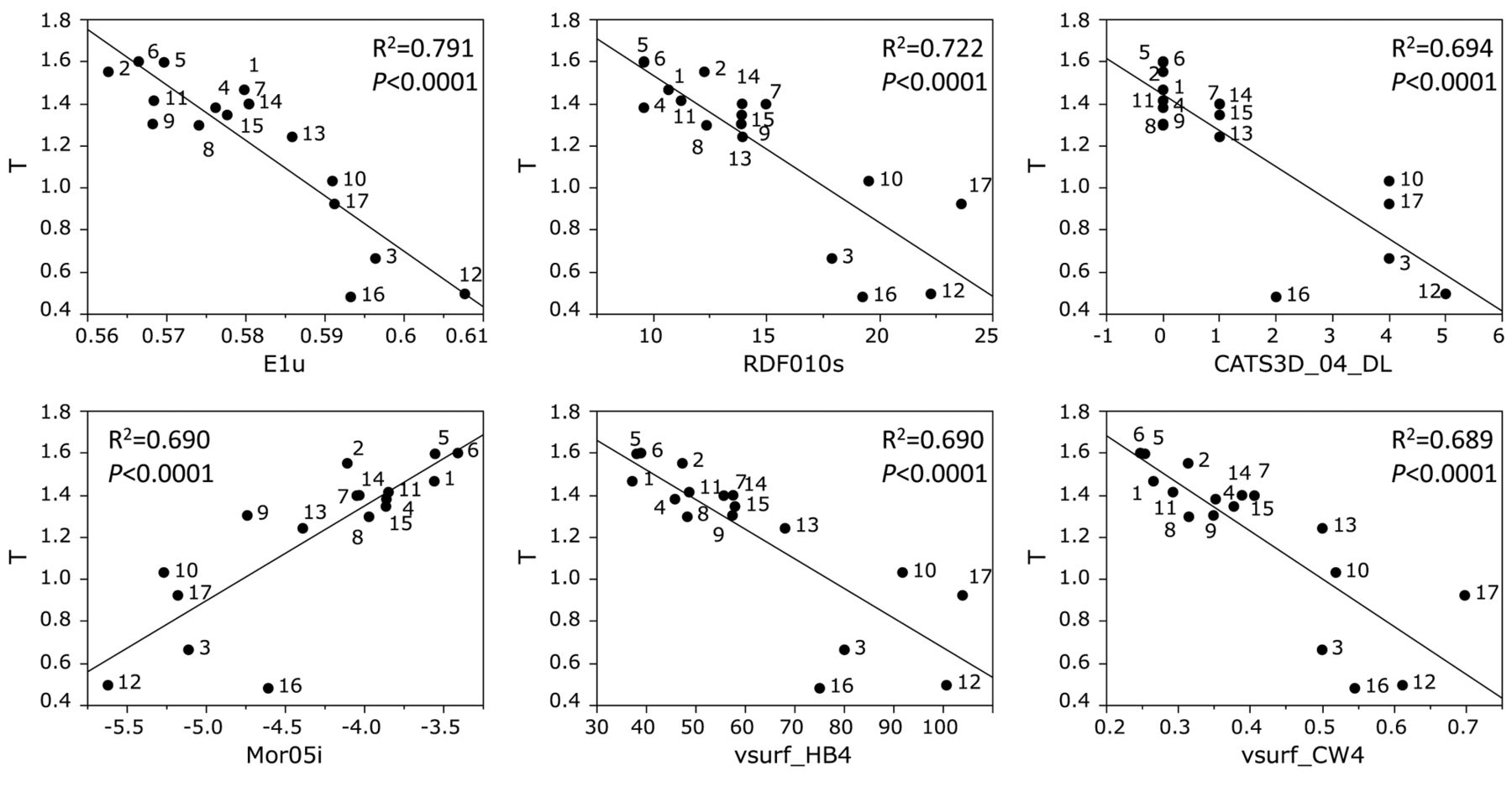
Determination of coefficient between chemical descriptors and cytotoxicity of aurones against tumor cells (defined as T). The mean (pCC50 i.e., the −log CC50) values for tumor cell lines were defined as T.
Descriptors that showed significant correlation with cytotoxicity against tumor cells (T) and normal cells (N), and tumor-specificity (T-N).
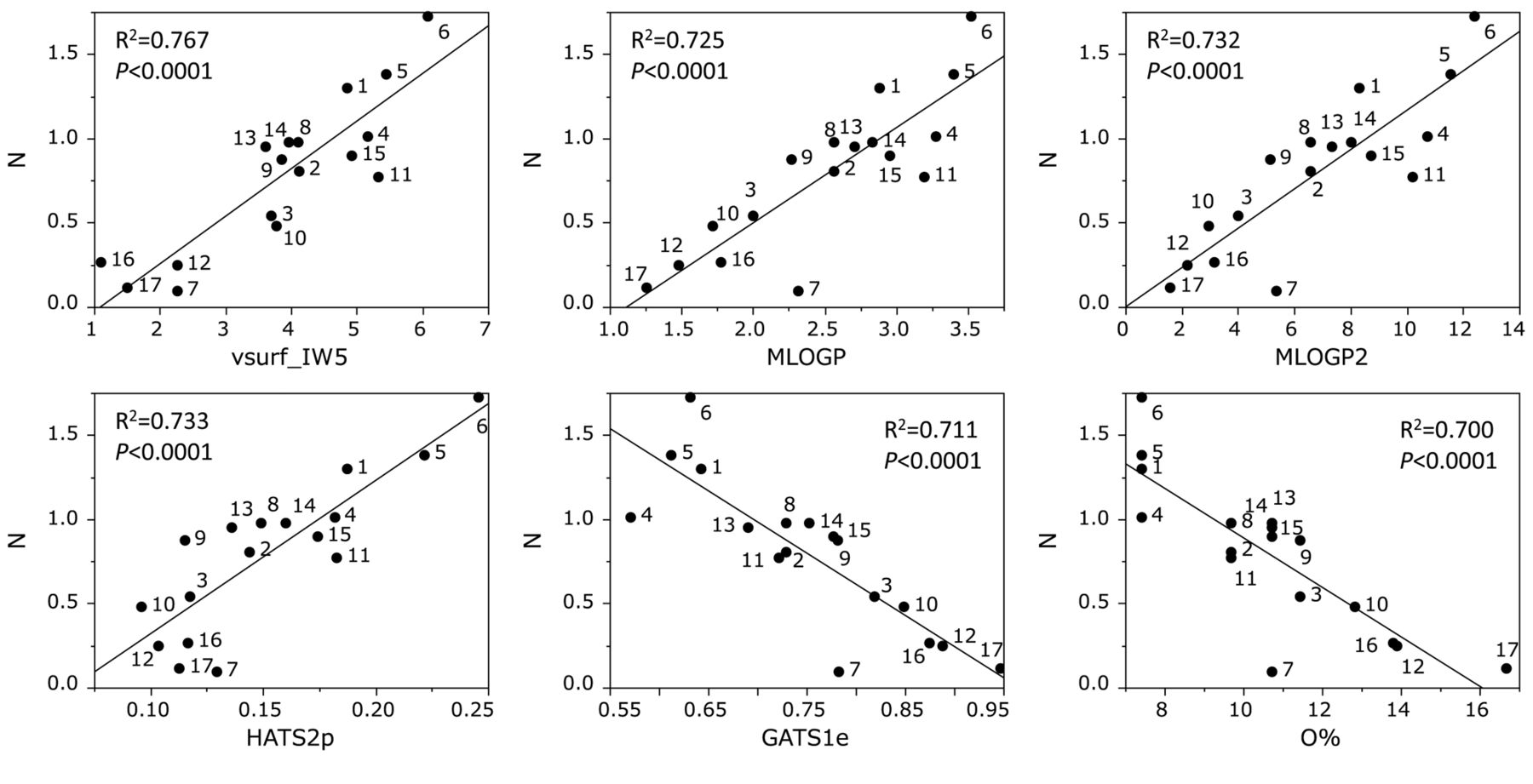
Determination of coefficient between chemical descriptors and cytotoxicity of aurones against normal cells (defined as N). The mean (pCC50 i.e., the −log CC50) values for normal cells were defined as N.

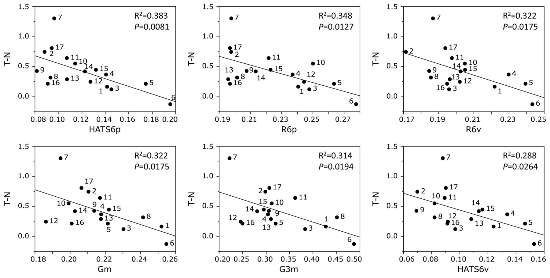
Determination of coefficient between chemical descriptors and tumor specificity of aurones (defined as T–N).
Cytotoxicity of aurones against human normal oral mesenchymal cells was correlated with vsurf_IW5 (hydrophilicity) (r2=0.767, p<0.0001), MLOGP (lipophilicity) (r2=0.725, p<0.0001), MLOGP2 (lipophilicity) (r2=0.732, p<0.0001), HATS2p (polarizability) (r2=0.733, p<0.0001), GATS1e (electronegatibvity) (r2=0.711, p<0.0001), O % (percentage of O atoms) (r2=0.700, p<0.0001) (Figure 3).
Tumor specificity of piperic acid esters was correlated with HATS6p (polarizability) (r2=0.383, p=0.0081), R6p (polarizability) (r2=0.348, p=0.0127), R6v (molecular volume) (r2=0.322, p=0.0175), Gm (mass) (r2=0.322, p=0.0175), G3m (mass) (r2=0.314, p=0.0194), HATS6v (molecular volume) (r2=0.288, p=0.0264) (Figure 4).

Substituted groups that significantly affected the cytotoxicity against OSCC cell lines (T) and normal oral mesenchymal cells (N) and tumor specificity (T-N).
Discussion
The present study demonstrated that sixteen out of seventeen aurones showed relatively higher cytotoxicity and tumor specificity. Among them, [7] showed the highest TS value (D/B=>9.7, C/A=>11.0) and PSE values (D/B2×100=>23.3, C/A2×100=>30.0) (Table I). It should be noted that TS values of [7] was comparable with those of doxorubicin (D/B=11.7, C/A=2.5) and higher than 5-FU (D/B=><2.5, C/A=>7.0). PSE value of [7] was also higher than that of 5-FU (D/B2×100=><0.6, C/A2×100=>4.8) albeit much lower than those of doxorubicin (D/B2×100=4329.4, C/A2×100=552.1) (Table I).
QSAR analysis demonstrated that tumor specificity of aurones were rather correlated with HATS6p, R6p, R6v, Gm, G3m, HATS6v that reflects molecular shape, size and polarizability (Figure 4). However, chemical descriptors that were correlated with cytotoxicity against normal cells (vsurf_IW5, MLOGP, MLOGP2, HATS2p, CATS1e, O% (Figure 3) and tumor cells (E1u, RDF10s, CATS3D_04_DL, Mor05i, vsurf, vsurf_CW4) (Figure 2) were quite different with each other. This suggests that modification of the backbone structure of aurones can produce more selective compounds.
We calculated the possible contribution of substituted groups to the expression of cytotoxicity against OSCC cell lines and normal oral mesenchymal cells and tumor-specificity (Table III). Most of the substituents listed did not affect these activities (p=0.0503-0.9838) except for hydrogen (p=0.0027), OCH3 (p=0.0066) at R3, or X at R2 (p=0.0376) in determining cytotoxicity against tumor cells (upper panel in Figure 5), and OH (p=0.0038) and X (p=0.0121) at R2 and H (p=0.0202) at R3 in determining cytotoxicity against normal cells (lower panel in Figure 5). From these data, tumor specificity of aurones was rather correlated with molecular shape, size and polarizability (Figure 5).
In conclusion, compound 7 is a potential lead compound for synthesizing more potent compounds targeted to OSCC cells.
Substituted groups that affect the cytotoxicity against OSCC cell lines (T) and normal oral mesenchymal cells (N) and tumor specificity (T-N).
Acknowledgements
This work was partially supported by KAKENHI from the Japan Society for the Promotion of Science (JSPS) (15K08111, 16K11519). The annual license of the statistical software, JMP Pro, was supported by the grant-in-aid of the oncology specialists' promotion program by the Ministry of Education, Culture, Sports, Science and Technology, Japan.
Footnotes
This article is freely accessible online.
Conflicts of Interest
The Authors wish to confirm that there are no known conflicts of interest associated with this publication and there has been no significant financial support for this work that could have influenced its outcome.
- Received August 21, 2017.
- Revision received September 11, 2017.
- Accepted September 12, 2017.
- Copyright© 2017, International Institute of Anticancer Research (Dr. George J. Delinasios), All rights reserved


