
Main menu

  • Home
  • Current Issue
  • Archive
  • Info for
    • Authors
    • Subscribers
    • Advertisers
    • Editorial Board
  • Other Publications
    • In Vivo
    • Cancer Genomics & Proteomics
    • Cancer Diagnosis & Prognosis
  • More
    • IIAR
    • Conferences
    • 2008 Nobel Laureates
  • About Us
    • General Policy
    • Contact
  • Other Publications
    • Anticancer Research
    • In Vivo
    • Cancer Genomics & Proteomics

User menu

  • Register
  • Subscribe
  • My alerts
  • Log in
  • My Cart

Search

  • Advanced search
Anticancer Research
  • Other Publications
    • Anticancer Research
    • In Vivo
    • Cancer Genomics & Proteomics
  • Register
  • Subscribe
  • My alerts
  • Log in
  • My Cart
Anticancer Research

Advanced Search

  • Home
  • Current Issue
  • Archive
  • Info for
    • Authors
    • Subscribers
    • Advertisers
    • Editorial Board
  • Other Publications
    • In Vivo
    • Cancer Genomics & Proteomics
    • Cancer Diagnosis & Prognosis
  • More
    • IIAR
    • Conferences
    • 2008 Nobel Laureates
  • About Us
    • General Policy
    • Contact
  • Visit us on Facebook
  • Follow us on Linkedin
Research ArticleClinical Studies

Synchronous Quadruple Primary Neoplasms: Colon Adenocarcinoma, Collision Tumor of Neuroendocrine Tumor and Schwann Cell Hamartoma and Sessile Serrated Adenoma of the Appendix

MARSHALL W. MEEKS, SHANE GRACE, YONGXIN CHEN, JAMES PETTERCHAK, EDWARD BOLESTA, YIHUA ZHOU and JIN-PING LAI
Anticancer Research August 2016, 36 (8) 4307-4311;
MARSHALL W. MEEKS
1Department of Pathology, Saint Louis University School of Medicine, Saint Louis, MO, U.S.A.
  • Find this author on Google Scholar
  • Find this author on PubMed
  • Search for this author on this site
SHANE GRACE
1Department of Pathology, Saint Louis University School of Medicine, Saint Louis, MO, U.S.A.
  • Find this author on Google Scholar
  • Find this author on PubMed
  • Search for this author on this site
YONGXIN CHEN
1Department of Pathology, Saint Louis University School of Medicine, Saint Louis, MO, U.S.A.
  • Find this author on Google Scholar
  • Find this author on PubMed
  • Search for this author on this site
JAMES PETTERCHAK
1Department of Pathology, Saint Louis University School of Medicine, Saint Louis, MO, U.S.A.
  • Find this author on Google Scholar
  • Find this author on PubMed
  • Search for this author on this site
EDWARD BOLESTA
1Department of Pathology, Saint Louis University School of Medicine, Saint Louis, MO, U.S.A.
  • Find this author on Google Scholar
  • Find this author on PubMed
  • Search for this author on this site
YIHUA ZHOU
2Department of Radiology, Saint Louis University School of Medicine, Saint Louis, MO, U.S.A.
  • Find this author on Google Scholar
  • Find this author on PubMed
  • Search for this author on this site
JIN-PING LAI
1Department of Pathology, Saint Louis University School of Medicine, Saint Louis, MO, U.S.A.
  • Find this author on Google Scholar
  • Find this author on PubMed
  • Search for this author on this site
  • For correspondence: jinpinglai@slu.edu
  • Article
  • Figures & Data
  • Info & Metrics
  • PDF
Loading

Abstract

Quadruple synchronous primary neoplasms are very rare with only three cases reported in the English-speaking literature to date. Collision tumors are also rare entities, especially of the appendix. We herein report a case of synchronous quadruple primary neoplasm in a 95-year-old female. She was diagnosed with colon adenocarcinoma, sessile serrated adenoma of the appendix and a collision tumor composed of a well-differentiated neuroendocrine tumor and Schwann cell hamartoma. Histological examination and immunohistochemistry supported these four lesions as separate entities. This case is unique because we report the diagnosis of quadruple synchronous primary, an extremely rare occurrence, in addition to a collision tumor of the appendix. We also provide a review of the literature for synchronous neoplasms and collision tumors.

  • Synchronous
  • multiple primary malignant neoplasms
  • neuroendocrine tumor
  • sessile serrated adenoma/polyp
  • schwannoma
  • collision tumor
  • colon adenocarcinoma

Multiple primary neoplasms (MPNs) in a single patient are a relatively rare occurrence. MPNs are classified as either synchronous or metachronous depending on the time of diagnosis. Synchronous cancers are diagnosed either simultaneously or within 6 months of each other and metachronous cancers are diagnosed at least 6 months apart. The prevalence of MPNs varies significantly between 0.73% and 16% (1, 2) depending on the source and synchronous cancer is much rarer than metachronous cancer. The prevalence of MPNs has grown in recent years and is expected to increase due to improved cancer survivorship and more sensitive cancer diagnostic techniques (3).

In this report, we describe a case of a 95-year-old Caucasian woman with synchronous quadruple primary neoplasms with two of the tumors being collision tumors located in the appendix. She was diagnosed with adenocarcinoma of the colon, collision tumor of neuroendocrine tumor and Schwann cell hamartoma in the appendix and sessile serrated adenoma of the appendix. To the best of our knowledge, this particular combination of synchronous malignancies has never been previously reported.

Case Report

A 95-year-old Caucasian woman presented with an episode of nausea, vomiting and abdominal distention. Her past medical history was significant for previous episodes of small bowel obstruction, type II diabetes mellitus, iron deficiency anemia, peripheral neuropathy and hypertension. She had no history of melena, hematochezia or diarrhea. Laboratory findings and clinical examination at this time were unremarkable. Her family history was non-contributory. Computed tomography (CT) revealed a large cecal mass (Figure 1A) causing obstruction of the terminal ilium and a mildly enlarged tail of the appendix measuring 8 mm in diameter (Figure 1B).

A colonoscopy with a biopsy from the cecal mass was subsequently performed revealing a moderately differentiated adenocarcinoma. Given the patient's good performance status, surgery was offered to the patient. A right hemicolectomy was then performed. Grossly, the cecum contained a fungating purple-pink mass measuring 3.7 cm in the greatest dimension that did not appear to involve the appendiceal orifice. The appendix was 4.7 cm in length with mildly enlarged at the tail measuring 1 cm in diameter.

Figure 1.
  • Download figure
  • Open in new tab
  • Download powerpoint
Figure 1.

Radiographic findings. A) Axial computed tomography (CT) image without contrast showing a cecal mass (large open arrow) causing obstruction of the terminal ilium (white solid arrow) and a normal sized proximal appendix measuring 6 mm in diameter (thin white arrow). B) Coronal CT image without contrast showing a mildly enlarged tail of the appendix measuring 8 mm in diameter (thin white arrow) and a cecal mass (large open arrow).

Microscopic examination of the resected cecum revealed a moderately differentiated adenocarcinoma, intestinal type (Figure 2A), with invasion through the mascularis propria into the serosa. Eight of fifteen regional lymph nodes were positive for metastatic adenocarcinoma. The tumors' cells were diffusely positive for cytokeratin-20 (CK20) and CDX2 and negative for synaptophysin and chromogranin. The immunohistochemistry of four mismatch repair proteins (MLH1, MSH2, MSH6 and PMS2) showed intact expression, suggesting that the patient did not have Lynch syndrome.

Microscopic examination of the resected appendix revealed a collision tumor (Figure 3A-B) of a well-differentiated neuroendocrine tumor (NET) (Figure 2B) and a Schwann cell hamartoma (Figure 2C) at the tip of the appendix. The NET cells were diffusely positive for synaptophysin (Figure 3D, left), chromogranin, CDX2 (Figure 3D, right) and partially positive for pan-cytokeratin but negative for CK7, CK20 and carcinoembryonic antigen (CEA). The Schwann cell hamartoma cells were diffusely positive for S100 (Figure 3C) but negative for CD117 and desmin. In addition, a separate sessile serrated adenoma that was 0.7 cm in the greatest dimension was also seen in the middle of the appendix (Figure 2D). The final histologic diagnoses of this right hemicolectomy include four primary neoplastic lesions of moderately differentiated adenocarcinoma of the cecum, sessile serrated adenoma of the appendix and a collision tumor composed of a well-differentiated neuroendocrine tumor and Schwann cell hamartoma. Following surgery, the patient was referred to palliative care since she was not a good candidate for systemic therapy given her age.

Discussion

The most commonly accepted criteria for the diagnosis of multiple primary neoplasms (MPNs) was proposed by Warren and Gates in 1932 (4): each tumor must be anatomically distinct from the others, they must have distinctively different histology and metastasis must be excluded. MPNs are known to occur in many different organ systems (3, 5) but are more commonly seen in the gynecologic (6, 7, 8) and gastrointestinal tracts (9, 10). This is most likely because they are derived from the same embryonic layer or tissue and, in the case of gynecologic malignancies, responsive to the same hormones.

The etiology of MPNs is still unclear at this time but, for some patients, could be explained by the presence of carcinogenetic risk factors. For instance, a heavy cigarette smoker will have an increased risk of developing lung, stomach, liver, kidney, uterine cervix and urinary bladder cancer (11). Other risk factors include previous cancer treatment, such as radiotherapy or chemotherapy, immunosuppression and hereditary predisposition (12). For patients without known risk factors, such as ours, MPNs may simply be due to advanced age as the risk of cancer increases with age.

The exact prevalence of patients diagnosed with at least two, non-metastatic, primary cancers varies significantly in the literature. This wide variation may be due to several factors, including different criteria to define MPNs depending on author. Furthermore, some reports are autopsy series which report higher frequency of MPNs compared to retrospective clinical case series. Demandante et al. (1) performed a literature review of over one million cancer patients diagnosed from 1966 to 2000 and found the overall occurrence rate of MPNs to be between 0.73% and 11.7%. In addition, a Surveillance, Epidemiology, and End Results (SEER) analysis revealed that second- or higher-order malignancies made up 16% of new cancer diagnoses (2). The incidence of MPNs will likely increase in the future given better access to care, an aging population and improved diagnostic techniques and treatment protocols that will lead patients living long enough to develop additional cancers (2).

Figure 2.
  • Download figure
  • Open in new tab
  • Download powerpoint
Figure 2.

Histological examination. A) The cecal tumor showed moderately differentiated adenocarcinoma. B) Histologic sections of the resected appendix demonstrated a collision tumor composed of well-differentiated neuroendocrine tumor (NET) cells with monotonous nuclei and characteristic “salt and pepper” chromatin along with a Schwann cell hamartoma (C) composed of benign-appearing spindle cells with elongated, tapering nuclei. D) Histologic sections from the distal 1/3 of the appendix also demonstrated a sessile serrated adenoma with characteristic ‘boot-shaped’, horizontally branching basal crypt architecture (Hematoxylin and eosin (H&E) staining; A, 200×; B and C, 400×; and D, 100×).

Depending on the time interval of diagnosis, MPNs can be synchronous or metachronous. Synchronous tumors are diagnosed simultaneously or within 6 months of one another, while metachronous tumors are diagnosed at least 6 months apart (4). Synchronous tumors are much rarer than metachronous tumors. A retrospective analysis at a Turkish oncology center revealed that, out of 130 MPNs cases, only 18.4% were synchronous (13). In addition, a Minneapolis Veterans Affair tumor registry from 2005 to 2009 revealed that, out of 506 patients with MPNs, 24.3% had synchronous tumors (14).

Triple synchronous MPNs are an extremely rare occurrence with only a few cases reported in the literature (6, 7, 8, 15, 16). To our knowledge, there have only been four cases (including the present case) of synchronous MPNs (3, 5, 17). In our case, three of the four neoplasms were located in the appendix. Of these, two were a collision tumor of neuroendocrine tumor and Schwann cell hamartoma.

Tumors located in the same organ with composite features of different histologic characteristics are divided into two distinct morphologic groups (18). Two histologically distinct tumors with a clear boundary between the two components is a collision tumor. In contrast, if there is intermingling between the components it is a composite (mixed) tumor. For our patient, there was no evidence of transition between the neuroendocrine and Schwann cell hamartoma components making it more consistent with a collision tumor than a composite tumor.

Figure 3.
  • Download figure
  • Open in new tab
  • Download powerpoint
Figure 3.

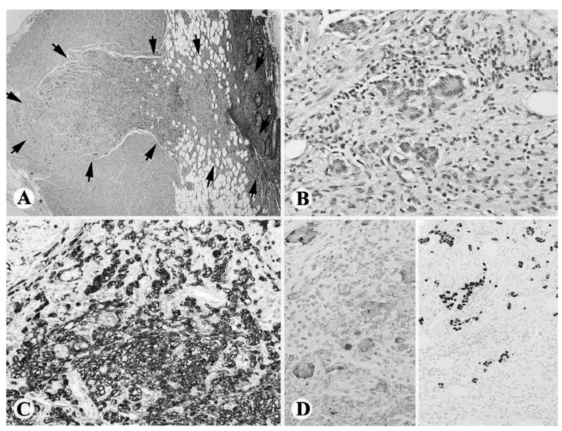
Collision tumor of the appendix. A) Low power view of the appendiceal collision tumor (outlined by black arrows, Hematoxylin and Eosin (H&E) staining, 40×). B) High power view of the collision tumor demonstrated distinctly different cell populations of spindle cell Schwann cell hamartoma cells and acinar arrangement of the round neuroendocrine tumor (NET) cells (H&E, 400×). C) The Schwann cell hamartoma cells were diffusely positive for S100 (400×). D) The NET cells were positive for synaptophysin (left) and CDX2 (right) (400×).

The etiology of collision and composite tumors is currently not fully understood. Most likely, collision tumors arise from cells of a different clonal origin (19), whereas composite tumors arise from a multipotent stem cell that is capable of bi-directional differentiation (20). While composite tumors of the gastrointestinal tract are rare, they appear to be most common in the appendix (19). Collison tumors of the appendix are even rarer with only four cases previously reported (19, 21, 22, 23). The histologic finding of collision tumors are relevant for the prognosis and treatment of the patient because usually the most aggressive component will determine disease progression (23).

Footnotes

  • Conflicts of Interests

    The Authors declare that they have no financial conflicts.

  • Received June 4, 2016.
  • Revision received June 24, 2016.
  • Accepted July 4, 2016.
  • Copyright© 2016 International Institute of Anticancer Research (Dr. John G. Delinassios), All rights reserved

References

  1. ↵
    1. Demandante CG,
    2. Troyer DA,
    3. Miles TP
    : Multiple primary malignant neoplasms: case report and a comprehensive review of the literature. Am J Clin Oncol 26: 79-83, 2003.
    OpenUrlCrossRefPubMed
  2. ↵
    1. Travis LB
    : The epidemiology of second primary cancers. Cancer Epidemiol Biomarkers Prev 15: 2020-2026, 2006.
    OpenUrlAbstract/FREE Full Text
  3. ↵
    1. Grace S,
    2. Muzaffar R,
    3. Veerapong J,
    4. Alkaade S,
    5. Poddar N,
    6. Phillips N,
    7. Guzman M,
    8. Batanian J,
    9. Vogler C,
    10. Lai JP
    : Synchronous quadruple primary neoplasms: glioblastoma, neuroendocrine tumor, schwannoma and sessile serrated adenoma in a patient with history of prostate cancer. Anticancer Res 35: 2121-2127, 2015.
    OpenUrlAbstract/FREE Full Text
  4. ↵
    1. Warren S,
    2. Gates O
    : Multiple primary malignant tumours: A survey of the literature and a statistical study. Am J Cancer 16: 1358-1414, 1932.
    OpenUrl
  5. ↵
    1. Kim JS,
    2. Chung CY,
    3. Park HC,
    4. Myung DS,
    5. Cho SB,
    6. Lee WS,
    7. Min JJ,
    8. Joo YE
    : Synchronous quadruple primary tumors of thyroid, breast, pancreas, and stomach: a case report. Anticancer Res 33: 2135-2138, 2013.
    OpenUrlAbstract/FREE Full Text
  6. ↵
    1. Isin Dogan Ekici A,
    2. Kucukali T,
    3. Coskun Salman M,
    4. Ayhan A
    : Triple simultaneous primary gynecological malignancies in a 56-year-old patient. Int J Gynecol Cancer 16: 1947-1950, 2006.
    OpenUrlCrossRefPubMed
  7. ↵
    1. Hale CS,
    2. Lee L,
    3. Mittal K
    : Triple synchronous primary gynecologic carcinomas: a case report and review of the literature. Int J Surg Pathol 19: 552-555, 2011.
    OpenUrlAbstract/FREE Full Text
  8. ↵
    1. Ree YS,
    2. Cho SH,
    3. Kim SR,
    4. Cho SH,
    5. Kim KT,
    6. Park MH
    : Synchronous primary endometrial and ovarian cancer with three different histologic patterns: A case report. Int J Gynecol Cancer 13: 678-682, 2003.
    OpenUrlPubMed
  9. ↵
    1. Kim JW,
    2. Han JW,
    3. Jung SY,
    4. Jung JP,
    5. Kim JW
    : Synchronous double primary malignant tumor of the gallbladder and liver: a case report. World J Surg Oncol 9: 84, 2011.
    OpenUrlCrossRefPubMed
  10. ↵
    1. Cheng HY,
    2. Chu CH,
    3. Chang WH,
    4. Hsu TC,
    5. Lin SC,
    6. Liu CC,
    7. Yang AM,
    8. Shih SC
    : Clinical analysis of multiple primary malignancies in the digestive system: a hospital-based study. World J Gastroenterol 11: 4215-4219, 2005.
    OpenUrlPubMed
  11. ↵
    1. Sasco AJ,
    2. Secretan MB,
    3. Straif K
    : Tobacco smoking and cancer: a brief review of recent epidemiological evidence. Lung Cancer 45(Suppl 2): S3-S9, 2004.
    OpenUrl
  12. ↵
    1. Angurana SL,
    2. Kapoor R,
    3. Kumar P,
    4. Khosla D,
    5. Kumar N,
    6. Sharma SC,
    7. Patel FD
    : Quadruple malignancy in a single patient: a case report and comprehensive review of literature. J Cancer Res Ther 6: 230-232, 2010.
    OpenUrlPubMed
  13. ↵
    1. Arpaci E,
    2. Tokluoglu S,
    3. Yetigyigit T,
    4. Alkis N
    : Multiple primary malignancies – a retrospective analysis at a single center in Turkey. Asian Pac J Cancer Prev 14: 769-773, 2013.
    OpenUrlPubMed
  14. ↵
    1. Powell S,
    2. Tarchand G,
    3. Rector T,
    4. Klein M
    : Synchronous and metachronous malignancies: analysis of the Minneapolis Veterans Affairs (VA) tumor registry. Cancer Causes Control 24: 1565-1573, 2013.
    OpenUrlPubMed
  15. ↵
    1. Lee JS,
    2. Moon W,
    3. Park SJ,
    4. Park MI,
    5. Kim KJ,
    6. Jang LL,
    7. Park MJ,
    8. Chun BK
    : Triple synchronous primary cancers of rectum, thyroid, and uterine cervix detected during the workup for hematochezia. Intern Med 49: 1745-1747, 2010.
    OpenUrlPubMed
  16. ↵
    1. Chong VH,
    2. Idros A,
    3. Telisinghe PU
    : Triple synchronous gastrointestinal malignancies: a rare occurrence. Singapore Med J 51: e176-177, 2010.
    OpenUrlPubMed
  17. ↵
    1. Atasever M,
    2. Yilmaz B,
    3. Dilek G,
    4. Akcay EY,
    5. Kelekci S
    : Synchronous primary carcinoma in 5 different organs of a female genital tract: an unusual case and review of the literature. Int J Gynecol Cancer 19: 802-807, 2009.
    OpenUrlCrossRefPubMed
  18. ↵
    1. Jain M,
    2. Das KK
    : Concurrent occurrence of terminal ileum carcinoid tumor and cecal adenocarcinoma: a collision tumor or composite tumor? Indian J Pathol Microbiol 52: 285-286, 2009.
    OpenUrlCrossRefPubMed
  19. ↵
    1. Baena-del-Valle J,
    2. Palau-Lazaro M,
    3. Mejia-Arango M,
    4. Otero J,
    5. Londono-Schimmer E,
    6. Cortes N,
    7. Pozo M,
    8. Herrera-Almario G,
    9. Arias F,
    10. Rodriguez-Urrego P
    : Well differentiated neuroendocrine tumor of the appendix and low-grade appendiceal mucinous neoplasm presenting as a collision tumor. Rev Esp Enferm Dig 107: 396-398, 2015.
    OpenUrlPubMed
  20. ↵
    1. Anagnostopoulos GK,
    2. Arvanitidis D,
    3. Sakorafas G,
    4. Pavlakis G,
    5. Kolilekas L,
    6. Arkoumani E,
    7. Stefanou D
    : Combined carcinoid-adenocarcinoma tumour of the anal canal. Scand J Gastroenterol 39: 198-200, 2004.
    OpenUrlPubMed
  21. ↵
    1. Tan HL,
    2. Tan GH,
    3. Teo M
    : Two rare cases of appendiceal collision tumours involving an appendiceal mucinous neoplasm and carcinoid. BMJ Case Rep 2016, 2016.
  22. ↵
    1. Dellaportas D,
    2. Vlahos N,
    3. Polymeneas G,
    4. Gkiokas G,
    5. Dastamani C,
    6. Carvounis E,
    7. Theodosopoulos T
    : Collision tumor of the appendix: mucinous cystadenoma and carcinoid. A case report. Chirurgia (Bucur) 109: 843-845, 2014.
    OpenUrlPubMed
  23. ↵
    1. Singh NG,
    2. Mannan AA,
    3. Kahvic M,
    4. Nur AM
    : Mixed adenocarcinoma-carcinoid (collision tumor) of the appendix. Med Princ Pract 20: 384-386, 2011.
    OpenUrlPubMed
PreviousNext
Back to top

In this issue

Anticancer Research: 36 (8)
Anticancer Research
Vol. 36, Issue 8
August 2016
  • Table of Contents
  • Table of Contents (PDF)
  • Index by author
  • Back Matter (PDF)
  • Ed Board (PDF)
  • Front Matter (PDF)
Print
Download PDF
Article Alerts
Sign In to Email Alerts with your Email Address
Email Article

Thank you for your interest in spreading the word on Anticancer Research.

NOTE: We only request your email address so that the person you are recommending the page to knows that you wanted them to see it, and that it is not junk mail. We do not capture any email address.

Enter multiple addresses on separate lines or separate them with commas.
Synchronous Quadruple Primary Neoplasms: Colon Adenocarcinoma, Collision Tumor of Neuroendocrine Tumor and Schwann Cell Hamartoma and Sessile Serrated Adenoma of the Appendix
(Your Name) has sent you a message from Anticancer Research
(Your Name) thought you would like to see the Anticancer Research web site.
CAPTCHA
This question is for testing whether or not you are a human visitor and to prevent automated spam submissions.
2 + 0 =
Solve this simple math problem and enter the result. E.g. for 1+3, enter 4.
Citation Tools
Synchronous Quadruple Primary Neoplasms: Colon Adenocarcinoma, Collision Tumor of Neuroendocrine Tumor and Schwann Cell Hamartoma and Sessile Serrated Adenoma of the Appendix
MARSHALL W. MEEKS, SHANE GRACE, YONGXIN CHEN, JAMES PETTERCHAK, EDWARD BOLESTA, YIHUA ZHOU, JIN-PING LAI
Anticancer Research Aug 2016, 36 (8) 4307-4311;

Citation Manager Formats

  • BibTeX
  • Bookends
  • EasyBib
  • EndNote (tagged)
  • EndNote 8 (xml)
  • Medlars
  • Mendeley
  • Papers
  • RefWorks Tagged
  • Ref Manager
  • RIS
  • Zotero
Reprints and Permissions
Share
Synchronous Quadruple Primary Neoplasms: Colon Adenocarcinoma, Collision Tumor of Neuroendocrine Tumor and Schwann Cell Hamartoma and Sessile Serrated Adenoma of the Appendix
MARSHALL W. MEEKS, SHANE GRACE, YONGXIN CHEN, JAMES PETTERCHAK, EDWARD BOLESTA, YIHUA ZHOU, JIN-PING LAI
Anticancer Research Aug 2016, 36 (8) 4307-4311;
del.icio.us logo Digg logo Reddit logo Twitter logo CiteULike logo Facebook logo Google logo Mendeley logo
  • Tweet Widget
  • Facebook Like
  • Google Plus One

Jump to section

  • Article
    • Abstract
    • Case Report
    • Discussion
    • Footnotes
    • References
  • Figures & Data
  • Info & Metrics
  • PDF

Related Articles

  • No related articles found.
  • PubMed
  • Google Scholar

Cited By...

  • Collision Tumors of the Gastrointestinal Tract: A Systematic Review of the Literature
  • Synchronous Pancreatic Ductal Adenocarcinoma and Hepatocellular Carcinoma: Report of a Case and Review of the Literature
  • Neuroendocrine Neoplasms of the Appendix: A Review of the Literature
  • Triple synchronous primary malignancies: a rare occurrence
  • Synchronous Type 1 Papillary Renal Cell Carcinoma in a Patient with Rectal Adenocarcinoma
  • Google Scholar

More in this TOC Section

  • Pelvic Recurrence After Curative Resection for Rectal Adenocarcinoma: Impact of Surgery on Survival
  • Glasgow Prognostic Score Predicts Survival and Recurrence Pattern in Patients With Hepatocellular Carcinoma After Hepatectomy
  • Small Bowel Lipomatosis: An Unusual Radiological Finding in Patients With Renal Cell Cancer on Pazopanib
Show more Clinical Studies

Similar Articles

Keywords

  • synchronous
  • multiple primary malignant neoplasms
  • neuroendocrine tumor
  • sessile serrated adenoma/polyp
  • Schwannoma
  • collision tumor
  • colon adenocarcinoma
Anticancer Research

© 2023 Anticancer Research

Powered by HighWire