
Main menu

  • Home
  • Current Issue
  • Archive
  • Info for
    • Authors
    • Subscribers
    • Advertisers
    • Editorial Board
  • Other Publications
    • In Vivo
    • Cancer Genomics & Proteomics
    • Cancer Diagnosis & Prognosis
  • More
    • IIAR
    • Conferences
    • 2008 Nobel Laureates
  • About Us
    • General Policy
    • Contact
  • Other Publications
    • Anticancer Research
    • In Vivo
    • Cancer Genomics & Proteomics

User menu

  • Register
  • Subscribe
  • My alerts
  • Log in
  • My Cart

Search

  • Advanced search
Anticancer Research
  • Other Publications
    • Anticancer Research
    • In Vivo
    • Cancer Genomics & Proteomics
  • Register
  • Subscribe
  • My alerts
  • Log in
  • My Cart
Anticancer Research

Advanced Search

  • Home
  • Current Issue
  • Archive
  • Info for
    • Authors
    • Subscribers
    • Advertisers
    • Editorial Board
  • Other Publications
    • In Vivo
    • Cancer Genomics & Proteomics
    • Cancer Diagnosis & Prognosis
  • More
    • IIAR
    • Conferences
    • 2008 Nobel Laureates
  • About Us
    • General Policy
    • Contact
  • Visit us on Facebook
  • Follow us on Linkedin
Research ArticleExperimental Studies

Color-coded Live Imaging of Heterokaryon Formation and Nuclear Fusion of Hybridizing Cancer Cells

ATSUSHI SUETSUGU, TAKURO MATSUMOTO, KOSUKE HASEGAWA, MIKI NAKAMURA, TAKAHIRO KUNISADA, MASAHITO SHIMIZU, SHIGETOYO SAJI, HISATAKA MORIWAKI, MICHAEL BOUVET and ROBERT M. HOFFMAN
Anticancer Research August 2016, 36 (8) 3827-3831;
ATSUSHI SUETSUGU
1Gifu University Graduate School of Medicine, Gifu, Japan
2AntiCancer, Inc., San Diego, CA, U.S.A.
3Department of Surgery, University of California San Diego, San Diego, CA, U.S.A.
  • Find this author on Google Scholar
  • Find this author on PubMed
  • Search for this author on this site
TAKURO MATSUMOTO
1Gifu University Graduate School of Medicine, Gifu, Japan
  • Find this author on Google Scholar
  • Find this author on PubMed
  • Search for this author on this site
KOSUKE HASEGAWA
1Gifu University Graduate School of Medicine, Gifu, Japan
  • Find this author on Google Scholar
  • Find this author on PubMed
  • Search for this author on this site
MIKI NAKAMURA
1Gifu University Graduate School of Medicine, Gifu, Japan
  • Find this author on Google Scholar
  • Find this author on PubMed
  • Search for this author on this site
TAKAHIRO KUNISADA
1Gifu University Graduate School of Medicine, Gifu, Japan
  • Find this author on Google Scholar
  • Find this author on PubMed
  • Search for this author on this site
MASAHITO SHIMIZU
1Gifu University Graduate School of Medicine, Gifu, Japan
  • Find this author on Google Scholar
  • Find this author on PubMed
  • Search for this author on this site
SHIGETOYO SAJI
1Gifu University Graduate School of Medicine, Gifu, Japan
  • Find this author on Google Scholar
  • Find this author on PubMed
  • Search for this author on this site
HISATAKA MORIWAKI
1Gifu University Graduate School of Medicine, Gifu, Japan
  • Find this author on Google Scholar
  • Find this author on PubMed
  • Search for this author on this site
MICHAEL BOUVET
3Department of Surgery, University of California San Diego, San Diego, CA, U.S.A.
  • Find this author on Google Scholar
  • Find this author on PubMed
  • Search for this author on this site
ROBERT M. HOFFMAN
2AntiCancer, Inc., San Diego, CA, U.S.A.
3Department of Surgery, University of California San Diego, San Diego, CA, U.S.A.
  • Find this author on Google Scholar
  • Find this author on PubMed
  • Search for this author on this site
  • For correspondence: all@anticancer.com
  • Article
  • Figures & Data
  • Info & Metrics
  • PDF
Loading

Abstract

Background/Aim: Fusion of cancer cells has been studied for over half a century. However, the steps involved after initial fusion between cells, such as heterokaryon formation and nuclear fusion, have been difficult to observe in real time. In order to be able to visualize these steps, we have established cancer-cell sublines from the human HT-1080 fibrosarcoma, one expressing green fluorescent protein (GFP) linked to histone H2B in the nucleus and a red fluorescent protein (RFP) in the cytoplasm and the other subline expressing RFP in the nucleus (mCherry) linked to histone H2B and GFP in the cytoplasm. Materials and Methods: The two reciprocal color-coded sublines of HT-1080 cells were fused using the Sendai virus. The fused cells were cultured on plastic and observed using an Olympus FV1000 confocal microscope. Results: Multi-nucleate (heterokaryotic) cancer cells, in addition to hybrid cancer cells with single-or multiple-fused nuclei, including fused mitotic nuclei, were observed among the fused cells. Heterokaryons with red, green, orange and yellow nuclei were observed by confocal imaging, even in single hybrid cells. The orange and yellow nuclei indicate nuclear fusion. Red and green nuclei remained unfused. Cell fusion with heterokaryon formation and subsequent nuclear fusion resulting in hybridization may be an important natural phenomenon between cancer cells that may make them more malignant. Conclusion: The ability to image the complex processes following cell fusion using reciprocal color-coded cancer cells will allow greater understanding of the genetic basis of malignancy.

  • Green fluorescent protein
  • red fluorescent protein
  • dual-color HT-1080
  • cell fusion
  • Sendai virus
  • heterokaryon
  • nuclear fusion
  • cell hybrid
  • imaging

Cell fusion has been subjected to extensive research over many decades (1-4). However, the steps of cell hybridization, including heterokaryon formation and nuclear fusion, have not been thoroughly observed in real time. Color-coded imaging of hybrid cancer cells expressing spectrally-distinct fluorescent proteins suggested that this method could be used to visualize the steps of cell hybridization (5).

In 1998, the laboratory of Geoff Wahl created a fusion gene of histone H2B and green fluorescent protein (GFP) to study nuclear dynamics in live cells (6). Subsequently, our laboratory created dual-color cells with H2B-GFP expressed in the nucleus and red fluorescent protein (RFP) expressed in the cytoplasm (7, 8), which enabled real-time simultaneous imaging of nuclear and cytoplasmic behavior.

The dual-color cancer cells have been used to demonstrate novel cellular behavior, including nuclear and cytoplasmic destruction during infection by tumor-targeting Salmonella typhimurium A1-R (9); nuclear and cytoplasmic deformation by cancer cells trafficking in narrow capillaries (10, 11); clasmocytosis (destruction of the cytoplasm) of cancer cells in vivo in real time in the liver, circulation and other organs (12, 13); nuclear-cytoplasmic behavior in real time of cells trafficking in blood vessels (14, 15) and lymphatic vessels (16); nuclear-cytoplasmic separation during cancer cell death (17); real-time imaging of apoptosis in vitro (7, 18) and in vivo (19-21); real-time imaging of mitosis in vitro (7) and in vivo (22); macrophage ingestion and digestion of cancer-cell nuclei and cytoplasm (23-25) and the role of nuclear rigidity in cancer-cell migration (10, 11, 26).

In the present study, we imaged cell and nulear fusion using reciprocally color-coded sublines of a human fibrosarcoma (HT-1080), one of which expresses GFP in the nucleus and RFP in the cytoplasm (HT-1080-H2B-GFP-RFP) and a sister subline expressing RFP in the nucleus and GFP in the cytoplasm (HT-1080-H2B-mCherry-GFP).

Materials and Methods

Cell lines and culture. HT-1080-H2B-GFP-RFP cells and HT-1080-H2B-mCherry-GFP (7-27) cells were maintained in RPMI 1640 medium supplemented with 10% heat-inactivated fetal bovine serum (FBS) and 1% penicillin and streptomycin (Gibco-BRL). The cell lines were cultured at 37°C with 5% CO2.

HVJ (Sendai virus)-mediated cell fusion. HT-1080-H2B-GFP-RFP cells were fused with HT-1080-H2B-mCherry-GFP cells using Sendai virus (Hemagglutinating Virus of Japan: HVJ) envelope, GenomONE-CF (Ishihara Sangyo Kaisha, Ltd, Osaka, Japan) according to the manufacturers' instructions. The cells were fused in suspension.

Imaging. An FV1000 confocal microscope, Olympus Corp. (Tokyo, Japan), was used in this study, with two-laser diodes (473 nm and 559 nm [28]). Scanning and image acquisition were controlled by Fluoview software (Olympus).

Results and Discussion

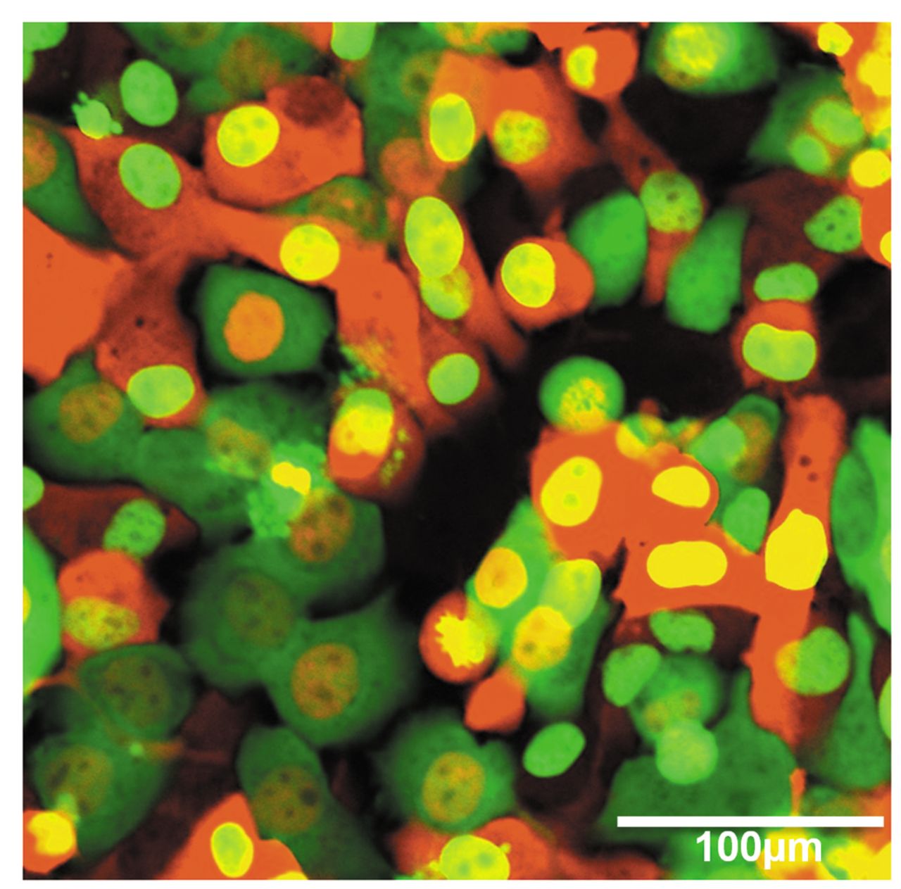
Imaging the steps of cell hybridization. Reciprocal color-coded sister sublines of dual-color HT-1080 human fibrosarcoma cells, HT-1080-H2B-mCherry-GFP and HT-1080-H2B-GFP-RFP, were used. Figure 1 shows images of co-cultured dual-color HT-1080-H2B-mCherry-GFP and HT-1080-H2B-GFP-RFP cells captured by FV1000 confocal microscopy. Spontaneous cell fusion was not observed in co-cultures of the reciprocal sister sublines.

Cell fusion was effected by HVJ. Highly unusual heterokaryons were observed. For example, in Figure 2A, a heterokaryon with 7 GFP-expressing nuclei is shown. This may represent a fusion of 7 HT-1080-H2B-GFP-RFP cells with each other, which maintained only the GFP-expressing nuclei.

Figure 2B shows a heterokaryon with 3 orange nuclei, 1 yellow nucleus and 5 green nuclei, resulting from multiple cell- and nuclear-fusion events. Various nuclei with 3 different fluorescence colors were observed. It appears the green nuclei are unfused; the yellow nucleus represents a fusion of a red and green nucleus and the orange nuclei may be the result of two red nuclei and one green nucleus fusing. Thus, we have demonstrated that it is possible to image heterokaryon formation and subsequent nuclear fusion after fusion of the reciprocal color-coded HT-1080 sublines.

Figure 2C shows two heterokaryons, one with two yellow nuclei and a green cytoplasm and another heterokaryon with two yellow nuclei that appears to have just divided along with multiple small green nuclei and a yellow cytoplasm. The results in Figure 2C suggest that it is possible for nuclei to fuse without cytoplasmic fusion as seen in the heterokaryon with fused and unfused nuclei with the green cytoplasm that appears not fused. It seems also possible for cytoplasms to fuse, in case of the latter heterokaryon, with a yellow cytoplasm, which also contains fused yellow nuclei as well as unfused nuclei.

Figure 1.
  • Download figure
  • Open in new tab
  • Download powerpoint
Figure 1.

Co-culture of HT-1080 human fibrosarcoma H2B-mCherry-GFP and HT-1080-H2B-GFP-RFP cells in a 1:1 ratio in vitro (bar=100 μm). In the co-culture, no cell fusion was observed. Images were captured with an Olympus FV1000 confocal microscope.

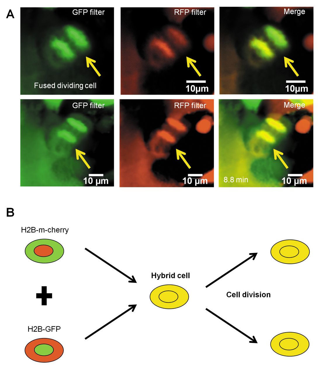
Imaging of a dividing fully hybridized cell. Figure 3A shows a fused HT-1080-H2B-mCherry-GFP–HT-1080-H2B-GFP-RFP hybrid cell undergoing mitosis of its fused yellow nucleus. This cell was observed in the process of mitosis. Figure 3B shows a schematic of this cell.

Future studies will use hybrid cells, as described in this report, to visualize color-coded chromosomes within them in order to observe chromosome loss, gain, and recombination in real time during cancer cell progression i order to thereby determine each chromosome's role in cancer-cell behavior in real time.

Figure 2.
  • Download figure
  • Open in new tab
  • Download powerpoint
Figure 2.

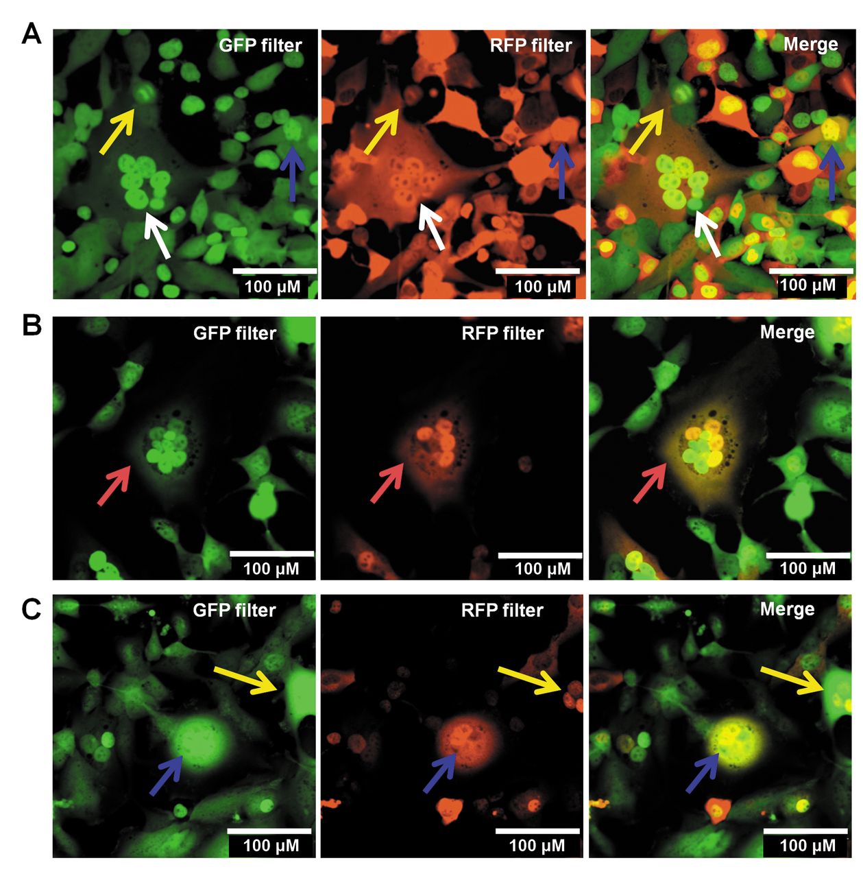
Imaging the steps of cell hybridization. Reciprocal color-coded sister sublines of human fibrosarcoma cells: HT-1080-H2B-mCherry-GFP cells and HT-1080-H2B-GFP-RFP cells were fused. A) A heterokaryon with 7 GFP-expressing nuclei is shown. B) A heterokaryon with 3 orange nuclei, 1 yellow nucleus and 5 green nuclei is shown. C) Two heterokaryons are shown, one with a green cytoplasm and 3 yellow nuclei and one with a yellow cytoplasm and yellow nuclei and multiple small green nuclei

Figure 3.
  • Download figure
  • Open in new tab
  • Download powerpoint
Figure 3.

Imaging of a dividing hybrid cell. A) A hybrid fused HT-1080-H2B-mCherry-GFP – HT-1080-H2B-GFP-RFP cell, with a yellow nucleus, was observed to divide. This cell was observed in the process of mitosis over 8.8 minutes. B) A schematic of Figure 3A.

Footnotes

  • This article is freely accessible online.

  • Conflicts of Interest

    None of the authors have any conflict of interest in regard to this study.

  • Dedication

    This paper is dedicated to the memory of A. R. Moossa, MD and Sun Lee, MD.

  • Received May 21, 2016.
  • Revision received June 13, 2016.
  • Accepted June 14, 2016.
  • Copyright© 2016 International Institute of Anticancer Research (Dr. John G. Delinassios), All rights reserved

References

  1. ↵
    1. Littlefield JW
    : Selection of Hybrids from Matings of Fibroblasts in vitro and Their Presumed Recombinants. Science 145(3633): 709-710, 1964.
    OpenUrlAbstract/FREE Full Text
    1. Harris H
    : Cell Fusion. Oxford: Clarendon Press, 1970 (ISBN 0-19-857344-8).
    1. Harris H
    : Suppression of malignancy in hybrid cells: the mechanism. J Cell Sci 79: 83-94, 1985.
    OpenUrlAbstract/FREE Full Text
  2. ↵
    1. Ephrussi B
    : Hybridization of somatic cells. Princeton University Press, Princeton, NJ, 1972 (ISBN: 978-0691646343).
  3. ↵
    1. Glinsky GV,
    2. Glinskii AB,
    3. Berezovskaya O,
    4. Smith BA,
    5. Jiang P,
    6. Li X-M,
    7. Yang M,
    8. Hoffman RM
    . Dual-color-coded imaging of viable circulating prostate carcinoma cells reveals genetic exchange between tumor cells in vivo, contributing to highly metastatic phenotypes. Cell Cycle 5: 191-197, 2006.
    OpenUrlPubMed
  4. ↵
    1. Kanda T,
    2. Sullivan KF,
    3. Wahl GM
    : Histone-GFP fusion protein enables sensitive analysis of chromosome dynamics in living mammalian cells. Curr Biol 8: 377-385, 1998.
    OpenUrlCrossRefPubMed
  5. ↵
    1. Yamamoto N,
    2. Jiang P,
    3. Yang M,
    4. Xu M,
    5. Yamauchi K,
    6. Tsuchiya H,
    7. Tomita K,
    8. Wahl GM,
    9. Moossa AR,
    10. Hoffman RM
    : Cellular dynamics visualized in live cells in vitro and in vivo by differential dual-color nuclear-cytoplasmic fluorescent-protein expression. Cancer Res 64: 4251-4256, 2004.
    OpenUrlAbstract/FREE Full Text
  6. ↵
    1. Jiang P,
    2. Yamauchi K,
    3. Yang M,
    4. Tsuji K,
    5. Xu M,
    6. Maitra A,
    7. Bouvet M,
    8. Hoffman RM
    : Tumor cells genetically labeled with GFP in the nucleus and RFP in the cytoplasm for imaging cellular dynamics. Cell Cycle 5: 1198-1201, 2006.
    OpenUrlCrossRefPubMed
  7. ↵
    1. Zhao M,
    2. Yang M,
    3. Li X-M,
    4. Jiang P,
    5. Baranov E,
    6. Li S,
    7. Xu M,
    8. Penman S,
    9. Hoffman RM
    : Tumor-targeting bacterial therapy with amino acid auxotrophs of GFP-expressing Salmonella typhimurium. Proc Natl Acad Sci USA 102: 755-760, 2005.
    OpenUrlAbstract/FREE Full Text
  8. ↵
    1. Yamauchi K,
    2. Yang M,
    3. Jiang P,
    4. Yamamoto N,
    5. Xu M,
    6. Amoh Y,
    7. Tsuji K,
    8. Bouvet M,
    9. Tsuchiya H,
    10. Tomita K,
    11. Moossa AR,
    12. Hoffman RM
    : Real-time in vivo dual-color imaging of intracapillary cancer cell and nucleus deformation and migration. Cancer Res 65: 4246-4252, 2005.
    OpenUrlAbstract/FREE Full Text
  9. ↵
    1. Hayashi K,
    2. Kimura H,
    3. Yamauchi K,
    4. Yamamoto N,
    5. Tsuchiya H,
    6. Tomita K,
    7. Kishimoto H,
    8. Hasegawa A,
    9. Bouvet M,
    10. Hoffman RM
    : Comparison of cancer-cell seeding, viability and deformation in the lung, muscle and liver, visualized by subcellular real-time imaging in the live mouse. Anticancer Res 31: 3665-3672, 2011.
    OpenUrlAbstract/FREE Full Text
  10. ↵
    1. Tsuji K,
    2. Yamauchi K,
    3. Yang M,
    4. Jiang P,
    5. Bouvet M,
    6. Endo H,
    7. Kanai Y,
    8. Yamashita K,
    9. Moossa AR,
    10. Hoffman RM
    : Dual-color imaging of nuclear-cytoplasmic dynamics, viability, and proliferation of cancer cells in the portal vein area. Cancer Res 66: 303-306, 2006.
    OpenUrlAbstract/FREE Full Text
  11. ↵
    1. Bouvet M,
    2. Tsuji K,
    3. Yang M,
    4. Jiang P,
    5. Moossa AR,
    6. Hoffman RM
    : In vivo color-coded imaging of the interaction of colon cancer cells and splenocytes in the formation of liver metastases. Cancer Res 66: 11293-11297, 2006.
    OpenUrlAbstract/FREE Full Text
  12. ↵
    1. Yamauchi K,
    2. Yang M,
    3. Jiang P,
    4. Xu M,
    5. Yamamoto N,
    6. Tsuchiya H,
    7. Tomita K,
    8. Moossa AR,
    9. Bouvet M,
    10. Hoffman RM
    : Development of real-time subcellular dynamic multicolor imaging of cancer cell-trafficking in live mice with a variable-magnification whole-mouse imaging system. Cancer Res 66: 4208-4214, 2006.
    OpenUrlAbstract/FREE Full Text
  13. ↵
    1. Yang M,
    2. Jiang P,
    3. Hoffman RM
    : Early reporting of apoptosis by real-time imaging of cancer cells labeled with green fluorescent protein in the nucleus and red fluorescent protein in the cytoplasm. Anticancer Res 35: 2539-2543, 2015.
    OpenUrlAbstract/FREE Full Text
  14. ↵
    1. Hayashi K,
    2. Jiang P,
    3. Yamauchi K,
    4. Yamamoto N,
    5. Tsuchiya H,
    6. Tomita K,
    7. Moossa AR,
    8. Bouvet M,
    9. Hoffman RM
    : Real-time imaging of tumor-cell shedding and trafficking in lymphatic channels. Cancer Res 67: 8223-8228, 2007.
    OpenUrlAbstract/FREE Full Text
  15. ↵
    1. Amoh Y,
    2. Hamada Y,
    3. Katsuoka K,
    4. Hoffman RM
    : In vivo imaging of nuclear-cytoplasmic deformation and partition during cancer cell death due to immune rejection. J Cell Biochem 113: 465-472, 2012.
    OpenUrlPubMed
  16. ↵
    1. Hu M,
    2. Zhao M,
    3. An C,
    4. Yang M,
    5. Li Q,
    6. Zhang Y,
    7. Suetsugu A,
    8. Tome Y,
    9. Yano S,
    10. Fu Y,
    11. Hoffman RM,
    12. Hu K
    : Real-time imaging of apoptosis induction of human breast cancer cells by the traditional Chinese medicinal herb tubeimu. Anticancer Res 32: 2509-2514, 2012.
    OpenUrlAbstract/FREE Full Text
  17. ↵
    1. Momiyama M,
    2. Suetsugu A,
    3. Chishima T,
    4. Bouvet M,
    5. Endo I,
    6. Hoffman RM
    : Subcellular real-time imaging of the efficacy of temozolomide on cancer cells in the brain of live mice. Anticancer Res 33: 103-106, 2013.
    OpenUrlAbstract/FREE Full Text
    1. Kimura H,
    2. Hayashi K,
    3. Yamauchi K,
    4. Yamamoto N,
    5. Tsuchiya H,
    6. Tomita K,
    7. Kishimoto H,
    8. Bouvet M,
    9. Hoffman RM
    : Real-time imaging of single cancer-cell dynamics of lung metastasis. J Cell Biochem 109: 58-64, 2010.
    OpenUrlPubMed
  18. ↵
    1. Momiyama M,
    2. Suetsugu A,
    3. Kimura H,
    4. Kishimoto H,
    5. Aki R,
    6. Yamada A,
    7. Sakurada H,
    8. Chishima T,
    9. Bouvet M,
    10. Endo I,
    11. Hoffman RM
    : Imaging the efficacy of UVC irradiation on superficial brain tumors and metastasis in live mice at the subcellular level. J Cell Biochem 114: 428-434, 2013.
    OpenUrlCrossRefPubMed
  19. ↵
    1. Momiyama M,
    2. Suetsugu A,
    3. Kimura H,
    4. Chishima T,
    5. Bouvet M,
    6. Endo I,
    7. Hoffman RM
    : Dynamic subcellular imaging of cancer cell mitosis in the brain of live mice. Anticancer Res 33: 1367-1371, 2013.
    OpenUrlAbstract/FREE Full Text
  20. ↵
    1. Hiroshima Y,
    2. Maawy A,
    3. Hassanein MK,
    4. Menen R,
    5. Momiyama M,
    6. Murakami T,
    7. Miwa S,
    8. Yamamoto M,
    9. Uehara F,
    10. Yano S,
    11. Mori R,
    12. Matsuyama R,
    13. Chishima T,
    14. Tanaka K,
    15. Ichikawa Y,
    16. Bouvet M,
    17. Endo I,
    18. Hoffman RM
    : The tumor-educated-macrophage increase of malignancy of human pancreatic cancer is prevented by zoledronic acid. PLoS One 9: e103382, 2014.
    OpenUrlCrossRefPubMed
    1. Yamauchi K,
    2. Tome Y,
    3. Yamamoto N,
    4. Hayashi K,
    5. Kimura H,
    6. Tsuchiya H,
    7. Tomita K,
    8. Bouvet M,
    9. Hoffman RM
    : Color-coded real-time subcellular fluorescence imaging of the interaction between cancer and host cells in live mice. Anticancer Res 32: 39-43, 2012.
    OpenUrlAbstract/FREE Full Text
  21. ↵
    1. Yang M,
    2. Li L,
    3. Jiang P,
    4. Moossa AR,
    5. Penman S,
    6. Hoffman RM
    : Dual-color fluorescence imaging distinguishes tumor cells from induced host angiogenic vessels and stromal cells. Proc Natl Acad Sci USA 100: 14259-14262, 2003.
    OpenUrlAbstract/FREE Full Text
  22. ↵
    1. Wolf K,
    2. Te Lindert M,
    3. Krause M,
    4. Alexander S,
    5. Te Riet J,
    6. Willis AL,
    7. Hoffman RM,
    8. Figdor CG,
    9. Weiss SJ,
    10. Friedl P
    : Physical limits of cell migration: Control by ECM space and nuclear deformation and tuning by proteolysis and traction force. J Cell Biol 201: 1069-1084, 2013.
    OpenUrlAbstract/FREE Full Text
  23. ↵
    1. Suetsugu A,
    2. Jiang P,
    3. Yang M,
    4. Yamamoto N,
    5. Moriwaki H,
    6. Saji S,
    7. Hoffman RM
    : The use of living cancer cells expressing green fluorescent protein in the nucleus and red fluorescence protein in the cytoplasm for real-time confocal imaging of chromosome and cytoplasmic dynamics during mitosis. Anticancer Res 35: 2545-2551, 2015.
    OpenUrlAbstract/FREE Full Text
  24. ↵
    1. Uchugonova A,
    2. Duong J,
    3. Zhang N,
    4. König K,
    5. Hoffman RM
    : The bulge area is the origin of nestin-expressing pluripotent stem cells of the hair follicle. J Cell Biochem 112: 2046-2050, 2011.
    OpenUrlCrossRefPubMed
PreviousNext
Back to top

In this issue

Anticancer Research: 36 (8)
Anticancer Research
Vol. 36, Issue 8
August 2016
  • Table of Contents
  • Table of Contents (PDF)
  • Index by author
  • Back Matter (PDF)
  • Ed Board (PDF)
  • Front Matter (PDF)
Print
Download PDF
Article Alerts
Sign In to Email Alerts with your Email Address
Email Article

Thank you for your interest in spreading the word on Anticancer Research.

NOTE: We only request your email address so that the person you are recommending the page to knows that you wanted them to see it, and that it is not junk mail. We do not capture any email address.

Enter multiple addresses on separate lines or separate them with commas.
Color-coded Live Imaging of Heterokaryon Formation and Nuclear Fusion of Hybridizing Cancer Cells
(Your Name) has sent you a message from Anticancer Research
(Your Name) thought you would like to see the Anticancer Research web site.
CAPTCHA
This question is for testing whether or not you are a human visitor and to prevent automated spam submissions.
10 + 10 =
Solve this simple math problem and enter the result. E.g. for 1+3, enter 4.
Citation Tools
Color-coded Live Imaging of Heterokaryon Formation and Nuclear Fusion of Hybridizing Cancer Cells
ATSUSHI SUETSUGU, TAKURO MATSUMOTO, KOSUKE HASEGAWA, MIKI NAKAMURA, TAKAHIRO KUNISADA, MASAHITO SHIMIZU, SHIGETOYO SAJI, HISATAKA MORIWAKI, MICHAEL BOUVET, ROBERT M. HOFFMAN
Anticancer Research Aug 2016, 36 (8) 3827-3831;

Citation Manager Formats

  • BibTeX
  • Bookends
  • EasyBib
  • EndNote (tagged)
  • EndNote 8 (xml)
  • Medlars
  • Mendeley
  • Papers
  • RefWorks Tagged
  • Ref Manager
  • RIS
  • Zotero
Reprints and Permissions
Share
Color-coded Live Imaging of Heterokaryon Formation and Nuclear Fusion of Hybridizing Cancer Cells
ATSUSHI SUETSUGU, TAKURO MATSUMOTO, KOSUKE HASEGAWA, MIKI NAKAMURA, TAKAHIRO KUNISADA, MASAHITO SHIMIZU, SHIGETOYO SAJI, HISATAKA MORIWAKI, MICHAEL BOUVET, ROBERT M. HOFFMAN
Anticancer Research Aug 2016, 36 (8) 3827-3831;
del.icio.us logo Digg logo Reddit logo Twitter logo CiteULike logo Facebook logo Google logo Mendeley logo
  • Tweet Widget
  • Facebook Like
  • Google Plus One

Jump to section

  • Article
    • Abstract
    • Materials and Methods
    • Results and Discussion
    • Footnotes
    • References
  • Figures & Data
  • Info & Metrics
  • PDF

Related Articles

  • No related articles found.
  • PubMed
  • Google Scholar

Cited By...

  • Linking endoplasmic reticulum stress to polyploidy in ovarian cancer cells
  • Human CardioChimeras: Creation of a Novel 'Next Generation Cardiac Cell
  • Google Scholar

More in this TOC Section

  • 5-Azacytidine (5-aza) Induces p53-associated Cell Death Through Inhibition of DNA Methyltransferase Activity in Hep3B and HT-29 Cells
  • Prognostic Value of WNT1, NOTCH1, PDGFRβ, and CXCR4 in Oral Squamous Cell Carcinoma
  • Hypoxia-adapted Multiple Myeloma Stem Cells Resist γδ-T-Cell-mediated Killing by Modulating the Mevalonate Pathway
Show more Experimental Studies

Similar Articles

Keywords

  • green fluorescent protein
  • red fluorescent protein
  • dual-color HT-1080
  • cell fusion
  • Sendai virus
  • heterokaryon
  • nuclear fusion
  • cell hybrid
  • imaging
Anticancer Research

© 2023 Anticancer Research

Powered by HighWire