
Main menu

  • Home
  • Current Issue
  • Archive
  • Info for
    • Authors
    • Editorial Policies
    • Subscribers
    • Advertisers
    • Editorial Board
  • Other Publications
    • In Vivo
    • Cancer Genomics & Proteomics
    • Cancer Diagnosis & Prognosis
  • More
    • IIAR
    • Conferences
    • 2008 Nobel Laureates
  • About Us
    • General Policy
    • Contact
  • Other Publications
    • Anticancer Research
    • In Vivo
    • Cancer Genomics & Proteomics

User menu

  • Register
  • Subscribe
  • My alerts
  • Log in
  • My Cart

Search

  • Advanced search
Anticancer Research
  • Other Publications
    • Anticancer Research
    • In Vivo
    • Cancer Genomics & Proteomics
  • Register
  • Subscribe
  • My alerts
  • Log in
  • My Cart
Anticancer Research

Advanced Search

  • Home
  • Current Issue
  • Archive
  • Info for
    • Authors
    • Editorial Policies
    • Subscribers
    • Advertisers
    • Editorial Board
  • Other Publications
    • In Vivo
    • Cancer Genomics & Proteomics
    • Cancer Diagnosis & Prognosis
  • More
    • IIAR
    • Conferences
    • 2008 Nobel Laureates
  • About Us
    • General Policy
    • Contact
  • Visit us on Facebook
  • Follow us on Linkedin
Research ArticleExperimental Studies

Imaging Nuclear–Cytoplasmic Dynamics in Primary and Metastatic Colon Cancer in Nude Mice

KOSUKE HASEGAWA, ATSUSHI SUETSUGU, MIKI NAKAMURA, TAKURO MATSUMOTO, HITOMI AOKI, TAKAHIRO KUNISADA, MICHAEL BOUVET, MASAHITO SHIMIZU and ROBERT M. HOFFMAN
Anticancer Research May 2016, 36 (5) 2113-2117;
KOSUKE HASEGAWA
1Department of Gastroenterology, Gifu University Graduate School of Medicine, Gifu, Japan
  • Find this author on Google Scholar
  • Find this author on PubMed
  • Search for this author on this site
ATSUSHI SUETSUGU
1Department of Gastroenterology, Gifu University Graduate School of Medicine, Gifu, Japan
2AntiCancer, Inc., San Diego, CA, U.S.A.
3Department of Surgery, University of California San Diego, San Diego, CA, U.S.A.
  • Find this author on Google Scholar
  • Find this author on PubMed
  • Search for this author on this site
  • For correspondence: all@anticancer.com asue@gifu-u.ac.jp
MIKI NAKAMURA
1Department of Gastroenterology, Gifu University Graduate School of Medicine, Gifu, Japan
  • Find this author on Google Scholar
  • Find this author on PubMed
  • Search for this author on this site
TAKURO MATSUMOTO
1Department of Gastroenterology, Gifu University Graduate School of Medicine, Gifu, Japan
  • Find this author on Google Scholar
  • Find this author on PubMed
  • Search for this author on this site
HITOMI AOKI
1Department of Gastroenterology, Gifu University Graduate School of Medicine, Gifu, Japan
  • Find this author on Google Scholar
  • Find this author on PubMed
  • Search for this author on this site
TAKAHIRO KUNISADA
1Department of Gastroenterology, Gifu University Graduate School of Medicine, Gifu, Japan
  • Find this author on Google Scholar
  • Find this author on PubMed
  • Search for this author on this site
MICHAEL BOUVET
3Department of Surgery, University of California San Diego, San Diego, CA, U.S.A.
  • Find this author on Google Scholar
  • Find this author on PubMed
  • Search for this author on this site
MASAHITO SHIMIZU
1Department of Gastroenterology, Gifu University Graduate School of Medicine, Gifu, Japan
  • Find this author on Google Scholar
  • Find this author on PubMed
  • Search for this author on this site
ROBERT M. HOFFMAN
2AntiCancer, Inc., San Diego, CA, U.S.A.
3Department of Surgery, University of California San Diego, San Diego, CA, U.S.A.
  • Find this author on Google Scholar
  • Find this author on PubMed
  • Search for this author on this site
  • For correspondence: all@anticancer.com asue@gifu-u.ac.jp
  • Article
  • Figures & Data
  • Info & Metrics
  • PDF
Loading

Article Figures & Data

Figures

  • Figure 1.
    • Download figure
    • Open in new tab
    • Download powerpoint
    Figure 1.

    A: Left panel: Low-magnification images of dual-color HCT-116 colon cancer cells expressing green fluorescent protein (GFP) in the nucleus and red fluorescent protein (RFP) in the cytoplasm in vitro (Bar=20 μm). HCT-116 dual-color cells were cultured in RPMI-1640 medium supplemented with 10% heat-inactivated FBS and 1% penicillin and streptomycin. Yellow arrows indicate mitotic cells. Right panel: High-magnification images of a mitotic HCT-116 dual-color cell. Metaphase chromosomes are clearly visualized (Bar=5 μm). Images of live cells were captured with an Olympus FV1000 confocal microscope. B: Bright-field and fluorescence low-magnification images of primary and metastatic tumors in nude mice. Green arrows indicate liver metastasis. Blue arrows indicate splenic tumor (injection site). Yellow arrow indicates retroperitoneal metastasis (Bar=10 μm). All images were captured with an Olympus SZX7 microscope. C: High-magnification images of dual-color HCT-116 cells in each organ. Upper panel: Liver metastasis. Middle panel: Splenic tumor. Lower panel: Retroperitoneal metastasis.

  • Figure 2.
    • Download figure
    • Open in new tab
    • Download powerpoint
    Figure 2.

    High-magnification images of cells cultured from tumors in each organ. A: Liver metastasis. B: Splenic tumor. C: Retroperitoneal metastasis. In each tissue, mitotic cells and multi-nucleated cells ware observed. The inset contains multi-nucleated cells with high-magnification images of these cells shown in the adjacent panel. Blue arrows indicate multi-nucleated cells. White arrows indicate vacuolar degeneration. Multi-nucleated cells suggest the possibility of cancer-cell fusion. Images were captured with an Olympus FV1000 confocal microscope.
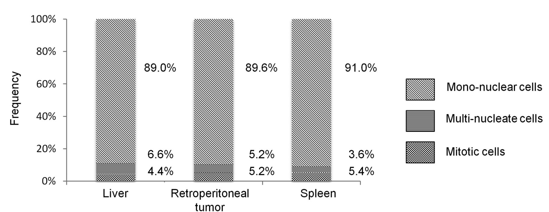
  • Figure 3.
    • Download figure
    • Open in new tab
    • Download powerpoint
    Figure 3.

    Five randomly-selected, low-magnification visual fields were quantified for the number of mono-nucleate, mitotic, and multi-nucleated HCT-116 cancer cells. The frequency of each cell type in the liver metastasis, spleen tumor, and retroperitoneal metastasis was plotted in the bar graphs. There were no significant differences in the ratios of the cell types between the different tumor sites.

PreviousNext
Back to top

In this issue

Anticancer Research: 36 (5)
Anticancer Research
Vol. 36, Issue 5
May 2016
  • Table of Contents
  • Table of Contents (PDF)
  • Index by author
  • Back Matter (PDF)
  • Ed Board (PDF)
  • Front Matter (PDF)
Print
Download PDF
Article Alerts
Sign In to Email Alerts with your Email Address
Email Article

Thank you for your interest in spreading the word on Anticancer Research.

NOTE: We only request your email address so that the person you are recommending the page to knows that you wanted them to see it, and that it is not junk mail. We do not capture any email address.

Enter multiple addresses on separate lines or separate them with commas.
Imaging Nuclear–Cytoplasmic Dynamics in Primary and Metastatic Colon Cancer in Nude Mice
(Your Name) has sent you a message from Anticancer Research
(Your Name) thought you would like to see the Anticancer Research web site.
CAPTCHA
This question is for testing whether or not you are a human visitor and to prevent automated spam submissions.
1 + 4 =
Solve this simple math problem and enter the result. E.g. for 1+3, enter 4.
Citation Tools
Imaging Nuclear–Cytoplasmic Dynamics in Primary and Metastatic Colon Cancer in Nude Mice
KOSUKE HASEGAWA, ATSUSHI SUETSUGU, MIKI NAKAMURA, TAKURO MATSUMOTO, HITOMI AOKI, TAKAHIRO KUNISADA, MICHAEL BOUVET, MASAHITO SHIMIZU, ROBERT M. HOFFMAN
Anticancer Research May 2016, 36 (5) 2113-2117;

Citation Manager Formats

  • BibTeX
  • Bookends
  • EasyBib
  • EndNote (tagged)
  • EndNote 8 (xml)
  • Medlars
  • Mendeley
  • Papers
  • RefWorks Tagged
  • Ref Manager
  • RIS
  • Zotero
Reprints and Permissions
Share
Imaging Nuclear–Cytoplasmic Dynamics in Primary and Metastatic Colon Cancer in Nude Mice
KOSUKE HASEGAWA, ATSUSHI SUETSUGU, MIKI NAKAMURA, TAKURO MATSUMOTO, HITOMI AOKI, TAKAHIRO KUNISADA, MICHAEL BOUVET, MASAHITO SHIMIZU, ROBERT M. HOFFMAN
Anticancer Research May 2016, 36 (5) 2113-2117;
Reddit logo Twitter logo Facebook logo Mendeley logo
  • Tweet Widget
  • Facebook Like
  • Google Plus One

Jump to section

  • Article
    • Abstract
    • Materials and Methods
    • Results and Discussion
    • Footnotes
    • References
  • Figures & Data
  • Info & Metrics
  • PDF

Related Articles

  • No related articles found.
  • PubMed
  • Google Scholar

Cited By...

  • Imaging the Role of Multinucleate Pancreatic Cancer Cells and Cancer-Associated Fibroblasts in Peritoneal Metastasis in Mouse Models
  • Google Scholar

More in this TOC Section

  • TIG1 Inhibits the mTOR Signaling Pathway in Malignant Melanoma Through the VAC14 Protein
  • Novel α-Trifluoromethyl Chalcone Exerts Antitumor Effects Against Prostate Cancer Cells
  • Differential Effects of Anti-PD-1/PD-L1 Checkpoint Inhibitors on Adhesion Molecules and Cytokine Secretion by THP-1 Monocytes
Show more Experimental Studies

Similar Articles

Keywords

  • Dual color cancer cells
  • GFP
  • RFP
  • mitosis
  • colon cancer
  • nude mice
  • liver metastasis
  • imaging
Anticancer Research

© 2023 Anticancer Research

Powered by HighWire