
Main menu

  • Home
  • Current Issue
  • Archive
  • Info for
    • Authors
    • Subscribers
    • Advertisers
    • Editorial Board
  • Other Publications
    • In Vivo
    • Cancer Genomics & Proteomics
    • Cancer Diagnosis & Prognosis
  • More
    • IIAR
    • Conferences
    • 2008 Nobel Laureates
  • About Us
    • General Policy
    • Contact
  • Other Publications
    • Anticancer Research
    • In Vivo
    • Cancer Genomics & Proteomics

User menu

  • Register
  • Subscribe
  • My alerts
  • Log in
  • My Cart

Search

  • Advanced search
Anticancer Research
  • Other Publications
    • Anticancer Research
    • In Vivo
    • Cancer Genomics & Proteomics
  • Register
  • Subscribe
  • My alerts
  • Log in
  • My Cart
Anticancer Research

Advanced Search

  • Home
  • Current Issue
  • Archive
  • Info for
    • Authors
    • Subscribers
    • Advertisers
    • Editorial Board
  • Other Publications
    • In Vivo
    • Cancer Genomics & Proteomics
    • Cancer Diagnosis & Prognosis
  • More
    • IIAR
    • Conferences
    • 2008 Nobel Laureates
  • About Us
    • General Policy
    • Contact
  • Visit us on Facebook
  • Follow us on Linkedin
Research ArticleExperimental Studies

Long Non-coding RNA, PANDA, Contributes to the Stabilization of p53 Tumor Suppressor Protein

YOJIRO KOTAKE, KYOKO KITAGAWA, TATSUYA OHHATA, SATOSHI SAKAI, CHIHARU UCHIDA, HIROYUKI NIIDA, MADOKA NAEMURA and MASATOSHI KITAGAWA
Anticancer Research April 2016, 36 (4) 1605-1611;
YOJIRO KOTAKE
1Department of Biological and Environmental Chemistry, Faculty of Humanity-Oriented Science and Engineering, Kinki University, Iizuka, Fukuoka, Japan
  • Find this author on Google Scholar
  • Find this author on PubMed
  • Search for this author on this site
  • For correspondence: ykotake@fuk.kindai.ac.jp
KYOKO KITAGAWA
2Department of Molecular Biology, Hamamatsu University School of Medicine, Higashi-ku, Hamamatsu, Shizuoka, Japan
  • Find this author on Google Scholar
  • Find this author on PubMed
  • Search for this author on this site
TATSUYA OHHATA
2Department of Molecular Biology, Hamamatsu University School of Medicine, Higashi-ku, Hamamatsu, Shizuoka, Japan
  • Find this author on Google Scholar
  • Find this author on PubMed
  • Search for this author on this site
SATOSHI SAKAI
2Department of Molecular Biology, Hamamatsu University School of Medicine, Higashi-ku, Hamamatsu, Shizuoka, Japan
  • Find this author on Google Scholar
  • Find this author on PubMed
  • Search for this author on this site
CHIHARU UCHIDA
3Laboratory for Promotion of Advanced Research, Advanced Research Facilities & Services, Preeminent Medical Photonics Education & Research Center, Hamamatsu University School of Medicine, Higashi ku, Hamamatsu, Shizuoka, Japan
  • Find this author on Google Scholar
  • Find this author on PubMed
  • Search for this author on this site
HIROYUKI NIIDA
2Department of Molecular Biology, Hamamatsu University School of Medicine, Higashi-ku, Hamamatsu, Shizuoka, Japan
  • Find this author on Google Scholar
  • Find this author on PubMed
  • Search for this author on this site
MADOKA NAEMURA
1Department of Biological and Environmental Chemistry, Faculty of Humanity-Oriented Science and Engineering, Kinki University, Iizuka, Fukuoka, Japan
  • Find this author on Google Scholar
  • Find this author on PubMed
  • Search for this author on this site
MASATOSHI KITAGAWA
2Department of Molecular Biology, Hamamatsu University School of Medicine, Higashi-ku, Hamamatsu, Shizuoka, Japan
  • Find this author on Google Scholar
  • Find this author on PubMed
  • Search for this author on this site
  • Article
  • Figures & Data
  • Info & Metrics
  • PDF
Loading

Abstract

Background: P21-associated noncoding RNA DNA damage-activated (PANDA) is induced in response to DNA damage and represses apoptosis by inhibiting the function of nuclear transcription factor Y subunit alpha (NF-YA) transcription factor. Herein, we report that PANDA affects regulation of p53 tumor-suppressor protein. Materials and Methods: U2OS cells were transfected with PANDA siRNAs. At 72 h post-transfection, cells were subjected to immunoblotting and quantitative reverse transcription-polymerase chain reaction. Results: Depletion of PANDA was associated with decreased levels of p53 protein, but not p53 mRNA. The stability of p53 protein was markedly reduced by PANDA silencing. Degradation of p53 protein by silencing PANDA was prevented by treatment of MG132, a proteasome inhibitor. Moreover, depletion of PANDA prevented accumulation of p53 protein, as a result of DNA damage, induced by the genotoxic agent etoposide. Conclusion: These results suggest that PANDA stabilizes p53 protein in response to DNA damage, and provide new insight into the regulatory mechanisms of p53.

  • Long non-coding RNA
  • PANDA
  • p53
  • protein degradation

Recent mass -scale transcriptome analysis by tiling array and RNA sequencing found loci for more than 10,000 long non-coding RNAs (lncRNAs) in the human genome (1). Although the functions of only a few dozen lncRNAs have been elucidated, several lncRNAs have been found to mediate cell-fate determination, including differentiation, proliferation, senescence, apoptosis, and cancer development (2-5). Moreover, the expression of multiple lncRNAs has been shown to fluctuate according to different biological conditions, such as DNA damage, oncogenic stimuli, and cell differentiation, suggesting the involvement of lncRNAs in biological processes (6-8).

P21-associated ncRNA DNA damage -activated (PANDA) has been identified as an lncRNA induced in response to DNA damage, using super high-resolution tiling array analysis (6). PANDA is located about 5 kb upstream of the transcription start site (TSS) of cyclin-dependent kinase (CDK) inhibitor p21. The genomic DNA of PANDA is composed of one exon and transcribed in the opposite direction of p21, with a 1.5 kb transcription product. The genomic region that includes p21 and PANDA is regulated by tumor suppressor p53 (9), which binds upstream of the p21 TSS and activates both p21 and PANDA transcription in response to DNA damage. PANDA associates with subunit A of nuclear transcription factor Y (NF-Y). NF-Y, a trimeric complex that includes subunits A, B, and C, induces transcription of the apoptosis gene, fas cell surface death receptor (FAS), by binding to its promoter (10). PANDA represses transcription of apoptosis activators, such as apoptotic peptidase activating factor 1 (APAF1), B-cell cll/lymphoma 2-interacting killer (BIK), FAS and LDD, by blocking NF-YA from binding to their promoters, thus repressing apoptosis (6).

The tumor-suppressor protein, p53, controls cell proliferation in response to oncogenic insults and DNA damage (11, 12). Activated p53 triggers cell-cycle arrest, cellular senescence and apoptosis by directly binding to promoters of numerous genes and regulating their transcription, thus preventing DNA-damaged or -stressed cells from proliferating (13). Disruption of the p53 pathway is thought to be a key step in carcinogenesis; and many studies have shown that p53 is frequently mutated in a wide range of human cancer types (14-16). Normally, p53 protein levels are kept low via ubiquitin–proteasome system, which is mediated mainly by murine double minute 2 (MDM2), a ubiquitin ligase (17-19). When cellular DNA is damaged, p53 is phosphorylated through the ataxia–telangiectasia mutated (ATM)/ataxia–telangiectasia and radiation sensitive 3-related (ATR) pathways (20), which disrupts the association of the MDM2-p53 complex, leading to stabilization of p53 protein. p53 is also regulated at translational (21) and transcriptional (22) levels.

p53 has been shown to induce expression of numerous lncRNAs, including lincRNA-regulation of reprogramming (RoR) (23), lincRNA-p21 (7) and PANDA (6). lincRNA-RoR associates with heterogeneous nuclear ribonucleoprotein-I (hnRNP-I) and suppresses p53 translation, which indicates that p53 and lincRNA-RoR form an auto-regulatory feedback loop (23). lincRNA-p21 associates with and recruits hnRNAP-K to target gene promoters and represses transcription, leading to induction of apoptosis (7). Herein, we show that PANDA induced by DNA damage affects stabilization of p53 protein.

Materials and Methods

Cell culture. We used U2OS (human osteosarcoma) cells that were cultured in Dulbecco's modified Eagle's medium (DMEM; Invitrogen, Carlsbad, CA, USA) supplemented with 10% fetal bovine serum (FBS; GIBCO, Grand Island, NY, USA) in a 37°C incubator with 5% CO2. The cells were exposed to proteosome inhibitor (10 μM) (PEPTIDE INSTITUTE, INC., Osaka, Japan), cycloheximide (100 μg/ml) (SIGMA-ALDRICH, Tokyo, Japan), etoposide (50 μM) (Wako, Osaka, Japan) or vehicle (dimethyl sulfoxide; SIGMA-ALDRICH), as indicated in Figures 2 and 4.

RNA interference (RNAi). U2OS cells were transfected with siRNA oligonucleotides that targeted human PANDA or NF-YA mRNA, using Lipofectamine RNAiMAX (Invitrogen), according to the manufacturer's protocol. Nucleotide sequences of siRNAs for human PANDA were: #1, 5’-AAUGUGUGCACGUAACAGAUU-3’ and #2, 5’-GGGCAUGUUUUCACAGAGGUU-3’. PANDA siRNA#1 was used in Figures 1, 2, 3, and 4. PANDA siRNA#2 was used in Figure 1. Validated siRNA oligonucleotides targeting human NF-YA were obtained from Ambion (siRNA ID: s9528, s9529, s9530; Austin, TX, USA) .

Immunoblotting. Treated cells were lysed with cell lysis buffer (50 mM Tris-HCl pH 7.5, 300 mM NaCl, and 0.3% Triton-X 100) supplemented with protease inhibitors (10 mg/l antipain, 10 mg/l leupeptin, 10 mg/l pepstatin, 10 mg/l trypsin inhibitor, 10 mg/l E64, and 2.5 mg/l chymostatin; PEPTIDE INSTITUTE, INC., Osaka, Japan) and phosphatase inhibitors (1 mM EDTA pH 8.0, 2.5 mM EGTA pH 8.0, 10 mM b-glycerophosphate, 1 mM NaF, and 0.1 mM sodium orthovanadate). The lysates were incubated on ice for 15 min and then centrifuged at 20,600 × g for 15 min at 4°C. The supernatant of each lysate was subjected to sodium dodecyl sulphate-polyacrylamide gel electrophoresis. The separated proteins were transferred to an Immobilon-PSQ transfer membrane (Merk Millipore, Darmstad, Germany) and subjected to western blot. The following antibodies were used for immunoblotting: anti-p53 (DO-1; Santa Cruz Biotechnology, Santa Cruz, CA, USA), anti-NF-YA (H-209; Santa Cruz Biotechnology) anti-α-tubulin (SIGMA-ALDRICH). Immune complexes were detected with horseradish peroxidase-conjugated secondary antibodies and an enhanced chemiluminescence system (PerkinElmer, Branchburg, NJ, USA).

Reverse transcription (RT) and quantitative polymerase chain reaction (q-PCR). Total RNA was extracted by an RNeasy Plus kit (Qiagen, Hilden, Germany), according to the manufacturer's protocol. We treated 10 μg of total RNA with Turbo DNase (Ambion); 1.6 μg of DNase-treated total RNA was subjected to RT reactions with oligo(dT)20 primers and SuperScript First-Strand Synthesis System for RT-PCR (Invitrogen); cDNA thus produced was subjected to PCR with Platinum Taq DNA Polymerase (Invitrogen) or q-PCR with QuantiTect SYBR Green PCR Master Mix (Qiagen) and 200 nM gene-specific primers. Primer sequences were: 5’-AGACCCCAGTGGCACCTGAC-3’ and 5’-GGGCAGAACTTGGCATGATG-3’ for PANDA; 5’-CCTCAGCATCTTATCCGAGTGG-3’ and 5’-TGGATGGTGGTACAGTCAGAGC-3’ for p53; and 5’-GCAAATTCCATGGCACCGT-3’ and 5’-TCGCCCCACTTGATTTTGG-3’ for glyceraldehyde-3-phosphate dehydrogenase (GAPDH). q-PCR assays were performed using an Mx3000P Real-Time Q-PCR System (Agilent Technologies, Santa Clara, CA, USA) and Rotor-Gene 3000 system (Corbett Research, Sydney, Australia). Relative expression levels of p53 mRNA were normalized to those of GAPDH mRNA.

Results

Several lncRNAs, including X inactive specific transcript (XIST) (24), KCNQ1 overlapping transcript 1 (KCNQ1OT1) (25, 26), antisense IGF2R RNA (AIRN) (27), antisense non-coding RNA in the INK4 locus (ANRIL)/p15 antisense (2, 28-31) and HOXA transcript at the distal tip (HOTTIP) (32), have been shown to affect gene transcription in cis. Therefore, we first examined the involvement of PANDA in regulating p21 expression. We knocked down human PANDA using PANDA-specific siRNA in U2OS cells in which PANDA expression was detectable. RT-PCR assay confirmed that this siRNA effectively reduced PANDA expression (Figure 1A). Immunoblotting assays showed that a substantial decrease in p21 protein was associated with the reduction in PANDA level (Figure 1B). Interestingly, silencing PANDA also reduced the level of p53 protein, which is an activator of p21 transcription (Figure 1B). q-RT-PCR assay showed that p53 mRNA levels were not reduced by silencing PANDA (Figure 1C). These results suggest that PANDA affects p53 protein expression in a trans manner that influences p21 expression.

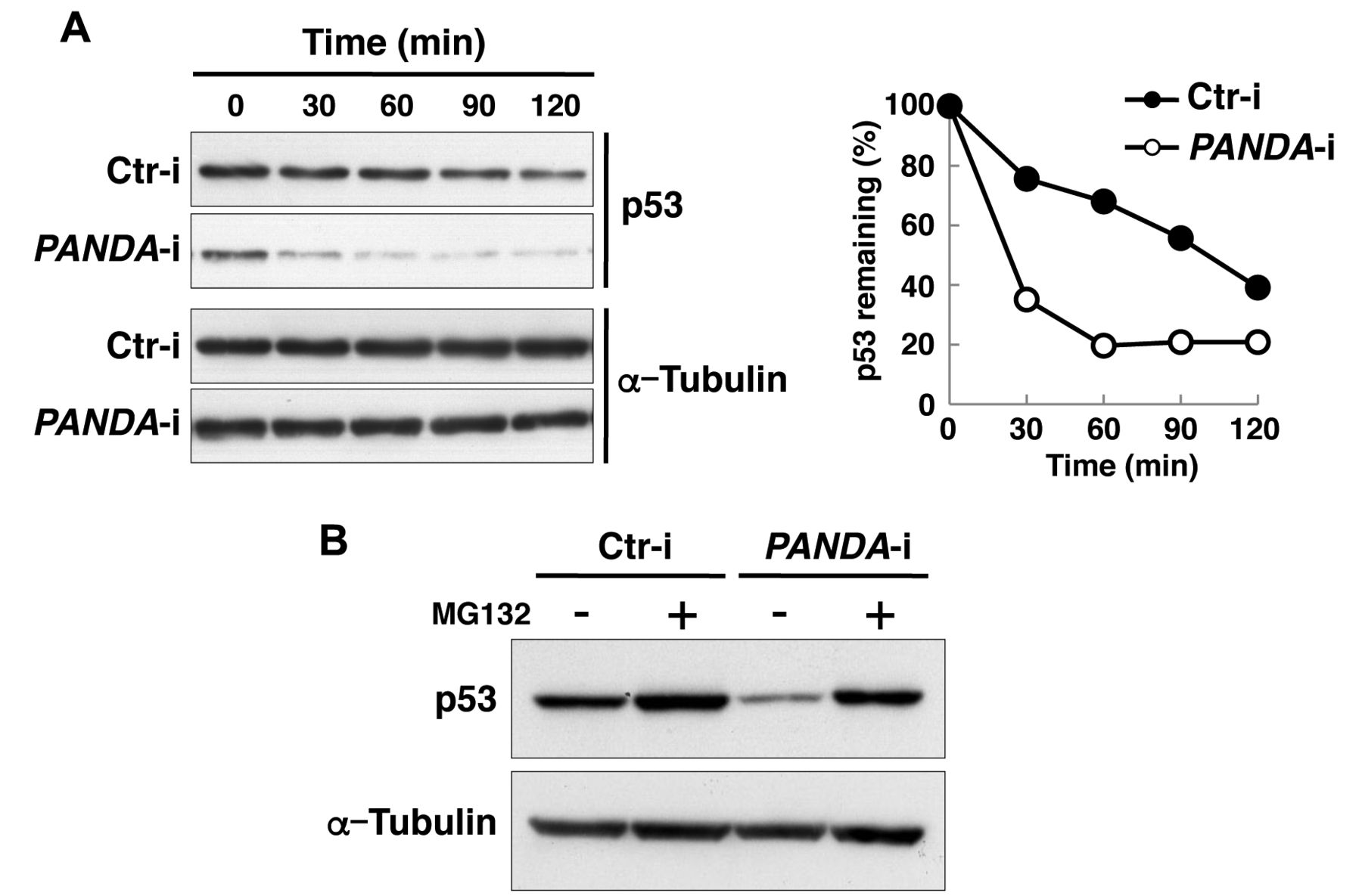
As PANDA depletion reduces p53 protein level, we investigated whether PANDA helps stabilize p53 protein. We measured the half-life of p53 protein in U2OS cells transfected with control and PANDA-targeting siRNA. At 72 h post-transfection with siRNAs, cells were treated with cycloheximide, a protein synthesis inhibitor, for 0, 30, 60, 90 or 120 min and subjected to immunoblot assays. We found p53 protein to be markedly less stable in PANDA-silenced cells (Figure 2A). Treatment with MG132, a proteasome inhibitor, prevented degradation of p53 protein in PANDA-knockout cells (Figure 2B), which indicates that proteolysis of p53 in PANDA-knockout cells is mediated by proteasomes. These results suggest that PANDA contributes to stabilization of p53 protein.

Figure 1.
  • Download figure
  • Open in new tab
  • Download powerpoint
Figure 1.

Silencing P21 associated ncRNA DNA damage-activated (PANDA) reduces p53 protein levels. A: U2OS cells were transfected with siRNAs targeting PANDA or control siRNA (Ctr-i) and harvested at 72 h after transfection. The efficiency of PANDA silencing was determined by reverse transcription-polymerase chain reaction (RT-PCR). B: Cells treated as in (A) were subjected to immunoblotting with antibodies against p53, p21 or α-tubulin. C: The effect of PANDA silencing on p53 mRNA levels was determined by quantitative RT-PCR. Data are expressed relative to corresponding values for U2OS cells transfected with control siRNA (Ctr-i). Mean value and standard deviations were calculated from three representative experiments.

Figure 2.
  • Download figure
  • Open in new tab
  • Download powerpoint
Figure 2.

Effect of silencing P21 associated ncRNA DNA damage-activated (PANDA) on p53 stability. A: At 72 h post-transfection with siRNAs targeting PANDA or control siRNA (Ctr-i), cells were incubated with cycloheximide (100 μg/ml) for the indicated times. Cell extracts were then immunoblotted with antibodies against p53 or α-tubulin (left panel). Band intensity was measured and normalized to that of α-tubulin. The amount of p53 at 0 min was defined as 100% (right panel). B: At 72 h post-transfection with siRNAs that target PANDA or control siRNA (Ctr-i), U2OS cells were incubated with or without MG132, a proteasome inhibitor (10 μM) for 4 h, after which cell extracts were subjected to immunoblotting as in (A).

Figure 3.
  • Download figure
  • Open in new tab
  • Download powerpoint
Figure 3.

P21 associated ncRNA DNA damage-activated (PANDA) regulates p53 protein in an nuclear transcription factor Y subunit alpha (NF-YA)-independent manner. A: U2OS cells were transfected with siRNAs targeting PANDA with or without NF-YA, or with control siRNA (Ctr-i). At 72 h after transfection, cells were harvested and subjected to reverse transcription-polymerase chain reaction to determine the efficiency of PANDA silencing. B: Cells treated as in (A) were immunoblotted with antibodies against p53, NF-YA or α-tubulin.

Figure 4.
  • Download figure
  • Open in new tab
  • Download powerpoint
Figure 4.

P21 associated ncRNA DNA damage-activated (PANDA) is involved in the accumulation of p53 protein as a result of DNA damage. A: At 54 h post-transfection by siRNAs targeting PANDA or control siRNA (Ctr-i), U2OS cells were incubated with or without etoposide (50 μM) for 18 h and harvested. Levels of PANDA and glyceraldehyde-3-phosphate dehydrogenase (GAPDH) mRNA were determined by reverse transcription-polymerase chain reaction. B: Cell lysates were immunoblotted with antibodies against p53 or α-tubulin.

As PANDA has been shown to repress transcription of apoptosis activators by associating with and inhibiting NF-YA (6), we next examined whether PANDA stabilizes p53 protein through NF-YA. To examine this, we knocked-down both PANDA and NF-YA. RT-PCR and immunoblotting assay confirmed decreased expression of both PANDA and NF-YA (Figure 3A and B). Knocking-down both PANDA and NF-YA also reduced p53 protein to the same level as when knocking-down PANDA alone (Figure 3B), which indicates that PANDA stabilizes p53 protein in an NF-YA-independent manner.

In response to DNA damage, the ATM/ATR pathway phosphorylates p53 protein. This phosphorylation blocks ubiquitination of p53 by MDM2, leading to stabilization of p53 protein that can arrest the cell cycle or induce apoptosis (20). RT-PCR and immunoblotting assay showed that DNA damage induced by treatment with etoposide (topoisomerase II inhibitor) increased the levels of PANDA (Figure 4A) and p53 protein (Figure 4B). Silencing PANDA reduced DNA damage-induced p53 accumulation (Figure 4B). These data suggest that PANDA induced by DNA damage is required in order for p53 protein to accumulate.

Discussion

Several lncRNAs are implicated in regulating transcription, translation and nucleus structure (3, 4, 33). PANDA has been shown to repress transcription of apoptosis activators by inhibiting NF-YA-mediated transcription (6). In this study, we showed a new function of PANDA: stabilization of p53 protein. Reducing PANDA levels resulted in a decrease of p53 protein, but not p53 mRNA level, due to proteolysis of p53 protein by proteasomes. This indicates that PANDA regulates p53 protein post-translationally. p53 degradation in PANDA-knockdown cells was also seen in cells depleted of NF-YA, which suggests that PANDA regulates p53 protein in an NF-YA-independent manner.

The mechanism by which PANDA stabilizes p53 protein is yet to be determined in detail. p53 protein is reportedly ubiquitinated by several ubiquitin ligases, including MDM2 (17-19), p53-induced protein with a RING-H2 domain (PIRH2) (34), ARF-binding protein 1 (ARF-BP1) (35), and constitutive photomorphogenic 1 (COP1) (36), leading to its degradation by proteasome. Because silencing PANDA did not increase expression levels of these ubiquitin ligases (data not shown), PANDA may stabilize p53 protein by repressing the enzymatic activity rather than the expression of these ubiquitin ligases. Phosphorylation of p53 protein by ATM/ATR pathway leads to stabilization of p53 protein by disrupting the association of the MDM2–p53 complex (20). Additionally, the tumor-suppressor protein ARF binds to and antagonizes MDM2 activity, resulting in more stable p53 protein (37-39). The involvement of PANDA in these pathways is an important issue that required further investigation.

We and Hung et al. reported that forced expression of an oncogenic form of small GTPase RAS (called oncogenic RAS) or treatment by doxorubicin (which causes DNA damage) increases PANDA expression (6). In this study, we also showed that treatment with etoposide (which also causes DNA damage) increased PANDA expression. Induction of PANDA by doxorubicin treatment has been shown to require p53 (6). Oncogenic RAS and DNA damage signals may induce PANDA expression through p53.

We demonstrated that silencing PANDA inhibits accumulation of p53 protein induced by DNA damage, which indicates that PANDA is required for p53 to accumulate in response to DNA damage. Our results also imply a regulatory loop between p53 and PANDA, with p53 acting as an upstream inducer of PANDA and PANDA stabilizing p53 protein in positive feedback.

Because the p53 pathway affects cell-fate determination, such as differentiation, apoptosis, senescence and malignant transformation, any lncRNAs that are involved in this pathway, might be pivotal to these processes. Indeed, lincRNA-RoR, which suppresses p53 translation, modulates reprogramming of human-induced pluripotent stem cells (40). Metastasis-associated in lung adenocarcinoma transcript 1 (MALAT1), which represses p53 expression, significantly contributes to malignant lung cancer phenotypes (41-43). lincRNA-p21 induced by p53 affects regulation of the Warburg effects on cancer cells and functions as an oncogene to promote tumor growth (7, 44). PANDA may also contribute to such processes as cancer development, through its place in the p53 pathway. Elucidating the function of PANDA thus warrants further investigation.

Acknowledgements

The Authors thank Chihiro Murasaki and Mika Matsumoto for their technical assistance. We also thank our laboratory members for their helpful discussions. This work was supported in part by grants from the Ministry of Education, Culture, Sports, Science and Technology of Japan 26430127 (Y. Kotake), 25290044 (M. Kitagawa), 26640073 (M. Kitagawa) and Takeda Science Foundation (Y. Kotake).

  • Received November 25, 2015.
  • Revision received February 22, 2016.
  • Accepted February 24, 2016.
  • Copyright© 2016 International Institute of Anticancer Research (Dr. John G. Delinassios), All rights reserved

References

  1. ↵
    1. Necsulea A,
    2. Soumillon M,
    3. Warnefors M,
    4. Liechti A,
    5. Daish T,
    6. Zeller U,
    7. Baker JC,
    8. Grutzner F,
    9. Kaessmann H
    : The evolution of lncRNA repertoires and expression patterns in tetrapods. Nature 505: 635-640, 2014.
    OpenUrlCrossRefPubMed
  2. ↵
    1. Kotake Y,
    2. Nakagawa T,
    3. Kitagawa K,
    4. Suzuki S,
    5. Liu N,
    6. Kitagawa M,
    7. Xiong Y
    : Long non-coding RNA ANRIL is required for the PRC2 recruitment to and silencing of p15(INK4B) tumor - suppressor gene. Oncogene 30: 1956-1962, 2011.
    OpenUrlCrossRefPubMed
  3. ↵
    1. Kitagawa M,
    2. Kotake Y,
    3. Ohhata T
    : Long non-coding RNAs involved in cancer development and cell fate determination. Curr Drug Targets 13: 1616-1621, 2012.
    OpenUrlPubMed
  4. ↵
    1. Kitagawa M,
    2. Kitagawa K,
    3. Kotake Y,
    4. Niida H,
    5. Ohhata T
    : Cell-cycle regulation by long non-coding RNAs. Cell Mol Life Sci 70: 4785-4794, 2013.
    OpenUrlCrossRefPubMed
  5. ↵
    1. Flynn RA,
    2. Chang HY
    : Long noncoding RNAs in cell-fate programming and reprogramming. Cell Stem Cell 14: 752-761, 2014.
    OpenUrlCrossRefPubMed
  6. ↵
    1. Hung T,
    2. Wang Y,
    3. Lin MF,
    4. Koegel AK,
    5. Kotake Y,
    6. Grant GD,
    7. Horlings HM,
    8. Shah N,
    9. Umbricht C,
    10. Wang P,
    11. Kong B,
    12. Langerod A,
    13. Borresen-Dale AL,
    14. Kim SK,
    15. van de Vijver M,
    16. Sukumar S,
    17. Whitfield ML,
    18. Kellis M,
    19. Xiong Y,
    20. Wong DJ,
    21. Chang HY
    : Extensive and coordinated transcription of noncoding RNAs within cell-cycle promoters. Nat Genet 43: 621-629, 2011.
    OpenUrlCrossRefPubMed
  7. ↵
    1. Huarte M,
    2. Guttman M,
    3. Feldser D,
    4. Garber M,
    5. Koziol MJ,
    6. Kenzelmann-Broz D,
    7. Khalil AM,
    8. Zuk O,
    9. Amit I,
    10. Rabani M,
    11. Attardi LD,
    12. Regev A,
    13. Lander ES,
    14. Jacks T,
    15. Rinn JL
    : A large intergenic noncoding RNA induced by p53 mediates global gene repression in the p53 response. Cell 142: 409-419, 2010.
    OpenUrlCrossRefPubMed
  8. ↵
    1. Mizutani R,
    2. Wakamatsu A,
    3. Tanaka N,
    4. Yoshida H,
    5. Tochigi N,
    6. Suzuki Y,
    7. Oonishi T,
    8. Tani H,
    9. Tano K,
    10. Ijiri K,
    11. Isogai T,
    12. Akimitsu N
    : Identification and characterization of novel genotoxic stress-inducible nuclear long noncoding RNAs in mammalian cells. PLoS One 7: e34949, 2012.
    OpenUrlCrossRefPubMed
  9. ↵
    1. Wei CL,
    2. Wu Q,
    3. Vega VB,
    4. Chiu KP,
    5. Ng P,
    6. Zhang T,
    7. Shahab A,
    8. Yong HC,
    9. Fu Y,
    10. Weng Z,
    11. Liu J,
    12. Zhao XD,
    13. Chew JL,
    14. Lee YL,
    15. Kuznetsov VA,
    16. Sung WK,
    17. Miller LD,
    18. Lim B,
    19. Liu ET,
    20. Yu Q,
    21. Ng HH,
    22. Ruan Y
    : A global map of p53 transcription-factor binding sites in the human genome. Cell 124: 207-219, 2006.
    OpenUrlCrossRefPubMed
  10. ↵
    1. Morachis JM,
    2. Murawsky CM,
    3. Emerson BM
    : Regulation of the p53 transcriptional response by structurally diverse core promoters. Genes Dev 24: 135-147, 2010.
    OpenUrlAbstract/FREE Full Text
  11. ↵
    1. Sherr CJ,
    2. McCormick F
    : The RB and p53 pathways in cancer. Cancer Cell 2: 103-112, 2002.
    OpenUrlCrossRefPubMed
  12. ↵
    1. Campisi J
    : Senescent cells, tumor suppression, and organismal aging: good citizens, bad neighbors. Cell 120: 513-522, 2005.
    OpenUrlCrossRefPubMed
  13. ↵
    1. Riley T,
    2. Sontag E,
    3. Chen P,
    4. Levine A
    : Transcriptional control of human p53-regulated genes. Nat Rev Mol Cell Biol 9: 402-412, 2008.
    OpenUrlCrossRefPubMed
  14. ↵
    1. Hollstein M,
    2. Sidransky D,
    3. Vogelstein B,
    4. Harris CC
    : p53 mutations in human cancers. Science 253: 49-53, 1991.
    OpenUrlAbstract/FREE Full Text
    1. Levine AJ,
    2. Momand J,
    3. Finlay CA
    : The p53 tumour - suppressor gene. Nature 351: 453-456, 1991.
    OpenUrlCrossRefPubMed
  15. ↵
    1. Arfaoui A,
    2. Douik H,
    3. Kablouti G,
    4. Chaaben AB,
    5. Handiri N,
    6. Zid Z,
    7. Ouni N,
    8. Zouiouch F,
    9. Ayari F,
    10. Mamoughli T,
    11. Bouassida J,
    12. Abazza H,
    13. Harzallaha L,
    14. Guemira F
    : Role of p53 codon 72 SNP in breast cancer risk and anthracycline resistance. Anticancer Res 35: 1763-1769, 2015.
    OpenUrlAbstract/FREE Full Text
  16. ↵
    1. Kubbutat MH,
    2. Jones SN,
    3. Vousden KH
    : Regulation of p53 stability by Mdm2. Nature 387: 299-303, 1997.
    OpenUrlCrossRefPubMed
    1. Haupt Y,
    2. Maya R,
    3. Kazaz A,
    4. Oren M
    : Mdm2 promotes the rapid degradation of p53. Nature 387: 296-299, 1997.
    OpenUrlCrossRefPubMed
  17. ↵
    1. Honda R,
    2. Tanaka H,
    3. Yasuda H
    : Oncoprotein MDM2 is a ubiquitin ligase E3 for tumor suppressor p53. FEBS Lett 420: 25-27, 1997.
    OpenUrlCrossRefPubMed
  18. ↵
    1. Meek DW
    : Tumour suppression by p53: A role for the DNA damage response? Nat Rev Cancer 9: 714-723, 2009.
    OpenUrlCrossRefPubMed
  19. ↵
    1. Takagi M,
    2. Absalon MJ,
    3. McLure KG,
    4. Kastan MB
    : Regulation of p53 translation and induction after DNA damage by ribosomal protein L26 and nucleolin. Cell 123: 49-63, 2005.
    OpenUrlCrossRefPubMed
  20. ↵
    1. Wang S,
    2. El-Deiry WS
    : p73 or p53 directly regulates human p53 transcription to maintain cell -cycle checkpoints. Cancer Res 66: 6982-6989, 2006.
    OpenUrlAbstract/FREE Full Text
  21. ↵
    1. Zhang A,
    2. Zhou N,
    3. Huang J,
    4. Liu Q,
    5. Fukuda K,
    6. Ma D,
    7. Lu Z,
    8. Bai C,
    9. Watabe K,
    10. Mo YY
    : The human long non-coding RNA-RoR is a p53 repressor in response to DNA damage. Cell Res 23: 340-350, 2013.
    OpenUrlCrossRefPubMed
  22. ↵
    1. Zhao J,
    2. Sun BK,
    3. Erwin JA,
    4. Song JJ,
    5. Lee JT
    : Polycomb proteins targeted by a short repeat RNA to the mouse X chromosome. Science 322: 750-756, 2008.
    OpenUrlAbstract/FREE Full Text
  23. ↵
    1. Pandey RR,
    2. Mondal T,
    3. Mohammad F,
    4. Enroth S,
    5. Redrup L,
    6. Komorowski J,
    7. Nagano T,
    8. Mancini-Dinardo D,
    9. Kanduri C
    : Kcnq1ot1 antisense noncoding RNA mediates lineage-specific transcriptional silencing through chromatin-level regulation. Mol Cell 32: 232-246, 2008.
    OpenUrlCrossRefPubMed
  24. ↵
    1. Terranova R,
    2. Yokobayashi S,
    3. Stadler MB,
    4. Otte AP,
    5. van Lohuizen M,
    6. Orkin SH,
    7. Peters AH
    : Polycomb group proteins EZH2 and RNF2 direct genomic contraction and imprinted repression in early mouse embryos. Dev Cell 15: 668-679, 2008.
    OpenUrlCrossRefPubMed
  25. ↵
    1. Nagano T,
    2. Mitchell JA,
    3. Sanz LA,
    4. Pauler FM,
    5. Ferguson-Smith AC,
    6. Feil R,
    7. Fraser P
    : The Air noncoding RNA epigenetically silences transcription by targeting G9a to chromatin. Science 322: 1717-1720, 2008.
    OpenUrlAbstract/FREE Full Text
  26. ↵
    1. Yap KL,
    2. Li S,
    3. Munoz-Cabello AM,
    4. Raguz S,
    5. Zeng L,
    6. Mujtaba S,
    7. Gil J,
    8. Walsh MJ,
    9. Zhou MM
    : Molecular interplay of the noncoding RNA ANRIL and methylated histone H3 lysine 27 by polycomb CBX7 in transcriptional silencing of INK4A. Mol Cell 38: 662-674, 2010.
    OpenUrlCrossRefPubMed
    1. Yu W,
    2. Gius D,
    3. Onyango P,
    4. Muldoon-Jacobs K,
    5. Karp J,
    6. Feinberg AP,
    7. Cui H
    : Epigenetic silencing of tumour -suppressor gene p15 by its antisense RNA. Nature 451: 202-206, 2008.
    OpenUrlCrossRefPubMed
    1. Kotake Y,
    2. Naemura M,
    3. Murasaki C,
    4. Inoue Y,
    5. Okamoto H
    : Transcriptional Regulation of the p16 tumor-suppressor gene. Anticancer Res 35: 4397-4401, 2015.
    OpenUrlAbstract/FREE Full Text
  27. ↵
    1. Naemura M,
    2. Murasaki C,
    3. Inoue Y,
    4. Okamoto H,
    5. Kotake Y
    : Long noncoding RNA ANRIL regulates proliferation of non-small cell lung cancer and cervical cancer cells. Anticancer Res 35: 5377-5382, 2015.
    OpenUrlAbstract/FREE Full Text
  28. ↵
    1. Wang KC,
    2. Yang YW,
    3. Liu B,
    4. Sanyal A,
    5. Corces-Zimmerman R,
    6. Chen Y,
    7. Lajoie BR,
    8. Protacio A,
    9. Flynn RA,
    10. Gupta RA,
    11. Wysocka J,
    12. Lei M,
    13. Dekker J,
    14. Helms JA,
    15. Chang HY
    : A long noncoding RNA maintains active chromatin to coordinate homeotic gene expression. Nature 472: 120-124, 2011.
    OpenUrlCrossRefPubMed
  29. ↵
    1. Batista PJ,
    2. Chang HY
    : Long noncoding RNAs: cellular address codes in development and disease. Cell 152: 1298-1307, 2013.
    OpenUrlCrossRefPubMed
  30. ↵
    1. Leng RP,
    2. Lin Y,
    3. Ma W,
    4. Wu H,
    5. Lemmers B,
    6. Chung S,
    7. Parant JM,
    8. Lozano G,
    9. Hakem R,
    10. Benchimol S
    : Pirh2, a p53-induced ubiquitin-protein ligase, promotes p53 degradation. Cell 112: 779-791, 2003.
    OpenUrlCrossRefPubMed
  31. ↵
    1. Chen D,
    2. Kon N,
    3. Li M,
    4. Zhang W,
    5. Qin J,
    6. Gu W
    : ARF-BP1/Mule is a critical mediator of the ARF tumor suppressor. Cell 121: 1071-1083, 2005.
    OpenUrlCrossRefPubMed
  32. ↵
    1. Dornan D,
    2. Wertz I,
    3. Shimizu H,
    4. Arnott D,
    5. Frantz GD,
    6. Dowd P,
    7. O'Rourke K,
    8. Koeppen H,
    9. Dixit VM
    : The ubiquitin ligase COP1 is a critical negative regulator of p53. Nature 429: 86-92, 2004.
    OpenUrlCrossRefPubMed
  33. ↵
    1. Pomerantz J,
    2. Schreiber-Agus N,
    3. Liegeois NJ,
    4. Silverman A,
    5. Alland L,
    6. Chin L,
    7. Potes J,
    8. Chen K,
    9. Orlow I,
    10. Lee HW,
    11. Cordon-Cardo C,
    12. DePinho RA
    : The INK4A tumor -suppressor gene product, p19ARF, interacts with MDM2 and neutralizes MDM2's inhibition of p53. Cell 92: 713-723, 1998.
    OpenUrlCrossRefPubMed
    1. Stott FJ,
    2. Bates S,
    3. James MC,
    4. McConnell BB,
    5. Starborg M,
    6. Brookes S,
    7. Palmero I,
    8. Ryan K,
    9. Hara E,
    10. Vousden KH,
    11. Peters G
    : The alternative product from the human CDKN2A locus, p14(ARF), participates in a regulatory feedback loop with p53 and MDM2. EMBO J 17: 5001-5014, 1998.
    OpenUrlAbstract/FREE Full Text
  34. ↵
    1. Zhang Y,
    2. Xiong Y,
    3. Yarbrough WG
    : ARF promotes MDM2 degradation and stabilizes p53: ARF–INK4A locus deletion impairs both the Rb and p53 tumor suppression pathways. Cell 92: 725-734, 1998.
    OpenUrlCrossRefPubMed
  35. ↵
    1. Loewer S,
    2. Cabili MN,
    3. Guttman M,
    4. Loh YH,
    5. Thomas K,
    6. Park IH,
    7. Garber M,
    8. Curran M,
    9. Onder T,
    10. Agarwal S,
    11. Manos PD,
    12. Datta S,
    13. Lander ES,
    14. Schlaeger TM,
    15. Daley GQ,
    16. Rinn JL
    : Large intergenic non-coding RNA-RoR modulates reprogramming of human induced pluripotent stem cells. Nat Genet 42: 1113-1117, 2010.
    OpenUrlCrossRefPubMed
  36. ↵
    1. Tripathi V,
    2. Shen Z,
    3. Chakraborty A,
    4. Giri S,
    5. Freier SM,
    6. Wu X,
    7. Zhang Y,
    8. Gorospe M,
    9. Prasanth SG,
    10. Lal A,
    11. Prasanth KV
    : Long noncoding RNA MALAT1 controls cell cycle progression by regulating the expression of oncogenic transcription factor B-MYB. PLoS Genet 9: e1003368, 2013.
    OpenUrlCrossRefPubMed
    1. Tano K,
    2. Mizuno R,
    3. Okada T,
    4. Rakwal R,
    5. Shibato J,
    6. Masuo Y,
    7. Ijiri K,
    8. Akimitsu N
    : MALAT-1 enhances cell motility of lung adenocarcinoma cells by influencing the expression of motility-related genes. FEBS Lett 584: 4575-4580, 2010.
    OpenUrlCrossRefPubMed
  37. ↵
    1. Gutschner T,
    2. Hammerle M,
    3. Eissmann M,
    4. Hsu J,
    5. Kim Y,
    6. Hung G,
    7. Revenko A,
    8. Arun G,
    9. Stentrup M,
    10. Gross M,
    11. Zornig M,
    12. MacLeod AR,
    13. Spector DL,
    14. Diederichs S
    : The noncoding RNA MALAT1 is a critical regulator of the metastasis phenotype of lung cancer cells. Cancer Res 73: 1180-1189, 2013.
    OpenUrlAbstract/FREE Full Text
  38. ↵
    1. Yang F,
    2. Zhang H,
    3. Mei Y,
    4. Wu M
    : Reciprocal regulation of HIF-1alpha and lincRNA-p21 modulates the Warburg effect. Mol Cell 53: 88-100, 2014.
    OpenUrlCrossRefPubMed
PreviousNext
Back to top

In this issue

Anticancer Research: 36 (4)
Anticancer Research
Vol. 36, Issue 4
April 2016
  • Table of Contents
  • Table of Contents (PDF)
  • Index by author
  • Back Matter (PDF)
  • Ed Board (PDF)
  • Front Matter (PDF)
Print
Download PDF
Article Alerts
Sign In to Email Alerts with your Email Address
Email Article

Thank you for your interest in spreading the word on Anticancer Research.

NOTE: We only request your email address so that the person you are recommending the page to knows that you wanted them to see it, and that it is not junk mail. We do not capture any email address.

Enter multiple addresses on separate lines or separate them with commas.
Long Non-coding RNA, PANDA, Contributes to the Stabilization of p53 Tumor Suppressor Protein
(Your Name) has sent you a message from Anticancer Research
(Your Name) thought you would like to see the Anticancer Research web site.
CAPTCHA
This question is for testing whether or not you are a human visitor and to prevent automated spam submissions.
5 + 1 =
Solve this simple math problem and enter the result. E.g. for 1+3, enter 4.
Citation Tools
Long Non-coding RNA, PANDA, Contributes to the Stabilization of p53 Tumor Suppressor Protein
YOJIRO KOTAKE, KYOKO KITAGAWA, TATSUYA OHHATA, SATOSHI SAKAI, CHIHARU UCHIDA, HIROYUKI NIIDA, MADOKA NAEMURA, MASATOSHI KITAGAWA
Anticancer Research Apr 2016, 36 (4) 1605-1611;

Citation Manager Formats

  • BibTeX
  • Bookends
  • EasyBib
  • EndNote (tagged)
  • EndNote 8 (xml)
  • Medlars
  • Mendeley
  • Papers
  • RefWorks Tagged
  • Ref Manager
  • RIS
  • Zotero
Reprints and Permissions
Share
Long Non-coding RNA, PANDA, Contributes to the Stabilization of p53 Tumor Suppressor Protein
YOJIRO KOTAKE, KYOKO KITAGAWA, TATSUYA OHHATA, SATOSHI SAKAI, CHIHARU UCHIDA, HIROYUKI NIIDA, MADOKA NAEMURA, MASATOSHI KITAGAWA
Anticancer Research Apr 2016, 36 (4) 1605-1611;
Reddit logo Twitter logo Facebook logo Mendeley logo
  • Tweet Widget
  • Facebook Like
  • Google Plus One

Jump to section

  • Article
    • Abstract
    • Materials and Methods
    • Results
    • Discussion
    • Acknowledgements
    • References
  • Figures & Data
  • Info & Metrics
  • PDF

Related Articles

  • No related articles found.
  • PubMed
  • Google Scholar

Cited By...

  • The lncRNA APOLO interacts with the transcription factor WRKY42 to trigger root hair cell expansion in response to cold
  • Stress induced Differential Expression of THAP9 & THAP9-AS1 in the S-phase of cell cycle
  • Long Noncoding RNA PANDA Positively Regulates Proliferation of Osteosarcoma Cells
  • Google Scholar

More in this TOC Section

  • SOX10 Inhibits T Cell Recognition by Inducing Expression of the Immune Checkpoint Molecule PD-L1 in A375 Melanoma Cells
  • The Application of Static Magnetic Stimulation Reduces Survival of SH-SY5Y Neuroblastoma Cells
  • The Prognostic Role of ACO2 in Renal Cell Carcinoma
Show more Experimental Studies

Similar Articles

Keywords

  • long non-coding RNA
  • PANDA
  • p53
  • protein degradation
Anticancer Research

© 2023 Anticancer Research

Powered by HighWire