


Abstract
Background: The aim of the current study was to examine the role of semaphorin 3C (SEMA3C) in breast cancer. Materials and Methods: SEMA3C transcripts expressed by breast tissues were determined using real-time polymerase chain reaction (PCR). Knock-down of SEMA3C was performed in MDA-MB-231 and MCF-7 breast cancer cell lines using anti-SEMA3C hammerhead ribozyme transgenes. The effect of SEMA3C knockdown on cancer cells was determined using in vitro cellular function assays. Results: Higher SEMA3C transcript levels were significantly associated with poor differentiation of cancer cells, and transcript levels were significantly reduced in oestrogen receptor-positive tumours. Knock-down of SEMA3C expression resulted in a decrease in cell proliferation, adhesion and invasion of breast cancer cells. Conclusion: Higher SEMA3C expression is correlated with tumour differentiation. Inhibition of SEMA3C reduces adhesion and invasion of breast cancer cells. This suggests that SEMA3C may play a significant role in morphological changes of cancer cells, leading to enhanced growth and dissemination.
Neoplastic diversity is demonstrated by the six hallmarks of cancer that include sustained proliferative signaling, the evasion of growth suppression, activation of invasion and metastasis, replicative immortality, angiogenesis and resistance of cell death (1). In light of these acquired capabilities, the role of various molecules and the cell signalling pathways in which they participate have been investigated, including the role of semaphorins in breast cancer progression.
Semaphorins are secretary or transmembrane-bound molecules that act as axon guidance cues in the nervous system (2). These molecules are grouped into eight classes depending on their structure and sequence similarity. Class 1 and 2 are found in invertebrates only, while members of class 3-7 are present in vertebrates only. The role of these glycoproteins, along with their receptors, in the immune system (3) and cardiovascular system (4) have also been established. All seven members of class 3 semaphorins are up to 100 kDa in size and are designated by the letters A-G.
The role of semaphorin class 3 (SEMA3) members in tumourigenesis and disease progression has been explored in several different cancer types. SEMA3A, SEMA3B and SEMA3F and their role in suppressing tumour growth, angiogenesis and metastasis have been reported in lung, breast and prostate cancer, respectively (5-7). Increased expression of SEMA3C and SEMA3E has also been shown to be correlated with increased cancer cell invasion and cell adhesiveness in both prostate (8) and lung (9) cancer.
The prognostic association of various SEMA3s has also been elucidated in several clinical studies. A higher vascular endothelial growth factor (VEGF): SEMA3 ratio is associated with poor prognosis in patients with ovarian cancer (10). Similarly, high SEMA3A levels in pancreatic cancer are associated with poor prognosis (11).
The aim of the present study was to determine the expression of SEMA3C in breast cancer and its correlation with clinical and pathological traits of the disease. The effects of SEMA3C on breast cancer cell adhesion, growth and invasion were also investigated using a range of in vitro cellular functional assays.
Materials and Methods
Identification of breast cancer cohort. Mammary tumours (n=106) along with adjacent normal background tissues (n=28) were collected immediately after surgery with written consent signed by patients. Tissue samples were stored in a freezer (−20°C) until further use. Sectioning of these tissues was performed using a cryostat. These patients were followed-up for 120 months after their surgery. The clinical, pathological features of the cohort are shown in Table I. All protocols were reviewed and approved by the Bro Taf Health Authority local research ethics committee (01/4303).
Extraction of RNA and synthesis of cDNA. At least 25 frozen sections (6 μm/section) from each tissue sample were homogenised in TRI® Reagent (Sigma-Aldrich, Poole, Dorset, UK). Total RNA was subsequently extracted from each sample. The RNA samples were quantified using a UV spectrophotometer. RNA was converted to cDNA using iScriptcDNA synthesis kit (Primer Design Ltd., Southampton, Hampshire, UK).
Detection of SEMA3C using conventional polymerase chain reaction (PCR). Primers were designed using Beacon Designer software (Version 2; Biosoft International, Palo Alto, CA, USA), to amplify regions of human SEMA3C that have no significant overlap with other known sequences. The primers used for SEMA3C amplification were: 5’atttcctatcagggcagaat’3 and 5’actgaacctgaccgtac aaatcctctgcattgagtcag'3. glyceraldehyde 3-phosphate dehydrogenase (GAPDH) was used as an internal control with the following set of primers, 5’-ctgagtacgtcgtggagtc and 5’-actgaacctgaccgtacagagatg atgacccttttg. The following conditions were used for amplification: 94°C for 5 min, 80 cycles of 94°C for 15 sec, 55°C for 25 sec and 72°C for 30 sec. After 2% agarose gel electrophoresis, amplified products were observed under UV trans-illumination using SYBR® staining.
Quantitative analysis of SEMA3C transcripts using real-time PCR. Expression of SEMA3C in these tissues was also quantified using a quantitative real-time PCR, based on Amplifluor™ technology as previously reported (12). Briefly, reaction conditions comprised of 94°C for 12 min, followed by 50 cycles of 94°C for 15 sec, 55°C for 40 sec and 72°C for 20 sec. GAPDH was used as an internal control. SEMA3C transcript levels were normalised against their corresponding GAPDH levels.
Cell lines and culture conditions. Two breast cancer cell lines (MDA-MB-231 and MCF-7) were purchased from the American Type Culture Collection (Manassas, VA, USA). These cells were cultured in Dulbecco's modified Eagle's medium (DMEM) supplemented with 10% foetal calf serum (PAA Laboratories Ltd, Farnborough, Hampshire, UK) and antibiotics (streptomycin and penicillin), at 37°C, with 5% CO2 and 95% humidity.
Preparation of anti-SEMA3C ribozyme transgenes and SEMA3C knockdown breast cancer cells. Hammerhead ribozymes with corresponding primers were designed based on the secondary structure of SEMA3C mRNA and synthesised using a touchdown PCR procedure following a previously reported method (13, 14). The synthesised ribozymes were then cloned into a mammalian expression plasmid vector (pEF6/V5-His TOPO®; Invitrogen, Ltd., Paisley, UK). MDA-MB-231 cells were transfected with the ribozyme constructs and empty plasmid vector (as a control) using electroporation. After selection for up to 2 weeks with DMEM supplemented with blasticidin (5 μg/ml), expression of SEMA3C was verified using conventional PCR. Cells transfected with the ribozyme and empty plasmid vectors were designated as MDA-MB-231SEMA3Ckd and MDA-MB-231pEF (control), respectively. Similarly, MCF-7 cells were transfected, verified and prepared in a similar fashion for subsequent experiments.
Expression of semaphoring 3C (SEMA3C) in breast cancer.
In vitro growth assay. The impact of SEMA3C on breast cancer cell proliferation was assessed using a colorimetric-based method (14). On the 1st, 3rd and 5th day of this assay, cells were fixed with 4% formalin solution. Staining was carried out using crystal violet dye. After rinsing with water, cells were treated with 10% acetic acid solution to solubilise the dye. The cell density in each well was determined by measuring absorbance at a wavelength of 540 nm using a Bio-Tek EL x800 multiple plate reader (Bio-Tek Instruments Inc, Winooski, VT, USA).
Adhesion assay. This assay was used to monitor the effect of SEMA3C knockdown on cell attachment to a basement membrane. A 96-well plate was pre-coated with 5 μg Matrigel™ basement membrane (BD Biosciences, Oxford, UK) per well and air dried in a ventilated hood. The plate was rehydrated before seeding 3x10⁴ cells into each well. After a 40-min incubation, non-adherent cells were washed off using phosphate-buffered saline (PBS), adhered cells were then fixed in 4% formalin solution before staining with 0.5% crystal violet. The adhered cells were then counted under an inverted microscope.
Invasion assay. The effect of SEMA3C on invasiveness of breast cancer cells was also assessed by using a transwell invasion assay. Briefly, transwell inserts with 8 μm pores were purchased from Greiner Bio-one (Germany). The inserts were used together with a 24-well tissue culture plate. Each insert was pre-coated with 50 μg Matrigel and air dried. Following a 40-min rehydration, 40x104 cells were loaded into each insert. After culture of 3 days, cells that had invaded through the matrix and migrated across the bottom membrane were fixed, stained and counted.

Expression of semaphorin 3C (SEMA3C) in oestrogen receptor (ER)-positive breast cancer. Data are the mean number of SEMA3C transcripts in tumours according to ER expression. Error bars represent the standard error of the mean.
Statistical analysis. Statistical evaluation was carried out using the Minitab statistical software package (Version 14; Minitab Ltd., Coventry, UK). Non-normally distributed data were assessed using Mann–Whitney test, whereas Student's t-test was used for normally distributed data where appropriate. A p-value of less than 0.05 was defined as statistically significant.
Results
Expression of SEMA3C in breast cancer and correlation with clinical and pathological characteristics of the disease. The expression of SEMA3C in the cohort of breast cancer tumours together with adjacent background mammary tissues was determined using real-time PCR. SEMA3C transcript levels tended to be increased in tumours compared to the adjacent normal mammary tissues, but did not reach a statistically significant level. The transcript levels of SEMA3C was significantly higher in poorly differentiated tumours compared to those in well-differentiated tumours, although an increased level was also seen for moderately differentiated tumours (Table I). When analyzed by TNM stage, more advanced tumours tended to express a lower level of SEMA3C. Although this difference reached statistical significance, caution should be applied in determining the clinical significance of this finding due to the small number of the most advanced tumours in the current cohort. A significantly lower SEMA3C transcript level was also seen in tumours obtained from patients who subsequently developed local recurrence (Table I). Another interesting observation was that SEMA3C expression was significantly lower in ERα-positive tumours compared to ERα-negative tumours (Figure 1). A similar trend was also seen for ERβ-positive tumours but this did not reach statistical significance.

Knock-down of semaphoring 3C (SEMA3Ckd) in breast cancer cell lines. A: Transfection of anti-SEMA3C ribozyme transgenes into two breast cancer cell lines resulted in knock-down of SEMA3C in both cell lines, which was verified using conventional polymerase chain reaction (PCR). B: The knock-down of SEMA3C in the cells transfected with anti-SEMA3C ribozyme was further confirmed using real-time PCR. Data are SEMA3C expression normalised against the housekeeping gene glyceraldehyde 3-phosphate dehydrogenase (GAPDH). Data are the mean±standard deviation. ***p<0.001.
Knockdown of SEMA3C in breast cancer cell lines. Two breast cancer cell lines, MDA-MB-231 and MCF-7, were transfected with anti-SEMA3C ribozyme transgenes. After selection with blasticidin, expression of SEMA3C in stably transfected cells was determined using conventional PCR. Reduced SEMA3C expression was seen in both cell lines transfected with anti-SEMA3C ribozymes in comparison with their corresponding controls (Figure 2A). The knockdown of SEMA3C was further confirmed using real-time PCR (Figure 2B). Over 90% reduction in SEMA3C transcripts was seen in both breast cancer cell lines as a result of transfection with anti-SEMA3C ribozyme transgenes.

Effect of SEMA3C knockdown (SEMA3Ckd) on cancer-cell attachment to extracellular matrix. Reduced SEMA3C expression resulted in reduced cancer cell attachment to Matrigel. Six repeats of each cell line were included in each experiment. Number of adhered cells was counted under a microscope at a magnification of ×100. Representative results from three independent experiments are shown. Data are the mean±standard deviation.
Effect of SEMA3C knockdown on breast cancer cell adhesion and proliferation. Knockdown of SEMA3C in both breast cancer cell lines (MDA-MB-231SEMA3Ckd and MCF-7SEMA3Ckd) resulted in decreased cell attachment to the basement membrane when compared with the respective controls (MDA-MB-231pEF and MCF-7pEF) (Figure 3). Similar levels of reduction were seen in the adhesiveness of both breast cancer cell lines. In both cancer cell lines, cell proliferation was inhibited after 5 days of culture following the knockdown of SEMA3C in comparison to their respective controls (Figure 4).
Effect of SEMA3C on cancer cell invasion. To examine the effect of SEMA3C knockdown on the invasiveness of breast cancer cells, we performed Transwell invasion assays. The MCF-7SEMA3Ckd cells exhibited a reduction of their invasion of approximately 30% compared to the MCF-7pEF control cells (Figure 5). A similar level of reduction of invasion was also observed in the MDA-MB-231 cells.
Discussion
SEMA3C acts as an axon guidance cue and is responsible for directional migration. In recent years, the role of SEMA3C in different types of cancer has been explored. Functional characterization of semaphorins and plexins in different types of cancer is a promising area of research to extend our understanding of metastasis and angiogenesis in cancer. In the current study, the role of SEMA3C in human breast cancer was investigated.

Influence of semaphorin 3C knock-down (SEMA3Ckd) on in-vitro proliferation of breast cancer cells. Reduced SEMA3C expression had an inhibitory effect on in-vitro cancer cell proliferation, as observed for both MCF-7 and MDA-MB-231 cell lines on the fifth day. Data are the mean±standard deviation.
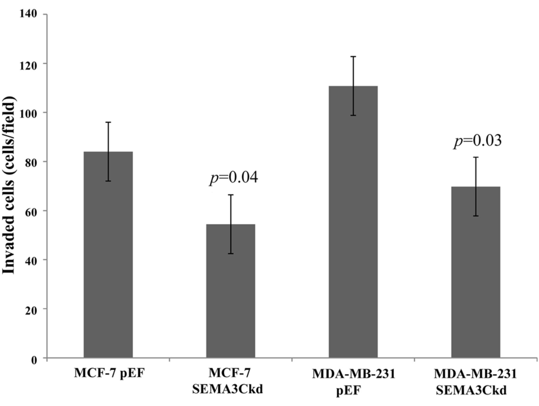
Semaphorin 3C knockdown (SEMA3Ckd) and invasiveness of breast cancer cells. Both MCF-7 and MDA-MB-231 SEMA3C knock-down cells exhibited reduced invasiveness. Number of invaded cells was counted under a microscope at a magnification of ×100. Three independent invasion assays were performed, representative data are shown. Data are the mean±standard deviation.
Our data suggest that SEMA3C expression is positively associated with tumour grade but is reduced in an advanced stage of disease and in tumours from patients who subsequently developed local recurrence. Interestingly, SEMA3C expression was lower in ER-positive tumours. In the current study, knockdown of SEMA3C was associated with reduced cell proliferation, in line with earlier findings investigating the impact of SEMA3C silencing in an in vivo murine model of gastric cancer (15). In addition, a previous study showed that overexpression of SEMA3C in PC-3 prostate cancer cells was associated with increased cell adhesion and invasion, in keeping with our findings reported here in (8, 15). Furthermore, SEMA3C overexpression was related to a strong down-regulation of E-cadherin expression (8). In contrast, forced overexpression of another class 3 semaphorin, SEMA3A, has been shown to reduce greatly PC-3 cancer cell adhesion and invasion (8).
The effect of oestrogen on the regulation of SEMA3C is an area that requires further research. A previous study has demonstrated that both SEMA3B and SEMA3F are significantly up-regulated in ovarian cancer cells in response to oestrogen exposure (16). Similarly, the regulation of neuropilins (NRPs), which are co-receptors for semaphorins, was shown to be influenced by oestrogen exposure. The expression of NRP1, which specifically acts as a receptor for SEMA3s, has been shown to be inversely related to the expression of B-cell lymphoma 2 homologous antagonist/ killer (BAK1), a proapoptotic regulator in mammary tumours. Suppression of BAK1 by 17β-estradiol stimulates an anti-apoptotic environment, increasing the survival probability of mammary epithelial cells (17).
The influence of SEMA3C on cancer-cell adhesive capacity was also studied using an in vitro model. We observed a marked reduction in breast cancer cell attachment to an artificial basement membrane when SEMA3C was knocked-down.
The current study focused on the expression of SEMA3C in breast cancer and its association with cancer-cell function and disease progression. Although our data suggest that SEMA3C may play a major role in breast cancer, its role in other types of cancer, or indeed the role of other SEMA3s in cancer is uncertain. The heterogeneous nature of this group of molecules, as well as local factors, such as expression of receptors and co-receptor interactions (plexins, neuropilins) and tissue-specific factors, mean that their role in cancer may be equally diverse. For example, some studies have suggested that angiogenic suppression of SEMA3A and SEMA3F is influenced by the inactivation of integrins (18), while in other reports, both these molecules were found to compete for the same VEGF-binding receptors (19, 20). The expression of SEMAA, SEMA3B and SEMA3F has mostly been associated with a reduction in cell adhesion, invasion and growth in prostate, breast, lung and ovarian cancer (8, 21-26).
Conclusion
The present study suggests that SEMA3C plays a role in the progression of breast cancer and may positively influence breast cancer cell adhesion, invasion and proliferation, as well as being associated with grade of disease and ER status. Interestingly, SEMA3C expression also appears to be negatively associated with tumour stage and risk of local recurrence, suggesting a complex role for this molecule in breast cancer.
Acknowledgements
The Authors wish to thank Cancer Research Wales and the Higher Education Commission of Pakistan (FAM) for providing funds for this research project.
- Received January 4, 2016.
- Revision received February 17, 2016.
- Accepted February 18, 2016.
- Copyright© 2016 International Institute of Anticancer Research (Dr. John G. Delinassios), All rights reserved
References
In this issue


Jump to section
Related Articles
Cited By...
- Estimating intraclonal heterogeneity and subpopulation changes from bulk expression profiles in CMap
- Estimating Intraclonal Heterogeneity and Subpopulation Changes from Perturbational Bulk Gene Expression Profiles in LINCS L1000 CMap by Premnas
- Proteotranscriptomics Reveal Signaling Networks in the Ovarian Cancer Microenvironment