


Abstract
Aim: Eudragit® E 100 (EE100) was used to improve the transfection efficiency of polyethylenimine (PEI). Materials and Methods: Mobility of PEI–DNA complexes with and without EE100 were visualized by agarose gel electrophoresis and their transfection efficiencies were investigated in KB human oral carcinoma cells by flow cytometry. 3-(4,5-Dimethylthiazol-2-yl)-2,5-diphenyl tetrazolium bromide (MTT) assay was used to determine the viability of transfected cells. Results: Gel electrophoresis illustrated formation of complete complexes at N/P ratios above 5. PEI had the highest transfection efficiency at an N/P ratio of 15, whereas in combination with EE100, the transfection efficiency was highest at an N/P ratio of 7.5. High concentrations of EE100 in combination with PEI were found to reduce cell viability. Conclusion: The results show a synergistic action of EE100 in transfection of DNA at low N/P ratios compared to PEI alone.
- Gene therapy
- gene delivery
- cationic polymer
- gene transfer
- polyethylenimine
- Eudragit
- KB cells
- PEI–DNA complex
According to the American Cancer Society in 2015, more than half a million Americans are expected to die of cancer (1). One of the most promising ways to understand cancer at the molecular level is to use gene therapy that selectively targets and destroys tumor cells. Gene delivery is achieved via viral or non-viral vectors. Using viral vectors has shown high transfection efficiency of genes. However, intensive investigation has revealed several safety risks (immunogenicity, oncogenicity, inflammatory potential) (2, 3) and complications in production (4). To overcome the deficiencies of viral delivery, non-viral gene delivery vectors have been developed. Non-viral vectors are easy to produce, highly stable, and have less immunogenicity and toxicity when compared to viral vectors.
Prominent among these vectors is the cationic polymer, polyethylenimine (PEI). PEI has a high capacity to delivery large DNA payloads (5), however, the transfection efficiency of non-viral vectors using PEl is low compared to viral vectors (6). PEI is cationic in nature and is considered to be an effective polymer for gene delivery (7, 8). It is commercially available as ExGen 500, a sterile solution consisting of linear PEI in water (9). Through electrostatic interaction, the positively charged amino groups on PEI form a complex with the negatively charged phosphates groups on DNA (4). By interacting non-specifically with glycoproteins, proteoglycans, or sulfated proteoglycans, the PEI–DNA complexes formed may enter into cells through absorptive or fluid-phase endocytosis (5). However, use of PEI has drawbacks such as limited transfection, DNA compaction and toxicity, limiting its usage (10). Nevertheless, transfection results show promise with PEI and research is continuing to improve its function as a transfection agent by itself or in combination (11, 12). One such approach was explored by our laboratory using Eudragit® E 100 (EE100).
EE100 is a cationic polymer containing dimethylaminoethyl methacrylate, butyl methacrylate, and methyl methacrylate (13). As far as we are aware, there is no research implementing EE100 as a transfection agent in combination with PEI. However, poly(2-dimethylaminoethyl methacrylate) (pDMAEMA), a polycationic synthetic polymer, has been studied as a transfection agent capable of forming complexes with DNA (14-18). At physiological pH, pDMAEMA becomes protonated and transfects cells in a similar way to PEI (16). However, akin to PEI, pDMAEMA has potent cytotoxicity, limiting its therapeutic application (14-18). Therefore, because of the structural similarity of EE100 to pDMAEMA (Figure 1) and in order to avoid the toxicity associated with high concentrations of transfection agent (PEI, pDMAEMA), we propose the use of a combination of transfection agents (PEI and EE100) at lower concentrations. Eudragit features many of the advantages of pDMAEMA and PEI without the added cytotoxic effect. By taking advantage of the inherent lytic activity due to hydrophobic moieties of the polymer and proton-sponge effect during endosomal acidification, Eudragit can increase transfection efficiency. The present study was therefore designed to verify enhanced green fluorescent protein (GFP) transfection with combinations of EE100 and PEI compared to PEI alone.
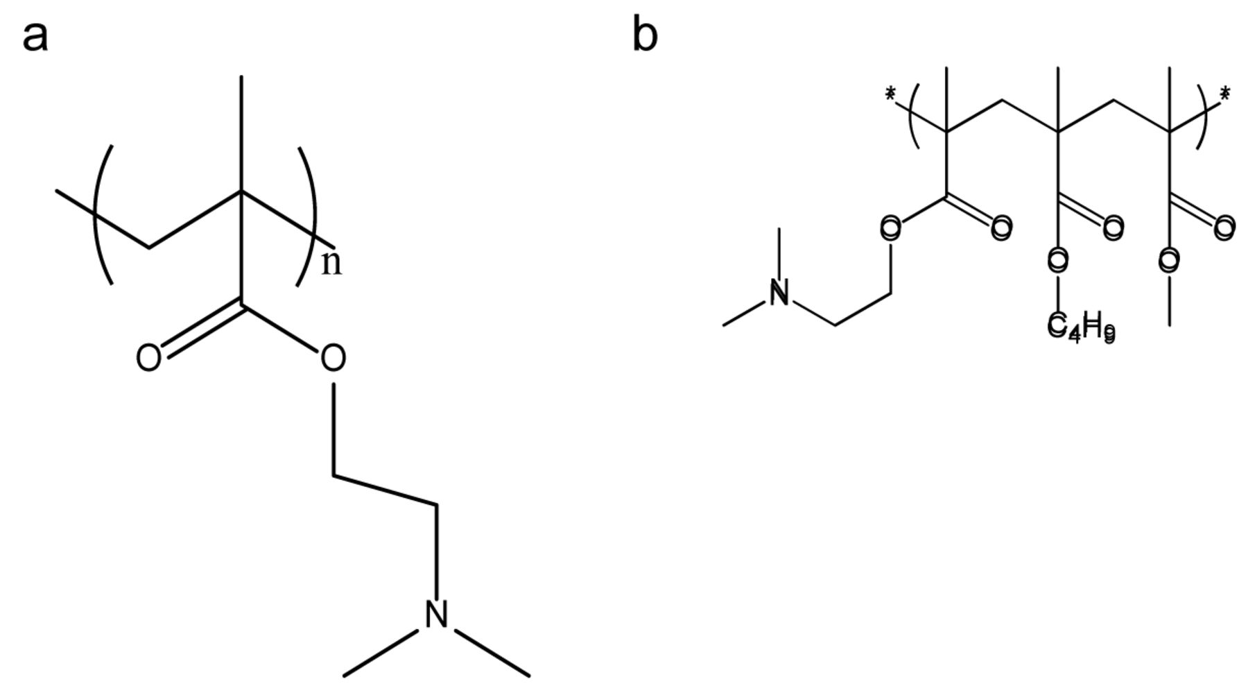
Structures of poly(2-dimethylaminoethyl methacrylate) (a) and Eudragit® E 100 (b).
Materials and Methods
Materials. Polyethylenimine (PEI, branched, MW 25 kDa) was purchased from Aldrich (Milwaukee, WI, USA). Agarose was purchased from Sigma (St Louis, MO, USA). Endotoxin-free Plasmid Maxiprep Kit was purchased from Qiagen (Santa Clarita, CA, USA). Enhanced GFP plasmid (pEGFP) DNA was obtained from Clontech (Mountain View, CA, USA). RPMI-1640, fetal bovine serum (FBS), Trypsin-EDTA, and penicillin-streptomycin were purchased from Gibco BRL (Rockville, MD, USA). Luciferase assay reagent was purchased from Promega (Madison, WI, USA). 3-(4,5-Dimethylthiazol-2-yl)-2,5-diphenyl tetrazolium bromide (MTT) was purchased from Sigma. KB human cervical carcinoma cells were obtained from the American Type Culture Collection (Rockville, MD USA). All other chemicals were of cell culture and molecular biology quality.
Complex formation between PEI and plasmid DNA. PEI and DNA complexes were formed as described by Wanlop et al. (19). Briefly, different N/P ratios of PEI (i.e. 5, 7.5, 10, 15, 20) were prepared by dissolving PEI in HEPES buffer (20 mM HEPES, pH 7.4). pEGFP plasmid DNA solution (0.5 mg/ml) was added to different N/P ratios of PEI to form PEI–pEGFP complexes. The formed mixture was gently vortexed for 5 s before leaving it at room temperature for 15 min. A stock solution of EE100 was prepared in ethanol and added in a 0.25:1 w/w ratio to PEI solution prior to mixing with DNA solution to attain the required N/P ratio. For experiments containing EE100 alone, equivalent concentrations were used.
Gel retardation. Gel retardation analysis was used to assess the formation of electrostatic complexes between cationic polymers and plasmid DNA. It was performed with slight modification to the method outlined by Palocci et al. (20). Briefly, a 0.8% agarose gel was prepared in 1× TBE buffer with 0.5 μg/ml ethidium bromide. Polymer samples were combined with 1 μg of pEGFP plasmid at N/P ratios of 5 and 10. Samples of PEI, EE100, and PEI-EE100 were run against free GFP plasmid. Electrophoresis was conducted at 100 V, 90 mA for 45 min. Gels were photographed under UV light with a Bio-Rad 170-8126 Universal Hood with Quantity One v4.5.2 Software.
Cell culture. KB (a subline of HeLa) cells were grown in RPMI-1640 medium containing glutamine. The medium was supplemented with 10% FBS, 50 μg/ml streptomycin, and 50 μg/ml penicillin. Cells were grown in an incubator at 100% relative humidity, 37°C, and 5% CO2.
In vitro transfection. KB cells were seeded in a 24-well plate at a density of 5×10⁵ cells/well in 500 μl of RPMI-1640 medium containing glutamine. The cells were incubated for 24 h at 37°C in a 5% CO2 environment. After 24 h, the medium was replaced with 200 μl growth medium containing either PEI, PEI in combination with EE100, or EE100 alone with GFP, at different N/P ratios (0, 5, 7.5, 10, 15 or 20). After 4 h of incubation, the transfection medium was replaced with fresh growth medium and the cells were incubated for another 24 h at 37°C in a 5% CO2 environment, before being analyzed for GFP expression. In all transfection protocols, the formulations used were combined with transfection vectors in serum-free medium and incubated for about 15 min at room temperature prior to mixing with cells.
GFP expression levels were analyzed by flow cytometry similar to the methods described by Zhao et al. (21). The cells were washed with PBS (pH 7.4) three times before detaching with trypsin-EDTA (0.25%) solution. The cells were then centrifuged and fixed with 500 μl of 4% formalin in phosphate-buffered saline (PBS). The cell suspension in 4% formalin was introduced into a Becton Dickinson FACS Calibur cytometer (Becton Dickinson, Franklin Lakes, NJ, USA) to determine the level of GFP expression. For each cell sample, 1×10⁵ events were collected. The data were analyzed using BD CellQuest Pro software (Becton Dickinson). Untreated cells and cells treated with GFP alone were used as controls. All transfection experiments were performed in triplicate.
Evaluation of cytotoxicity. Cytotoxicity studies were carried out using MTT assay as described previously (15). Briefly, KB cells were seeded in a 96-well plate at a density of 105 cells/well in 200 μl of growth medium. The cells were incubated for 24 h at 37°C under 5% CO2 atmosphere. After 24 h, the cells were washed with PBS (pH 7.4), followed by addition of 15 μl of transfection vectors containing either PEI, PEI in combination with EE100, or EE100 alone. The cells were incubated with medium containing formulations for 4 h before being replaced with 200 μl of fresh medium and further incubated for 24 h at 37°C under an atmosphere with 5% CO2. Non-treated cells and cells treated with GFP alone were used as controls and incubated for the same amount of time. After 24 h, the cells were treated with 20 μl of 5 mg/ml concentrated MTT solution and incubated for 2 h. Viable cells formed formazan crystals and these were dissolved by adding 100 μl dimethyl sulfoxide to each well. The resultant colored product was read at 550 nm using a microplate reader (340 ATTC; SLT Lab Instruments, Salzburg, Austria).
Statistical analysis. Statistical significance of differences in transfection efficiency and cell viability were examined using Student's t-test. The significance level was set at p<0.05.
Results
Gel retardation studies. Figure 2 shows the gel retardation study which was used to examine the formation of complexes between transfection agents and GFP. As shown in Figure 2, combination of EE100 with PEI led to stronger retardation of GFP when compared to PEI alone. In comparison to the naked DNA band, PEI formed complexes with GFP at an N/P ratio of 10, whereas the mixture of EE100 and PEI complexed with DNA showed significant retardation starting from N/P of 5.0. EE100 alone did not lead to any significant retardation of GFP.
Transfection studies. Transfection data obtained using flow cytometry is shown in Figure 3. Based on the number of cells showing transfection, the graph was plotted between the cells transfected and the N/P ratio. The flow cytometric results show no transfection of GFP with EE100 alone. Conversely, the combination of EE100 and PEI led to significant transfection, even at low N/P ratios of 5.0 and 7.5, when compared to PEI alone. The transfection of GFP with EE100 and PEI combinations at N/P ratios of 5.0 and 7.5 show a 15-fold increase when compared to PEI alone. However, at N/P ratios of 10 and 15, PEI alone increased transfection when compared to other formulations.
Cytotoxicity studies. Cell viability experiments were carried out to evaluate the effect of combinations of transfection agents on KB cells. Non-treated cells acted as a control, with a cell viability of 100%. Figure 4 shows the effect of transfected PEI–DNA complex with and without EE100 on KB cells. Formulations containing PEI in combination with GFP did not exhibit significant cytotoxicity. On the other hand, combinations of PEI with EE100 showed no significant cytotoxicity at N/P 7.5, but with higher concentrations of polymer, there was an increase in cytotoxicity. This increase in cytotoxicity further explains the reduction in transfection at the indicated N/Ps.
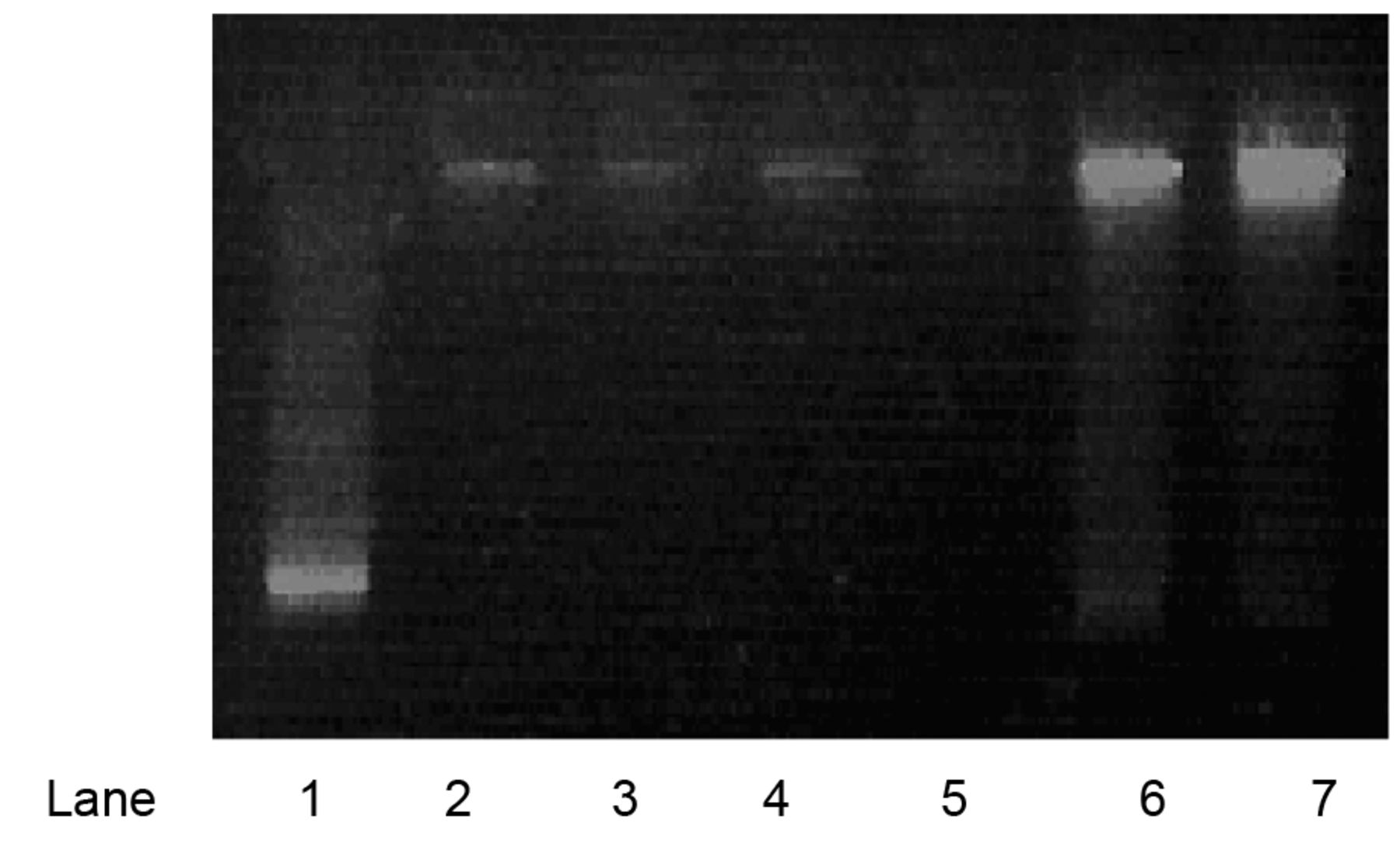
Gel retardation analysis of Polyethyleneimine (PEI)–DNA complexes. Lane 1: green fluorescent protein (GFP) (control); lane 2,3: PEI–DNA complexes; lanes 4,5: Eudragit E 100 (EE100) + PEI–DNA complexes; Lane 6, 7: EE100–DNA complexes; N/P ratios 5 and 10, respectively).
Discussion
Among the different materials used as non-viral vectors, polymers stand out because of their ease of preparation, purification, chemical modification, and stability (9). One prominent candidate among polymers is PEI. Use of PEI is restricted because of limited transfection efficiency, short duration of gene expression, and cytotoxic effects (10). Hence, several chemical and structural modifications have been carried out to enhance the activity of PEI. One such technique developed in our lab was to combine PEI with EE100. Previously, other forms of Eudragit have been applied towards the purposes of gene delivery including Eudragit L100, RS100, and RL100 (22-24). While Eudragit was originally conceived for the purposes of modulating enteric release properties of orally administered drugs, it shows promise as a promoter of transfection. Eudragit is able to combine with other cationic lipid and polymer components such as PEI, chitosan, and dioctadecyl-dimethylammonium bromide and form nanoparticles of small size and moderate zeta potential (22-24). The tertiary and quaternary groups on the various forms Eudragit allow for electrostatic complexation with negatively charged plasmids, oligonucleotides, or siRNA.

Transfection efficiency in KB cells of DNA at different N/P ratios of Polyethyleneimine (PEI), Eudragit E 100 (EE100) + PEI, and EE100. Results are the means of three separate experiments. Error bars represent standard deviations. *Significantly different from that of naked DNA control and that with PEI–DNA complex.

Effect of transfection agents (PEI, EE100+PEI, EE100) on the viability of KB cells. Results are the means of three separate experiments. Error bars represent standard deviations. *Significantly different from that of PEI–DNA complex (p<0.05; ANOVA).
The goal of the present study was to determine whether or not PEI-EE100 can efficiently bind to plasmid DNA and deliver it to cancer cells. From our study, we were able to show efficient condensation of pEGFP with PEI-EE100 at lower N/P ratio than either component alone. However, it is noted that EE100 by itself was not able to form complexes with GFP because it is less cationic in nature. It is suggested that a variant of Eudragit such as Eudragit RS100 may be better-suited for transfection due to the presence of quaternary amine groups which may better condense plasmid DNA than EE100.
Based on the preliminary data, we found enhanced transfection using EE100 in combinations with PEI, providing new insight on this polymer as a complementary transfection promoter in gene delivery. In the addition, this polymer combination has the potential of delivering DNA with reduced cytotoxic effects relative to PEI. However, flow cytometric assays showed decreasing transfection with increases in N/P ratio above 10 with EE100 and PEI combination, explained by increased cytotoxicity. This is further supported by cytotoxicity data, where cell viability decreased at higher N/P ratios for EE100 and PEI combination. The combinations at lower N/P ratio lead to higher transfection than with use of PEI alone and no significant cytotoxicity, which are promising factors in the development of gene-delivery systems.
Conclusion
Preliminary data indicate that the combination of EE100 and PEI show synergism, with higher transfection at lower N/P ratios, indicating that less PEI is required to perform the same action and with less toxicity. However, the toxicity of this combination at higher N/Ps is a major concern and further experiments will be carried out to affirm the feasibility of EE100 or related forms of Eudragit in combinations with PEI as transfection agents.
Acknowledgements
The Authors are grateful for the guidance and support of Drs. Bo Yu, Yicheng Mao, and Chenguang Zhou in this project. This work was made possible by the National Institute of Health Grant R01 CA135243 and R21 CA131832.
- Received October 15, 2015.
- Revision received November 13, 2015.
- Accepted November 17, 2015.
- Copyright© 2016 International Institute of Anticancer Research (Dr. John G. Delinassios), All rights reserved
References
In this issue


Jump to section
Related Articles
Cited By...
- No citing articles found.