


Article Figures & Data
Figures
- Figure 1.
Analysis of cell proliferation and angiogenesis following treatment with itraconazole or bevacizumab. Cell proliferation was measured in response to different concentrations of itraconazole and bevacizumab in HT-29 cells (A), MKN-28 cells (B), cancer-associated fibroblasts (CAFs) (C), and human umbilical vein endothelial cells (HUVECs) (D, E). The proliferation of HUVECs was also measured in the presence of different concentrations of vascular endothelial growth factor (VEGF) alone (F) or in combination with (G) 10 μg/mL bevacizumab or (H) 1 μM itraconazole. Proliferation is plotted as the percentage relative to the number of cells in the control untreated sample. Bars indicate the mean±SEM.
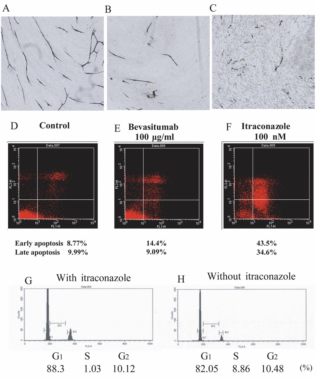
- Figure 2.
Induction of apoptosis by itraconazole. Upper panels: Representative images of angiogenesis assays following treatment of human umbilical vein endothelial cells (HUVECs) with bevacizumab or itraconazole. HUVECs in the control (A), after treatment with 100 μg/mL bevacizumab (B), and after treatment with 1 μM itraconazole (C). Middle and lower panels: Annexin assays were performed in the control (D) and in cells (E) treated with bevacizumab or itraconazole (F) to assess the induction of apoptosis. The cell cycle distribution was also measured by flow cytometry in HUVECStreated with itraconazole (G) or left untreated (control) (H).
- Figure 3.
Phosphorylation of ribosomal S6 kinase (RSK), mitogen-activated protein kinase (MAPK), and S6 in human umbilical vein endothelial cells (HUVECs) and analysis of angiogenic factors secreted by MKN-28 cells and cancer-associated fibroblasts (CAFs) with or without itraconazole in vitro using proteome arrays. HUVECs (A) were treated with 1 or 100 μM itraconazole, and the phosphorylation of p44/42 MAPK, p90RSK, Akt, and S6 was analyzed by western blotting. A protein molecular weight marker was included in the first lane. MKN-28 cells (B): MKN-28 secreted interleukin-8 (IL-8), tissue inhibitors of metalloproteinase (TIMP)-1, TIMP-2, chemokine (C-X-C motif) ligand (CXCL)13, and urokinase plasminogen activator receptor (uPAR), which may be associated with bevacizumab resistance in MKN-28 cells. vascular endothelial growth factor (VEGF) (box 6) was not detected. Itraconazole did not influence the secretion of VEGF by MKN-28 cells. Boxes: 1, CXCL13; 2, IL-8; 3, TIMP-1; 4, TIMP-2; and 5, uPAR. The other spots in the image are positive controls. CAFs (C): Representative images of Proteosome Profiler Assay membanes probed with conditioned medium collected from itraconazole- or saline-treated cells. A: Positive control, B: negative control, C: plasminogen activator inhibitor-1, D: TIMP-4, E: IL-1β, F: VEGF, and G: monocyte chemoattractant protein 1 (MCP-1). Itraconazole treatment dramatically suppressed the secretion of MCP-1 (D).
- Figure 4.
Effects of bevacizumab and itraconazole on xenograft tumors in vivo. Xenograft tumors were established from HT-29, MKN-28, and MKN-45 cells, and mice bearing these tumors were treated with bevacizumab (BV), itraconazole (ITZ), or a combination of both agents for 5 weeks. A: Volume of HT-29 tumors, B: weight of HT-29 tumors, C: volume of MKN-28 tumors, and D: weight of MKN-28 tumors. E: Representative images of microvessels in control and treated MKN-28-derived tumors in mice. F: Microvessel density assays were performed as described in the Materials and Methods, and a representative graph of the data is shown. NS: Not significant.


In this issue


Jump to section
Related Articles
Cited By...
- Breaking the Crosstalk of the Cellular Tumorigenic Network in NSCLC by a Highly Effective Drug Combination
- Itraconazole targets cell cycle heterogeneity in colorectal cancer
- Molecular Effects of Stromal-Selective Targeting by uPAR-Retargeted Oncolytic Virus in Breast Cancer
- Severe Cardiotoxicity in a Patient with Colorectal Cancer Treated with Bevacizumab