
Main menu

  • Home
  • Current Issue
  • Archive
  • Info for
    • Authors
    • Editorial Policies
    • Subscribers
    • Advertisers
    • Editorial Board
    • Special Issues
  • Journal Metrics
  • Other Publications
    • In Vivo
    • Cancer Genomics & Proteomics
    • Cancer Diagnosis & Prognosis
  • More
    • IIAR
    • Conferences
    • 2008 Nobel Laureates
  • About Us
    • General Policy
    • Contact
  • Other Publications
    • Anticancer Research
    • In Vivo
    • Cancer Genomics & Proteomics

User menu

  • Register
  • Subscribe
  • My alerts
  • Log in
  • My Cart

Search

  • Advanced search
Anticancer Research
  • Other Publications
    • Anticancer Research
    • In Vivo
    • Cancer Genomics & Proteomics
  • Register
  • Subscribe
  • My alerts
  • Log in
  • My Cart
Anticancer Research

Advanced Search

  • Home
  • Current Issue
  • Archive
  • Info for
    • Authors
    • Editorial Policies
    • Subscribers
    • Advertisers
    • Editorial Board
    • Special Issues
  • Journal Metrics
  • Other Publications
    • In Vivo
    • Cancer Genomics & Proteomics
    • Cancer Diagnosis & Prognosis
  • More
    • IIAR
    • Conferences
    • 2008 Nobel Laureates
  • About Us
    • General Policy
    • Contact
  • Visit us on Facebook
  • Follow us on Linkedin
Research ArticleExperimental Studies

Resistance to Gemcitabine in the Pancreatic Cancer Cell Line KLM1-R Reversed by Metformin Action

BYRON BARON, YUFENG WANG, SHIN-ICHIRO MAEHARA, YOSHIHIKO MAEHARA, YASUHIRO KURAMITSU and KAZUYUKI NAKAMURA
Anticancer Research April 2015, 35 (4) 1941-1949;
BYRON BARON
1Department of Biochemistry and Functional Proteomics, Yamaguchi University Graduate School of Medicine, Ube, Japan
2Department of Anatomy and Cell Biology, Faculty of Medicine and Surgery, University of Malta, Msida, Malta
  • Find this author on Google Scholar
  • Find this author on PubMed
  • Search for this author on this site
  • For correspondence: angenlabs{at}gmail.com
YUFENG WANG
1Department of Biochemistry and Functional Proteomics, Yamaguchi University Graduate School of Medicine, Ube, Japan
  • Find this author on Google Scholar
  • Find this author on PubMed
  • Search for this author on this site
SHIN-ICHIRO MAEHARA
4Department of Surgery and Science, Graduate School of Medical Sciences, Kyushu University, Fukuoka, Japan
  • Find this author on Google Scholar
  • Find this author on PubMed
  • Search for this author on this site
YOSHIHIKO MAEHARA
4Department of Surgery and Science, Graduate School of Medical Sciences, Kyushu University, Fukuoka, Japan
  • Find this author on Google Scholar
  • Find this author on PubMed
  • Search for this author on this site
YASUHIRO KURAMITSU
1Department of Biochemistry and Functional Proteomics, Yamaguchi University Graduate School of Medicine, Ube, Japan
  • Find this author on Google Scholar
  • Find this author on PubMed
  • Search for this author on this site
KAZUYUKI NAKAMURA
1Department of Biochemistry and Functional Proteomics, Yamaguchi University Graduate School of Medicine, Ube, Japan
3Centre of Clinical Laboratories in Tokuyama Medical Association Hospital, Shunan-shi, Japan
  • Find this author on Google Scholar
  • Find this author on PubMed
  • Search for this author on this site
  • Article
  • Figures & Data
  • Info & Metrics
  • PDF
Loading

Abstract

Background/Aim. The pancreatic cancer cell line KLM1 can gain chemoresistance following gemcitabine (GEM) treatment. Metformin was found to be a useful sensitising agent towards GEM treatment following gain of chemoresistance. Materials and Methods: The proliferation of GEM-sensitive and -resistant cells was investigated over a range of metformin concentrations from 0.005 to 5 mM. The intra- and extra-cellular energetic profiles of these two cell types under metformin exposure were investigated through adenosine triphosphate (ATP) and L-lactate assays. Results: There was an unexpected decrease in intracellular L-lactate following gain of chemoresistance, despite observable medium acidification. At the biochemical level, a mar ked effect on phosphorylated proteins upstream of Akt, along the mTOR pathway, was observed at 6 h. These changes followed a time-dependent pattern linked closely to the changes in the energetic profile. Conclusion: Together, these results indicate that metformin indirectly blocks protein phosphorylation, including that of heat shock protein 27 (HSP27).

  • HSP27
  • metformin
  • pancreatic cancer

Metformin (N,N-Dimethylimidodicarbonimidic diamide), a biguanide, is a widely used drug by type II diabetics and has been linked to a lower cancer incidence as shown in diabetics treated with metformin compared to those on other diabetic drugs (1-5).

Metformin was reported to indirectly activate adenosine monohosphate (AMP)-activated protein kinase (AMPK) activity by disrupting complex I of the mitochondrial respiratory chain and subsequently adenosine triphosphate (ATP) synthesis (6-8), which leads to phosphorylation by liver kinase B1 (LKB1) (9). This brings about a reduction in the mammalian target of rapamycin (mTOR) (10-14) and an associated downstream signalling suppression, such as eukaryotic initiation factor 4E-binding protein 1 (4E-BP1), ribosomal protein S6 kinase 1 (S6K1) and human epidermal growth factor receptor 2 (HER-2) (9, 11, 13). The suppression of mTOR also modulates other targets (13, 15-23) including the activation of tuberous sclerosis complex 2 (TSC2, tuberin), which further suppresses mTOR activity (24). Dysregulation of mTOR is common in many cancers and mTOR is known to be central in the phosphatidy linositol-3-kinase/ protein kinase B/mTOR (PI3K/Akt/mTOR) signalling pathway (25, 26).

Metformin has been found to also enhance apoptosis induction via the tumor suppressor p53, which is activated by AMPK (27, 28). Additionally, it suppresses cyclin D1 and E2F1 expression leading to decreased cancer prolife ration (14, 20), as well as a variety of mitosis-related gene families of which tubulins, histones and aurora kinases are of particular interest (18).

This study was designed to understand the effect of metformin on the pancreatic cancer cell lines KLM1 and KLM1-R particularly focusing on the effect on heat shock protein 27 (HSP27) in these two cell lines. Although many cancer types have been tested with metformin, it has only been recently demonstrated to suppress pancreatic cancer proliferation in patients and cell lines (ASPC-1, BxPc-3, PANC-1 and SW1990) (29, 30). HSP27 is known to associate with Akt (31, 32), which is activated via PI3K and stalls apoptosis (33, 34) by phosphorylating pro-apoptotic factors, such as B-cell lymphoma-2 (BCL-2)-associated death protein (BAD), leading to a sequestration and inhibition by 14-3-3 proteins (35). HSP27 has been shown to have the ability to reduce apoptosis by blocking the truncated BH3 interacting domain death agonist (tBID), as well as reduce the SMAC and cytochrome C release from mitochondria (36).

Figure 1.
  • Download figure
  • Open in new tab
  • Download powerpoint
Figure 1.

Metformin brings about a marked decrease in the proliferation of the Gemcitabine-resistant pancreatic cell line KLM1-R, comparable to its Gemcitabine-sensitive parent cell line KLM1 within the first 48h of treatment.

Materials and Methods

Pancreatic cancer cell lines and culture conditions. The cell lines used were KLM1 (provided by the Department of Surgery and Science at Kyushyu University Graduate School of Medical Science), KLM1-R (gemcitabine resistant KLM1) (37) (provided by the Department of Surgery and Science at Kyushyu University Graduate School of Medical Science), MIA PaCa-2 (provided by the Institute of Development, Aging and Cancer at Tohoku University) and PK-59 (provided by the Institute of Development, Aging and Cancer at Tohoku University). They were cultured in Dulbecco's modified Eagle's medium (DMEM) (high glucose; Wako, Osaka, Japan) with 2mM L-glutamine and 10% FBS at 37°C, in a 5% CO2 atmosphere, at high relative humidity.

Proliferation assays. KLM1 and KLM1-R cells were cultured in 96-well plates and treated with metformin at varying concentrations (from 0.005-5 mM) in conjunction with 500 ng/ml gemcitabine (GEM; Eli Lilly, Indianapolis, IN, USA). Cell proliferation assays using 3-(4,5-dimethyl-2-yl)-5-(3-carboxymethoxyphenyl)-2-(4-sulfophenyl)-2H-tetrazolium, inner salt (MTS; CellTiter 96® Aqueous One Solution Cell Proliferation Assay; Promega, Madison, WI, USA) were carried out at 24, 48 and 72 h to determine the ability of these cell lines to multiply after exposure to metformin treatment. Absorbance readings at 490 nm were taken after 1 h and 2 h incubation with MTS reagent.

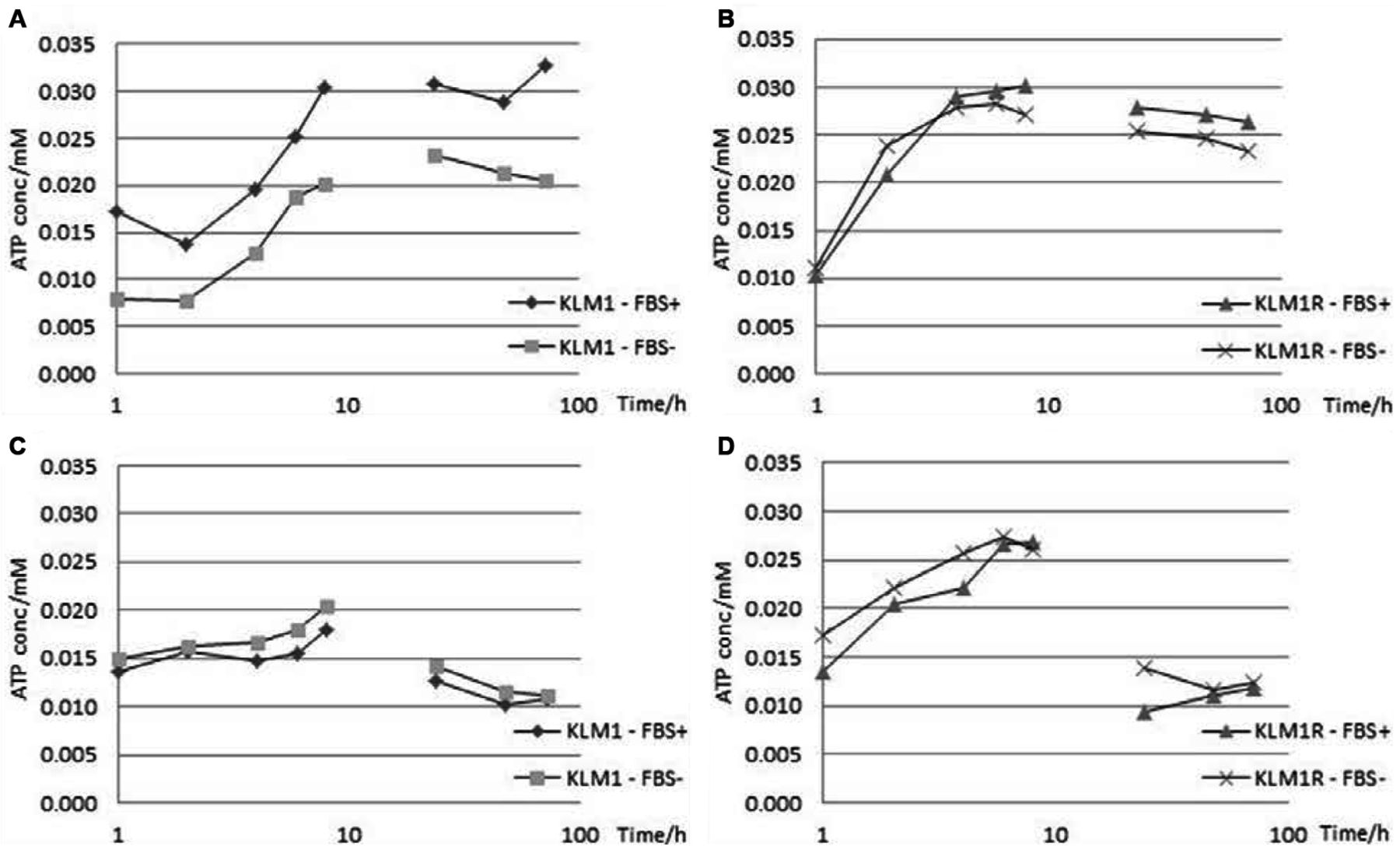
ATP and L-lactate assays. KLM1 and KLM1-R cells were cultured in DMEM with and without 10% FBS in 96-well plates and treated with 5 mM metformin for a period of 72 h. Intra- and extra-cellular ATP and L-lactate concentrations were measured at 2-h intervals for the first 8h and then at 24, 48 and 72 h following the start of the experiment using ATP and L-lactate Colorimetric/Fluorometric Assay Kits (BioVision Inc., Milpitas, CA, USA). Absorbance readings at 570 nm were taken after 1h incubation with the appropriate probe.

Figure 2.
  • Download figure
  • Open in new tab
  • Download powerpoint
Figure 2.

Metformin reduced cellular proliferation of KLM1 in a dose-dependent manner over the tested range of 0.005-5 mM.

Western blot analysis. KLM1-R cells were cultured in 6-well plates and treated with 5mM metformin for 6h. Cells were lysed using a lysis buffer containing 1 % NP-40, 1 mM sodium vanadate, 1 mM phenylmethanesulfonyl fluoride (PMSF), 10 mM sodium fluoride (NaF), 10 mM ethylenediaminetetraacetic acid (EDTA), 50 mM Tris, 165 mM NaCl, 10 μg/ml leupeptin and 10 μg/ml aprotinin and incubating the cell suspensions for 1h at 4°C with vigorous shaking. The samples were then spun down at 15,000 rpm for 30 min and the supernatants were stored at −80°C until use. After electrophoresis using 12% sodium dodecyl sulphate-polyacrylamide gel electrophoresis (SDS-PAGE), proteins were electroblotted onto polyvinylidene fluoride (PVDF) membranes (Immobilon-P; Millipore, Bedford, MA, USA) and blocked for 1 h at room temperature using TBS containing 5% skimmed milk. The primary antibodies used were a mouse monoclonal anti-human HSP27 antibody (sc-13132; 1:1000 dilution; Santa Cruz Biotechnology, Dallas, TX, USA), rabbit polyclonal anti-human pHSP27-Ser15 antibody (#2404; 1:1000 dilution; Cell Signalling Technology, Boston, MA, USA), rabbit polyclonal anti-human pHSP27-Ser78 antibody (#2405; 1:1000 dilution; Cell Signalling Technology, Boston, MA, USA), rabbit polyclonal anti-human pHSP27-Ser82 antibody (#2401; 1:1000 dilution; Cell Signalling Technology, Boston, MA, USA), rabbit polyclonal anti-human Akt antibody (#9272; 1:1000 dilution; Cell Signalling Technology, Boston, MA, USA), rabbit polyclonal anti-human pAkt-Ser473 antibody (#4058; 1:1000 dilution; Cell Signalling Technology, Boston, MA, USA), rabbit polyclonal anti-human AMPK-α1 antibody (#07-350; 1:1000 dilution; Merck Millipore, Hertfordshire, WD, UK), goat polyclonal anti-human pERK-Tyr204 antibody (sc-7976; 1:1000 dilution; Santa Cruz Biotechnology, Dallas, TX, USA), rabbit monoclonal anti-human Src antibody (#2109; 1:1000 dilution; Cell Signalling Technology, Boston, MA, USA), rabbit monoclonal anti-human pSrc-Tyr527 antibody (#2105; 1:1000 dilution; Cell Signalling Technology, Boston, MA, USA), rabbit polyclonal anti-human HSP70 antibody (SPA-812; 1:1000 dilution; Stressgen, San Diego, CA, USA), mouse monoclonal anti-human HSP90 antibody (sc-13119; 1:1000 dilution; Santa Cruz Biotechnology, Dallas, TX, USA), goat polyclonal anti-human GAPDH antibody (sc-20357; 1:1000 dilution; Santa Cruz Biotechnology, Dallas, TX, USA) and a goat polyclonal anti-human Actin antibody (sc-1616; dilution range 1:200; Santa Cruz Biotechnology, Dallas, TX, USA). For each primary antibody, membranes were incubated overnight at 4°C, with shaking. Membranes were then washed three times with TBS containing 0.05% Tween-20 and, finally, with TBS before being incubated with horseradish peroxidase-conjugated secondary antibody (dilution range 1:10,000; Jackson ImmunoResearch Lab., West Grove, PA, USA) for 2h at room temperature, with shaking. Development was carried out using chemifluorescence reagent (ImmunoStar LD; Wako, Osaka, Japan). Immunoreactive protein bands were detected using the Image Reader LAS-1000 Pro (Fujifilm Corporation, Tokyo, Japan).

Figure 3.
  • Download figure
  • Open in new tab
  • Download powerpoint
Figure 3.

The effect of metformin treatment is prolonged much beyond the administration period. Metformin was added at a dose of 5 mM for a period of 6 h (spike) or throughout the 72 h experiment duration (constant).

Results

Metformin brings about a marked decrease in proliferation of the pancreatic cell lines KLM1 and KLM1-R. To determine if metformin has an effect on pancreatic cancer, the cell lines KLM1 and KLM1-R, which are GEM-sensitive and resistant, respectively, were chosen since GEM is the main chemotherapeutic agent for this type of cancer. The initial test involved the combination of 500 ng/ml GEM and 5 mM metformin, which is the median of the therapeutic range of action. It is evident that both cell lines are sensitive to this combination to a similar extent. It seems to indicate that metformin sup presses biochemical pathways that nullify the GEM resistance mechanism in KLM1-R cells bringing their proliferation down to the same level as GEM-sensitive KLM1 cells (Figure 1).

The effect observed on cancer proliferation is dose-dependent within the tested range of 0.005-5 mM metformin. In order to understand the effectiveness of metformin over a range of concentrations, doses from 0.005 to 5 mM metformin (in combination with 500 ng/ml GEM) were tested following an incubation of 72 h. The reduction in cell proliferation was found to be directly proportional to the metformin concentration used (Figure 2).

Figure 4.
  • Download figure
  • Open in new tab
  • Download powerpoint
Figure 4.

The intracellular (4A, 4B) and extracellular (4C, 4D) L-lactate profiles for KLM1 and KLM1-R under the effect of metformin in medium supplemented with or lacking FBS over a period of 72 h sampled every 2 h for the first 8h and then every 24 h. L-lactate levels appear to undergo an increase intra-cellularly but a decrease extra-cellularly, with the intracellular L-lactate concentration presenting a link to FBS supplementation, most observable in KLM1.

Figure 5.
  • Download figure
  • Open in new tab
  • Download powerpoint
Figure 5.

The intracellular (5A, 5B) and extracellular (5C, 5D) ATP profiles for KLM1 and KLM1-R under the effect of metformin in medium supplemented with or lacking FBS over a period of 72 h sampled every 2 h for the first 8 h and then every 24 h. ATP levels appear to undergo a marked initial increase followed by a slow decrease and do not seem to be linked to FBS supplementation.

The effect of metformin treatment is prolonged way beyond the administration period. To study the half-life and effect of different metformin administration strategies, metformin was added at a dose of 5 mM for a period of 6hr (spike) or throughout the entire 72 h duration. The results showed no significant difference between the two set-ups indicating that the initial dose taken-up by cells is enough to reduce prolife ration even if the excess is removed (Figure 3). This suggests that metformin either binds tightly to its target and leads to a gradual decrease in cell proliferation or it is not metabolised and produces an effect through repeated interventions, or else, through some kind of positive feedback loop.

The energetics of KLM1 present a marked shift after becoming chemoresistant as observed through L-lactate and ATP levels. In all proliferation assays, including metformin, it was observed that the metformin-treated samples presented a decrease in pH from 7.4 to 6.1 compared to their equivalent metformin-free controls. For this reason, an assay for L-lactate (both intra- and extra-cellular) was carried-out. A number of interesting observations emerged both in terms of cellular dynamics and effects of experimental conditions.

A peak in intracellular L-lactate level was reached at 24 h, which was dependent on the FBS setting (i.e. presence or absence). However, KLM1-R cells appeared to be much less dependent on FBS and their peak L-lactate concentration was much lower (by almost 50%). Another noteworthy observation is the dip at 6 h, which was observable in both KLM1 and KLM1-R for both FBS settings. The magnitude of the change, however, was positively correlated with the FBS setting, being more pronounced in the presence of FBS (Figure 4A and 4B).

The extracellular L-lactate levels for both KLM1 and KLM1-R cells, however, were almost identical. The only observable difference was the peak obtained at 6 h in KLM, consistent with the dip observed intra-cellularly. This was absent in KLM1-R, possibly indicating that, instead of being secreted, it was being redirected to some biochemical pathway (Figure 4C and 4D).

At the end of the 72 h experimental period, the intracellular L-lactate concentration was higher than that at the start, while the extracellular L-lactate concentration was lower than the initial concentration. In all 4 conditions, the final concentrations of L-lactate show a tendency towards further decrease (Figure 4 A-D).

On the other hand, the peak in intracellular ATP level was reached somewhere between 8 and 24 h, which was dependent on the FBS setting in KLM1. However, KLM1-R cells presented almost overlapping profiles with little dependence on FBS. The other noteworthy observation is the dip at 2h, which was observable only in KLM, in the presence of FBS (Figure 5A and 5B).

Figure 6.
  • Download figure
  • Open in new tab
  • Download powerpoint
Figure 6.

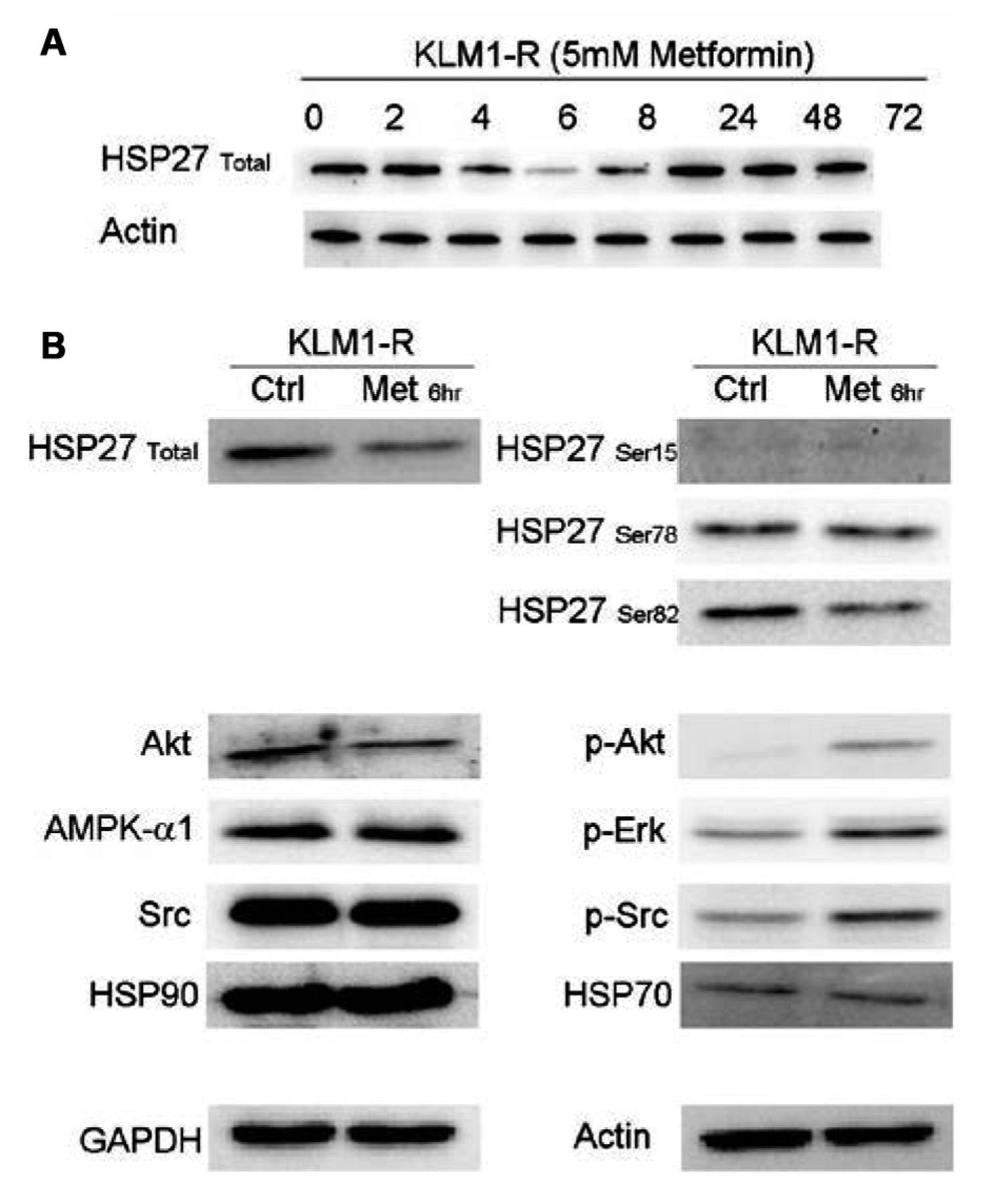
Western blotting experiments. A: HSP27 expression over the first 24 h following metformin administration. B: Expression levels of proteins upstream of and including Akt. The phosphorylation of proteins upstream of Akt is affected by metformin treatment.

The extracellular ATP levels for both KLM1 and KLM1-R cells reached a peak at 8 h. An observable difference was the peak obtained at 2 h in KLM1 in the presence of FBS, consistent with the dip observed intra-cellularly. By 24 h, the ATP level in both KLM1 and KLM1-R cells was lower than that at the start of the experiment (Figure 5C and 5D).

Similar to the L-lactate experiment, at the end of the 72 h experimental period, the intracellular ATP concentration was higher than that at the start, while the extracellular ATP concentration was lower than the initial concentration. In all 4 conditions, the final concentrations of ATP show a tendency towards further decrease (Figure 5 A-D).

Furthermore, the L-lactate and ATP profiles in KLM1 and KLM1-R were subsequently compared to those of GEM-sensitive MIA PaCa-2 and GEM-resistant PK-59 (data not shown). MIA PaCa-2 and PK-59 presented different profiles to KLM1 and KLM1-R, with PK-59 presenting only small fluctuations throughout the experiment. Since the mode of GEM resistance in KLM1-R and PK-59 is through different mechanisms, these results may indicate that the activity of metformin is less effective in PK-59 as this was reflected in the minor alterations observed in the energy profile.

The expression of various phosphorylated proteins upstream of Akt is altered by metformin. Western blotting was carried out for HSP27 over the entire duration of the metformin administration (72 h), sampling at the same time-points as the energetics experiment (Figure 6A). The fluctuation in HSP27 expression appeared to be directly correlated to the L-lactate profile. As the 6 h time-point appeared to be critical in the L-lactate profile, as well as in HSP27 expression, further western blotting analysis was carried out for Src, Erk, HSP27 and HSP90 proteins upstream of Akt (Figure 6B). KLM1-R cells treated with 5 mM metformin for 6 h were used for this experiment. At the 6-h time-point the total HSP27 and phosphrylated HSP27-Ser82 expression was reduced as was that for Akt. In contrast, pAkt, pERK and pSrc levels increased. It appears that, in general, the phosphorylation status of proteins in the mTOR pathway was affected and that this translated into increased sensitisation to GEM and decreased cellular proliferation.

Discussion

Based on previous reports of mTOR pathway involvement in metformin action (10-14) and the over-expression of HSP27 in KLM1 upon gaining resistance to GEM (38), the cell lines KLM1 (GEM-sensitive) and KLM1-R (GEM-resistant) were chosen to study the energetic and biochemical changes observed upon administration of metformin, particularly focusing on the effect on the role played by HSP27 in this type of treatment.

The data gathered fit-in with recently reported mechanistic explanations for the effects of metformin on various cancer types. Based on current understanding, the action of metformin can be described as being two-fold. The first is the inhibition of the mitochondrial electron transport chain complex 1 (ETC1), while the second is activation of AMPK. Via ETC1, metformin acts on AMP/ATP ratios through a decrease in oxidation of reduced nicotinamide adenine dinucleotide (NADH) to nicotinamide adenine dinucleotide+ (NAD+), required to maintain the function of the tricarboxylic acid cycle (TCA) cycle, and various other aspects of mitochondrial activity linked to oxidative stress (39-42). The AMPK pathway on the other hand can be activated in either an LKB1-dependent or independent manner and reduce protein synthesis via activation of tuberous sclerosis complex 1/2 (TSC1/2), which inhibits mTOR (43-45). Therefore, cells treated with metformin undergo a metabolic shift restricting glucose oxidation by mitochondria and opting for glycolysis with increases in purine biosynthesis and TCA cycle intermediates.

Figure 7.
  • Download figure
  • Open in new tab
  • Download powerpoint
Figure 7.

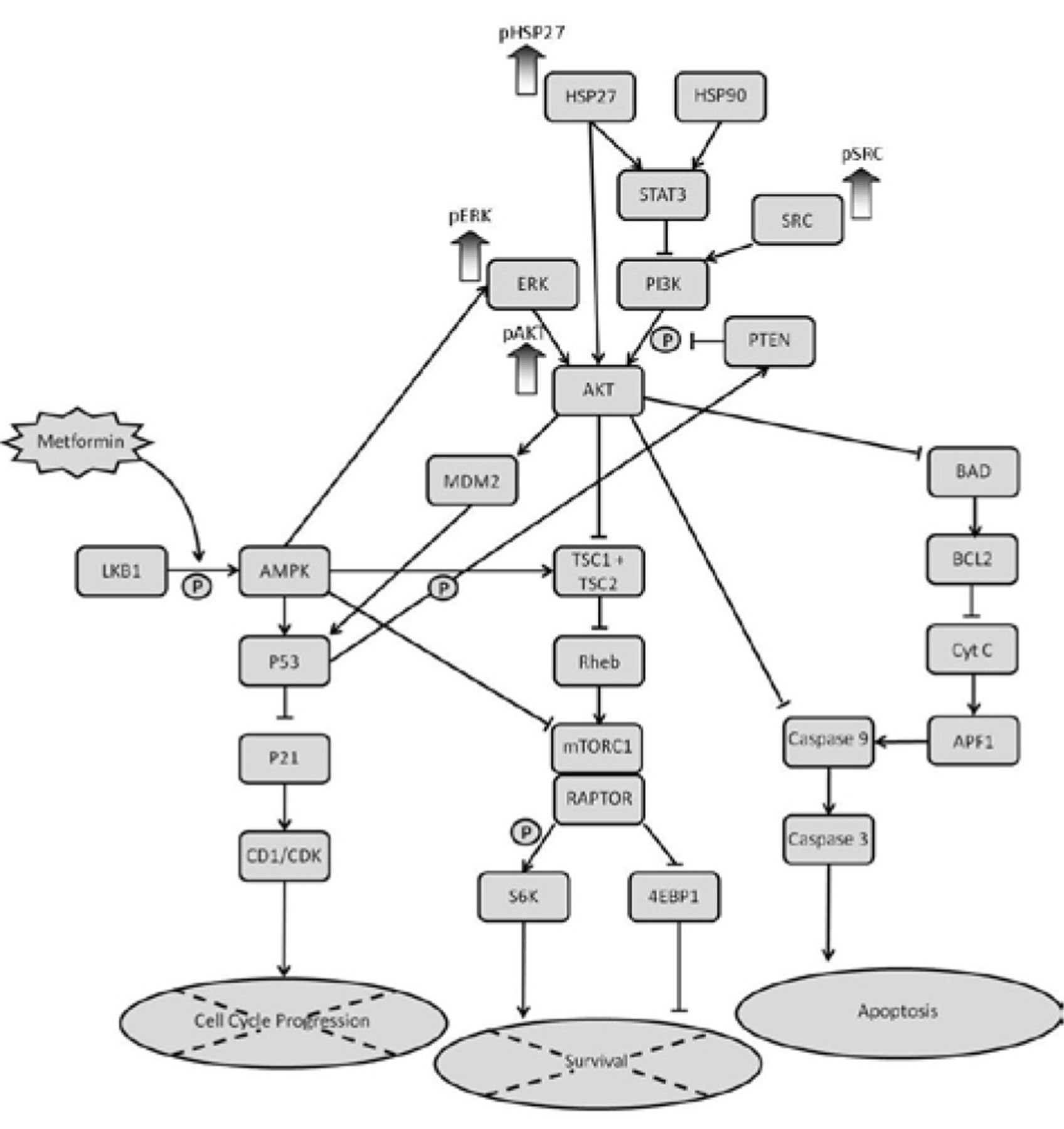
Schematic representation of the mTOR pathway and the proteins that are up-regulated in KLM1-R following treatment with metformin.

The results presented herein confirm that the anti-proliferative and pro-apoptotic effects of metformin do not alter total AMPK levels (although phosphorylation at Thr-172 was no probed for) (20, 43, 46-48). The transient changes observed in biochemistry were also previously reported as an adaptation to the early inhibitory effects of metformin on mitochondrial energetics with a redirection of carbon flux towards L-lactate and other biochemical intermediates (48).

The increase in pAkt level has been previously described as also being part of the above-mentioned adaptive mechanism. It fulfils its role as a key regulator of glucose metabolism by increasing the translocation of glucose trans porters, GLUT1 and GLUT4, to the plasma membrane to increase glucose uptake and aerobic glycolysis (49-51) together with other processes that affect apoptosis (48, 52-55).

It has also been shown that cells with high basal pAkt, glucose consumption or glycolysis are resistant to the early inhibitory effects of metformin and can, thus, avoid apoptosis (48). This does not seem to be an issue with KLM1-R cells, which, as shown by Western blotting, exhibit a very low pAkt expression level.

Recent research has shown that KLM1-R cells over-express SOX2, a stem cell marker, indicating that these cells arise from a small population within KLM1 after stress-induction by GEM (56). The action of metformin on cancer stem cells (CSCs), as reported for breast CSCs, appears to be mostly via depletion of nucleotide triphosphates (NTPs) rather than by exerting an effect on glycolytic and TCA cycle intermediates together with an inhibition of a signal transduction pathway that results in an inflammatory response (57, 58) dependent on nuclear factor-κB (NF-κB) and signal transducer and activator of transcription 3 (STAT3) (59, 60). The decreased NTP levels in CSCs reduce the availability of intermediates for energetics and biosynthesis of nucleic acids (DNA, RNA) and co-factors (e.g. flavin adenine dinucleotide (FAD), NADH, coenzyme A (CoA)) (42). This is corroborated in the current work in which it appears that CSCs in KLM1-R have a lower level of intracellular L-lactate (Figure 4B) compared to KLM1 (Figure 4A) in the presence of FBS.

Overall we corroborate the theory proposed by Scotland et al. (48) that metformin induces early inhibitory effects (peaking at 6h) on cell energetics via Akt signalling, which results in long-term (past 24 h) effects leading to inhibition of cell proliferation and apoptosis.

Our findings are, however, discordant with a previous report in pancreatic cancer (using 1 mM metformin for 16 h on PANC-1 and MiaPaCa-2), where metformin abolished mTORC1 activation without over-stimulating Akt phosphorylation on Ser-473 and prevented mitogen-stimulated ERK activation (61).

As a general note of caution, however, several recent publications agree that the metabolic effects of metformin may differ considerably between different cancer cell types and stage of cellular transformation, as well as undergo changes over time in response to cellular adaptation to the early inhibitory effects of metformin (42, 48, 61, 62). Our data, in addition, show that the micro-environment of the tumor also plays a role, as observed by the differences in energetic profiles in the presence or absence of FBS.

Acknowledgements

Western blot detection by Image Reader LAS-1000 Pro was carried-out at the Gene Research Centre of Yamaguchi University.

  • Received December 31, 2014.
  • Revision received January 20, 2015.
  • Accepted January 22, 2015.
  • Copyright© 2015 International Institute of Anticancer Research (Dr. John G. Delinassios), All rights reserved

References

  1. ↵
    1. Evans JM,
    2. Donnelly LA,
    3. Emslie-Smith AM,
    4. Alessi DR,
    5. Morris AD
    : Metformin and reduced risk of cancer in diabetic patients. BMJ 330: 1304-1305, 2005.
    OpenUrlFREE Full Text
    1. Bowker SL,
    2. Majumdar SR,
    3. Veugelers P,
    4. Johnson JA
    : Increased cancer-related mortality for patients with type 2 diabetes who use sulfonylureas or insulin. Diabetes Care 29: 254-258, 2006.
    OpenUrlAbstract/FREE Full Text
    1. Libby G,
    2. Donnelly LA,
    3. Donnan PT,
    4. Alessi DR,
    5. Morris AD,
    6. Evans JM
    : New users of metformin are at low risk of incident cancer: A cohort study among people with type 2 diabetes. Diabetes Care 32: 1620-1625, 2009.
    OpenUrlAbstract/FREE Full Text
    1. Jiralerspong S,
    2. Palla SL,
    3. Giordano SH,
    4. Meric-Bernstam F,
    5. Liedtke C,
    6. Barnett CM,
    7. Hsu L,
    8. Hung MC,
    9. Hortobagyi GN,
    10. Gonzalez-Angulo AM
    : Metformin and pathologic complete responses to neoadjuvant chemotherapy in diabetic patients with breast cancer. J Clin Oncol 27: 3297-3302, 2009.
    OpenUrlAbstract/FREE Full Text
  2. ↵
    1. Landman GW,
    2. Kleefstra N,
    3. van Hateren KJ,
    4. Groenier KH,
    5. Gans RO,
    6. Bilo HJ
    : Metformin associated with lower cancer mortality in type 2 diabetes ZODIAC-16. Diabetes Care 33: 322-326, 2010.
    OpenUrlAbstract/FREE Full Text
  3. ↵
    1. Kahn BB,
    2. Alquier T,
    3. Carling D,
    4. Hardie DG
    : AMP-activated protein knase: ancient energy gauge provides clues to modern understanding of metabolism. Cell Metab 1: 15-25, 2005.
    OpenUrlCrossRefPubMed
    1. Zhou G,
    2. Myers R,
    3. Li Y,
    4. Chen Y,
    5. Shen X,
    6. Fenyk-Melody J,
    7. Wu M,
    8. Ventre J,
    9. Doebber T,
    10. Fujii N,
    11. Musi N,
    12. Hirshman MF,
    13. Goodyear LJ,
    14. Moller DE
    : Role of AMP-activated protein kinase in mechanism of metformin action. J Clin Invest, 108: 1167-1174, 2001.
    OpenUrlCrossRefPubMed
  4. ↵
    1. El-Mir MY,
    2. Nogueira V,
    3. Fontaine E,
    4. Averet N,
    5. Rigoulet M,
    6. Leverve X
    : Dimethylbiguanide inhibits cell respiration via an indirect effect targeted on the respiratory chain complex I. J Biol Chem 275: 223-228, 2000.
    OpenUrlAbstract/FREE Full Text
  5. ↵
    1. Shaw RJ,
    2. Bardeesy N,
    3. Manning BD,
    4. Lopez L,
    5. Kosmatka M,
    6. DePinho RA,
    7. Cantley LC
    : The LKB1 tumor suppressor negatively regulates mTOR signaling. Cancer Cell 6: 91-99, 2004.
    OpenUrlCrossRefPubMed
  6. ↵
    1. Zakikhani M,
    2. Dowling R,
    3. Fantus IG,
    4. Sonenberg N,
    5. Pollak M
    : Metformin is an AMP kinase-dependent growth inhibitor for breast cancer cells. Cancer Res 66: 10269-10273, 2006.
    OpenUrlAbstract/FREE Full Text
  7. ↵
    1. Dowling RJ,
    2. Zakikhani M,
    3. Fantus IG,
    4. Pollak M,
    5. Sonenberg N
    : Metformin inhibits mammalian target of rapamycin-dependent translation initiation in breast cancer cells. Cancer Res 67: 10804-10812, 2007.
    OpenUrlAbstract/FREE Full Text
    1. Zakikhani M,
    2. Dowling RJ,
    3. Sonenberg N,
    4. Pollak MN
    : The effects of adiponectin and metformin on prostate and colon neoplasia involve activation of AMP-activated protein kinase. Cancer Prev Res 1: 369-375, 2008.
    OpenUrlAbstract/FREE Full Text
  8. ↵
    1. Vazquez-Martin A,
    2. Oliveras-Ferraros C,
    3. Menendez JA
    : The antidiabetic drug metformin suppresses HER2 (erbB-2) oncoprotein overexpression via inhibition of the mTOR effector p70S6K1 in human breast carcinoma cells. Cell Cycle 8: 88-96, 2009.
    OpenUrlCrossRefPubMed
  9. ↵
    1. Alimova IN,
    2. Liu B,
    3. Fan Z,
    4. Edgerton SM,
    5. Dillon T,
    6. Lind SE,
    7. Thor AD
    : Metformin inhibits breast cancer cell growth, colony formation and induces cell cycle arrest in vitro. Cell Cycle 8: 909-915, 2009.
    OpenUrlCrossRefPubMed
  10. ↵
    1. Buzzai M,
    2. Jones RG,
    3. Amaravadi RK,
    4. Lum JJ,
    5. DeBerardinis RJ,
    6. Zhao F,
    7. Viollet B,
    8. Thompson CB
    : Systemic treatment with the antidiabetic drug metformin selectively impairs p53-deficient tumor cell growth. Cancer Res 67: 6745-6752, 2007.
    OpenUrlAbstract/FREE Full Text
    1. Zhuang Y,
    2. Miskimins WK
    : Cell cycle arrest in Metformin treated breast cancer cells involves activation of AMPK, downregulation of cyclin D1, and requires p27Kip1 or p21Cip1. J Mol Signal 3(1): 1-11, 2008.
    OpenUrlCrossRefPubMed
    1. Liu B,
    2. Fan Z,
    3. Edgerton SM,
    4. Deng XS,
    5. Alimova IN,
    6. Lind SE,
    7. Thor AD
    : Metformin induces unique biological and molecular responses in triple negative breast cancer cells. Cell Cycle 8: 2031-2040, 2009.
    OpenUrlCrossRefPubMed
  11. ↵
    1. Oliveras-Ferraros C,
    2. Vazquez-Martin A,
    3. Menendez JA
    . Genome-wide inhibitory impact of the AMPK activator metformin on [kinesins, tubulins, histones, auroras and polo-like kinases] M-phase cell cycle genes in human breast cancer cells. Cell Cycle 8: 1633-1636, 2009.
    OpenUrlPubMed
    1. Vázquez-Martín A,
    2. Oliveras-Ferraros C,
    3. del Barco S,
    4. Martín-Castillo B,
    5. Menéndez JA
    : mTOR inhibitors and the anti-diabetic biguanide metformin: new insights into the molecular management of breast cancer resistance to the HER2 tyrosine ki nase inhibitor lapatinib (Tykerb). Clin Transl Oncol 11: 455-459, 2009.
    OpenUrlCrossRefPubMed
  12. ↵
    1. Ben Sahra I,
    2. Laurent K,
    3. Loubat A,
    4. Giorgetti-Peraldi S,
    5. Colosetti P,
    6. Auberger P,
    7. Tanti JF,
    8. Le Marchand-Brustel Y,
    9. Bost F
    : The antidiabetic drug metformin exerts an antitumoral effect in vitro and in vivo through a decrease of cyclin D1 level. Oncogene 27: 3576-3586, 2008.
    OpenUrlCrossRefPubMed
    1. Tomimoto A,
    2. Endo H,
    3. Sugiyama M,
    4. Fujisawa T,
    5. Hosono K,
    6. Takahashi H,
    7. Nakajima N,
    8. Nagashima Y,
    9. Wada K,
    10. Nakagama H,
    11. Nakajima A
    : Metformin suppresses intestinal polyp growth in ApcMin/+mice. Cancer Sci 99: 2136-2141, 2008.
    OpenUrlCrossRefPubMed
    1. Hardie DG
    : AMP-activated/SNF1 protein kinases: conserved guardians of cellular energy. Nat Rev Mol Cell Biol 8: 774-785, 2007.
    OpenUrlCrossRefPubMed
  13. ↵
    1. Towler MC,
    2. Hardie DG
    : AMP-activated protein kinase in metabolic control and insulin signaling. Circ Res 100: 328-341, 2007.
    OpenUrlAbstract/FREE Full Text
  14. ↵
    1. Inoki K,
    2. Zhu T,
    3. Guan KL
    : TSC2 mediates cellular energy response to control cell growth and survival. Cell 115: 577-590, 2003.
    OpenUrlCrossRefPubMed
  15. ↵
    1. Lee JW,
    2. Soung YH,
    3. Kim SY,
    4. Lee HW,
    5. Park WS,
    6. Nam SW,
    7. Kim SH,
    8. Lee JY,
    9. Yoo NJ,
    10. Lee SH
    : PIK3CA gene is frequently mutated in breast carcinomas and hepatocellular carcinomas. Oncogene 24: 1477-1480, 2005.
    OpenUrlCrossRefPubMed
  16. ↵
    1. Markman B,
    2. Atzori F,
    3. Perez-Garcia J,
    4. Tabernero J,
    5. Baselga J
    : Status of PI3K inhibition and biomarker development in cancer therapeutics. Ann Oncol 21: 683-691, 2010.
    OpenUrlAbstract/FREE Full Text
  17. ↵
    1. Thoreen CC,
    2. Sabatini DM
    : AMPK and p53 help cells through lean times. Cell Metab 1: 287-288, 2005.
    OpenUrlCrossRefPubMed
  18. ↵
    1. Ben Sahra I,
    2. Laurent K,
    3. Giuliano S,
    4. Larbret F,
    5. Ponzio G,
    6. Gounon P,
    7. Le Marchand-Brustel Y,
    8. Giorgetti-Peraldi S,
    9. Cormont M,
    10. Bertolotto C,
    11. Deckert M,
    12. Auberger P,
    13. Tanti JF,
    14. Bost F
    : Targeting cancer cell metabolism: the combination of metformin and 2-deoxyglucose induces p53-dependent apoptosis in prostate cancer cells. Cancer Res 70: 2465-2475, 2010.
    OpenUrlAbstract/FREE Full Text
  19. ↵
    1. Wang LW,
    2. Li ZS,
    3. Zou DW,
    4. Jin ZD,
    5. Gao J,
    6. Xu GM
    : Metformin induces apoptosis of pancreatic cancer cells. World J Gastroenterol 14: 7192-7198, 2008.
    OpenUrlCrossRefPubMed
  20. ↵
    1. Lee MS,
    2. Hsu CC,
    3. Wahlqvist ML,
    4. Tsai HN,
    5. Yu-Hung Chang YH,
    6. Huang YC
    : Type 2 diabetes increases and metformin reduces total, colorectal, liver and pancreatic cancer incidences in Taiwanese: a representative population prospective cohort study of 800,000 individuals. BMC Cancer 11: 20, 2011.
    OpenUrlCrossRefPubMed
  21. ↵
    1. Mearow KM,
    2. Dodge ME,
    3. Rahimtula M,
    4. Yegappan C
    : Stress-mediated signaling in PC12 cells—the role of the small heat shock protein, Hsp27, and Akt in protecting cells from heat stress and nerve growth factor withdrawal. J Neurochem 83: 452-462, 2002.
    OpenUrlCrossRefPubMed
  22. ↵
    1. Rane MJ,
    2. Pan Y,
    3. Singh S,
    4. Powell DW,
    5. Wu R,
    6. Cummins T,
    7. Chen Q,
    8. McLeish KR,
    9. Klein JB
    : Heat shock protein 27 controls apoptosis by regulating Akt activation. J Biol Chem 278: 27828-27835, 2003.
    OpenUrlAbstract/FREE Full Text
  23. ↵
    1. Downward J
    : Mechanisms and consequences of activation of protein kinase B/Akt. Curr Opin Cell Biol 10: 262-267, 1998.
    OpenUrlCrossRefPubMed
  24. ↵
    1. Concannon CG,
    2. Gorman AM,
    3. Samali A
    : On the role of Hsp27 in regulating apoptosis. Apoptosis 8(1): 61-70, 2003.
    OpenUrlCrossRefPubMed
  25. ↵
    1. Henshall DC,
    2. Araki T,
    3. Schindler CK,
    4. Lan JQ,
    5. Tiekoter KL,
    6. Taki W,
    7. Simon RP
    : Activation of Bcl-2-associated death protein and counter-response of Akt within cell populations during seizure-induced neuronal death. J Neurosci 22: 8458-8465, 2002.
    OpenUrlAbstract/FREE Full Text
  26. ↵
    1. Benn SC,
    2. Woolf CJ
    : Adult neuron survival strategies—slamming on the brakes. Nat Rev Neurosci 5(9): 686-700, 2004.
    OpenUrlCrossRefPubMed
  27. ↵
    1. Maehara S,
    2. Tanaka S,
    3. Shimada M,
    4. Shirabe K,
    5. Saito Y,
    6. Takahashi K,
    7. Maehara Y
    : Selenoprotein P, as a predictor for evaluating gemcitabine resistance in human pancreatic cancer cells. IJC 112(2): 184-189, 2004.
    OpenUrl
  28. ↵
    1. Mori-Iwamoto S,
    2. Kuramitsu Y,
    3. Ryozawa S,
    4. Mikuriya K,
    5. Fujimoto M,
    6. Maehara S,
    7. Maehara Y,
    8. Okita K,
    9. Nakamura K,
    10. Sakaida I
    : Proteomics finding heat shock protein 27 as a biomarker for resistance of pancreatic cancer cells to gemcitabine. Int J Oncol 31: 1345-1350, 2007.
    OpenUrlPubMed
  29. ↵
    1. Owen MR,
    2. Doran E,
    3. Halestrap AP
    : Evidence that metformin exerts its anti-diabetic effects through inhibition of complex 1 of the mitochondrial respiratory chain. Biochem J 348: 607-614, 2000.
    OpenUrlAbstract
    1. Guigas B,
    2. Detaille D,
    3. Chauvin C,
    4. Batandier C,
    5. De Oliveira F,
    6. Fontaine E,
    7. Leverve X
    : Metformin inhibits mitochondrial permeability transition and cell death: a pharmacological in vitro study. Biochem J 382: 877-884, 2004.
    OpenUrlAbstract/FREE Full Text
    1. Shaw RJ,
    2. Kosmatka M,
    3. Bardeesy N,
    4. Hurley RL,
    5. Witters LA,
    6. DePinho RA,
    7. Cantley LC
    : The tumor suppressor LKB1 kinase directly activates AMPactivated kinase and regulates apoptosis in response to energy stress. Proc Natl Acad Sci USA 101: 3329-3335, 2004.
    OpenUrlAbstract/FREE Full Text
  30. ↵
    1. Janzer A,
    2. German NJ,
    3. Gonzalez-Herrera KN,
    4. Asara JM,
    5. Haigis MC,
    6. Struhl K
    : Metformin and phenformin deplete tricarboxylic acid cycle and glycolytic intermediates during cell transformation and NTPs in cancer stem cells. PNAS 111(29): 10574-10579, 2014.
    OpenUrlAbstract/FREE Full Text
  31. ↵
    1. Kalender A,
    2. Selvaraj A,
    3. Kim SY,
    4. Gulati P,
    5. Brule S,
    6. Viollet B,
    7. Kemp BE,
    8. Bardeesy N,
    9. Dennis P,
    10. Schlager JJ,
    11. Marette A,
    12. Kozma SC,
    13. Thomas G
    : Metformin, independent of AMPK, inhibits mTORC1 in a rag GTPase-dependent manner. Cell Metab 11: 390-401, 2010.
    OpenUrlCrossRefPubMed
    1. Green AS,
    2. Chapuis N,
    3. Maciel TT,
    4. Willems L,
    5. Lambert M,
    6. Arnoult C,
    7. Boyer O,
    8. Bardet V,
    9. Park S,
    10. Foretz M,
    11. Viollet B,
    12. Ifrah N,
    13. Dreyfus F,
    14. Hermine O,
    15. Cruz Moura I,
    16. Lacombe C,
    17. Mayeux P,
    18. Bouscary D,
    19. Tamburini J
    : The LKB1/AMPK signaling pathway has tumor suppressor activity in acute myeloid leukemia through the repression of mTOR-dependent oncogenic mRNA translation. Blood 116: 4262-4273, 2010.
    OpenUrlAbstract/FREE Full Text
  32. ↵
    1. Grimaldi C,
    2. Chiarini F,
    3. Tabellini G,
    4. Ricci F,
    5. Tazzari PL,
    6. Battistelli M,
    7. Falcieri E,
    8. Bortul R,
    9. Melchionda F,
    10. Iacobucci I,
    11. Pagliaro P,
    12. Martinelli G,
    13. Pession A,
    14. Barata JT,
    15. McCubrey JA,
    16. Martelli AM
    : AMP-dependent kinase/mammalian target of rapamycin complex 1 signaling in T-cell acute lymphoblastic leukemia: therapeutic implications. Leukemia 26: 91-100, 2012.
    OpenUrlCrossRefPubMed
  33. ↵
    1. Tomic T,
    2. Botton T,
    3. Cerezo M,
    4. Robert G,
    5. Luciano F,
    6. Puissant A,
    7. Gounon P,
    8. Allegra M,
    9. Bertolotto C,
    10. Bereder JM,
    11. Tartare-Deckert S,
    12. Bahadoran P,
    13. Auberger P,
    14. Ballotti R,
    15. Rocchi S
    : Metformin inhibits melanoma development through autophagy and apoptosis mechanisms. Cell Death Dis 2: e199, 2011.
    OpenUrlCrossRefPubMed
    1. Ben Sahra I,
    2. Regazzetti C,
    3. Robert G,
    4. Laurent K,
    5. Le Marchand-Brustel Y,
    6. Auberger P,
    7. Tanti JF,
    8. Giorgetti-Peraldi S,
    9. Bost F
    : Metformin, independent of AMPK, induces mTOR inhibition and cell-cycle arrest through REDD1. Cancer Res 71: 4366-4372, 2011.
    OpenUrlAbstract/FREE Full Text
  34. ↵
    1. Scotland S,
    2. Saland E,
    3. Skuli N,
    4. de Toni F,
    5. Boutzen H,
    6. Micklow E,
    7. Senegas I,
    8. Peyraud R,
    9. Peyriga L,
    10. Theodoro F,
    11. Dumon E,
    12. Martineau Y,
    13. Danet-Desnoyers G,
    14. Bono F,
    15. Rocher C,
    16. Levade T,
    17. Manenti S,
    18. Junot C,
    19. Portais JC,
    20. Alet N,
    21. Recher C,
    22. Selak MA,
    23. Carroll M,
    24. Sarry JE
    : Mitochondrial energetic and AKT status mediate metabolic effects and apoptosis of metformin in human leukemic cells. Leukemia 27: 2129-2138, 2013.
    OpenUrlCrossRefPubMed
  35. ↵
    1. Barthel A,
    2. Okino ST,
    3. Liao J,
    4. Nakatani K,
    5. Li J,
    6. Whitlock JP Jr.,
    7. Roth RA
    : Regulation of GLUT1 gene transcription by the serine/threonine kinase Akt1. J Biol Chem 274: 20281-20286, 1999.
    OpenUrlAbstract/FREE Full Text
    1. Kohn AD,
    2. Summers SA,
    3. Birnbaum MJ,
    4. Roth RA
    : Expression of a constitutively active Akt Ser/Thr kinase in 3T3-L1 adipocytes stimulates glucose uptake and glucose transporter 4 translocation. J Biol Chem 271: 31372-31378, 1996.
    OpenUrlAbstract/FREE Full Text
  36. ↵
    1. Wieman HL,
    2. Wofford JA,
    3. Rathmell JC
    : Cytokine stimulation promotes glucose uptake via phosphatidylinositol-3 kinase/Akt regulation of Glut1 activity and trafficking. Mol Biol Cell 18: 1437-1446, 2007.
    OpenUrlAbstract/FREE Full Text
  37. ↵
    1. Majewski N,
    2. Nogueira V,
    3. Bhaskar P,
    4. Coy PE,
    5. Skeen JE,
    6. Gottlob K,
    7. Chandel NS,
    8. Thompson CB,
    9. Brooks Robey R,
    10. Hay N
    : Hexokinase mitochondria interaction mediated by Akt is required to inhibit apoptosis in the presence or absence of Bax and Bak. Mol Cell 16: 819-830, 2004.
    OpenUrlCrossRefPubMed
    1. Arzoine L,
    2. Zilberberg N,
    3. Ben-Romano R,
    4. Shoshan-Barmatz V
    : Voltage-dependent anion channel 1-based peptides interact with hexokinase to prevent its antiapoptotic activity. J Biol Chem 284: 3946-3955, 2009.
    OpenUrlAbstract/FREE Full Text
    1. McCubrey JA,
    2. Steelman LS,
    3. Abrams SL,
    4. Bertrand FE,
    5. Ludwig DE,
    6. Bäsecke J,
    7. Libra M,
    8. Stivala F,
    9. Milella M,
    10. Tafuri A,
    11. Lunghi P,
    12. Bonati A,
    13. Martelli AM
    : Targeting survival cascades induced by activation of Ras/Raf/MEK/ERK, PI3K/ PTEN/Akt/mTOR and Jak/STAT pathways for effective leukemia therapy. Leukemia 22: 708-722, 2008.
    OpenUrlCrossRefPubMed
  38. ↵
    1. Martelli AM,
    2. Evangelisti C,
    3. Chappell W,
    4. Abrams SL,
    5. Bäsecke J,
    6. Stivala F,
    7. Donia M,
    8. Fagone P,
    9. Nicoletti F,
    10. Libra M,
    11. Ruvolo V,
    12. Ruvolo P,
    13. Kempf CR,
    14. Steelman LS,
    15. McCubrey JA
    : Targeting the translational apparatus to improve leukemia therapy: roles of the PI3K/PTEN/Akt/mTOR pathway. Leukemia 25: 1064-1079, 2011.
    OpenUrlCrossRefPubMed
  39. ↵
    1. Nawata J,
    2. Kuramitsu Y,
    3. Wang Y,
    4. Kitagawa T,
    5. Tokuda K,
    6. Baron B,
    7. Akada J,
    8. Suenaga S,
    9. Kaino S,
    10. Maehara S,
    11. Maehara Y,
    12. Sakaida I,
    13. Nakamura K
    : Active hexose-correlated compound down-regulates Sex-determining region Y-box 2 of pancreatic cancer cells. Anticancer Res 34(9): 4807-4811, 2014.
    OpenUrlAbstract/FREE Full Text
  40. ↵
    1. Hirsch HA,
    2. Iliopoulos D,
    3. Tsichlis PN,
    4. Struhl K
    : Metformin selectively targets cancer stem cells, and acts together with chemotherapy to block tumor growth and prolong remission. Cancer Res 69(19): 7507-7511, 2009.
    OpenUrlAbstract/FREE Full Text
  41. ↵
    1. Hirsch HA,
    2. Iliopoulos D,
    3. Struhl K
    : Metformin inhibits the inflammatory response associated with cellular transformation and cancer stem cell growth. Proc Natl Acad Sci USA 110(3): 972-977, 2013.
    OpenUrlAbstract/FREE Full Text
  42. ↵
    1. Hirsch HA,
    2. Iliopoulos D,
    3. Joshi A,
    4. Zhang Y,
    5. Jaeger SA,
    6. Bulyk M,
    7. Tsichlis PN,
    8. Liu SX,
    9. Struhl K
    : A transcriptional signature and common gene networks link cancer with lipid metabolism and diverse human diseases. Cancer Cell 17(4): 348-361, 2010.
    OpenUrlCrossRefPubMed
  43. ↵
    1. Iliopoulos D,
    2. Hirsch HA,
    3. Struhl K
    : An epigenetic switch involving NF-kappaB, Lin28, Let-7 MicroRNA, and IL6 links inflammation to cell transformation. Cell 139(4): 693-706, 2009.
    OpenUrlCrossRefPubMed
  44. ↵
    1. Soares HP,
    2. Ni Y,
    3. Kisfalvi K,
    4. Sinnett-Smith J,
    5. Rozengurt E
    : Different Patterns of Akt and ERK Feedback Activation in Response to Rapamycin, Active-Site mTOR Inhibitors and Metformin in Pancreatic Cancer Cells. PLoS ONE 8(2): e57289, 2013.
    OpenUrlCrossRefPubMed
  45. ↵
    1. Zakikhani M,
    2. Blouin MJ,
    3. Piura E,
    4. Pollak MN
    : Metformin and rapamycin have distinct effects on the AKT pathway and proliferation in breast cancer cells. Breast Cancer Res Treat 123: 271-279, 2010.
    OpenUrlCrossRefPubMed
PreviousNext
Back to top

In this issue

Anticancer Research
Vol. 35, Issue 4
April 2015
  • Table of Contents
  • Table of Contents (PDF)
  • Index by author
  • Back Matter (PDF)
  • Ed Board (PDF)
  • Front Matter (PDF)
Print
Download PDF
Article Alerts
Sign In to Email Alerts with your Email Address
Email Article

Thank you for your interest in spreading the word on Anticancer Research.

NOTE: We only request your email address so that the person you are recommending the page to knows that you wanted them to see it, and that it is not junk mail. We do not capture any email address.

Enter multiple addresses on separate lines or separate them with commas.
Resistance to Gemcitabine in the Pancreatic Cancer Cell Line KLM1-R Reversed by Metformin Action
(Your Name) has sent you a message from Anticancer Research
(Your Name) thought you would like to see the Anticancer Research web site.
CAPTCHA
This question is for testing whether or not you are a human visitor and to prevent automated spam submissions.
6 + 11 =
Solve this simple math problem and enter the result. E.g. for 1+3, enter 4.
Citation Tools
Resistance to Gemcitabine in the Pancreatic Cancer Cell Line KLM1-R Reversed by Metformin Action
BYRON BARON, YUFENG WANG, SHIN-ICHIRO MAEHARA, YOSHIHIKO MAEHARA, YASUHIRO KURAMITSU, KAZUYUKI NAKAMURA
Anticancer Research Apr 2015, 35 (4) 1941-1949;

Citation Manager Formats

  • BibTeX
  • Bookends
  • EasyBib
  • EndNote (tagged)
  • EndNote 8 (xml)
  • Medlars
  • Mendeley
  • Papers
  • RefWorks Tagged
  • Ref Manager
  • RIS
  • Zotero
Reprints and Permissions
Share
Resistance to Gemcitabine in the Pancreatic Cancer Cell Line KLM1-R Reversed by Metformin Action
BYRON BARON, YUFENG WANG, SHIN-ICHIRO MAEHARA, YOSHIHIKO MAEHARA, YASUHIRO KURAMITSU, KAZUYUKI NAKAMURA
Anticancer Research Apr 2015, 35 (4) 1941-1949;
Twitter logo Facebook logo Mendeley logo
  • Tweet Widget
  • Facebook Like
  • Google Plus One

Jump to section

  • Article
    • Abstract
    • Materials and Methods
    • Results
    • Discussion
    • Acknowledgements
    • References
  • Figures & Data
  • Info & Metrics
  • PDF

Related Articles

Cited By...

  • (Ir)relevance of Metformin Treatment in Patients with Metastatic Pancreatic Cancer: An Open-Label, Randomized Phase II Trial
  • Google Scholar

More in this TOC Section

  • P53-armed Oncolytic Adenovirus Enhances the Efficacy of PD-1 Blockade in Neuroblastoma by Inducing Immunogenic Cell Death
  • Methoxy and Ketone Substitutions of Resveratrol Enhance Its Anticancer Activity by Increasing MEK and ERK Binding Affinity in Colorectal Cancer Cells
  • JI-CJ001 Enhances Paclitaxel Sensitivity in Gastric Cancer Cell Lines through Inhibition of HER2/mTOR Signaling and Induction of Autophagy
Show more Experimental Studies

Keywords

  • HSP27
  • metformin
  • pancreatic cancer
Anticancer Research

© 2026 Anticancer Research

Powered by HighWire