
Main menu

  • Home
  • Current Issue
  • Archive
  • Info for
    • Authors
    • Editorial Policies
    • Subscribers
    • Advertisers
    • Editorial Board
  • Other Publications
    • In Vivo
    • Cancer Genomics & Proteomics
    • Cancer Diagnosis & Prognosis
  • More
    • IIAR
    • Conferences
    • 2008 Nobel Laureates
  • About Us
    • General Policy
    • Contact
  • Other Publications
    • Anticancer Research
    • In Vivo
    • Cancer Genomics & Proteomics

User menu

  • Register
  • Subscribe
  • My alerts
  • Log in
  • Log out
  • My Cart

Search

  • Advanced search
Anticancer Research
  • Other Publications
    • Anticancer Research
    • In Vivo
    • Cancer Genomics & Proteomics
  • Register
  • Subscribe
  • My alerts
  • Log in
  • Log out
  • My Cart
Anticancer Research

Advanced Search

  • Home
  • Current Issue
  • Archive
  • Info for
    • Authors
    • Editorial Policies
    • Subscribers
    • Advertisers
    • Editorial Board
  • Other Publications
    • In Vivo
    • Cancer Genomics & Proteomics
    • Cancer Diagnosis & Prognosis
  • More
    • IIAR
    • Conferences
    • 2008 Nobel Laureates
  • About Us
    • General Policy
    • Contact
  • Visit us on Facebook
  • Follow us on Linkedin
Research ArticleExperimental Studies

HVEM Expression Contributes to Tumor Progression and Prognosis in Human Colorectal Cancer

TAKASHI INOUE, MASAYUKI SHO, SATOSHI YASUDA, SATOSHI NISHIWADA, SHINJI NAKAMURA, TAKESHI UEDA, NAOTO NISHIGORI, KEIJIRO KAWASAKI, SHINSAKU OBARA, TAKAYUKI NAKAMOTO, FUMIKAZU KOYAMA, HISAO FUJII and YOSHIYUKI NAKAJIMA
Anticancer Research March 2015, 35 (3) 1361-1367;
TAKASHI INOUE
1Department of Surgery, Nara Medical University, Kashihara, Nara, Japan
  • Find this author on Google Scholar
  • Find this author on PubMed
  • Search for this author on this site
MASAYUKI SHO
1Department of Surgery, Nara Medical University, Kashihara, Nara, Japan
  • Find this author on Google Scholar
  • Find this author on PubMed
  • Search for this author on this site
  • For correspondence: m-sho@naramed-u.ac.jp
SATOSHI YASUDA
1Department of Surgery, Nara Medical University, Kashihara, Nara, Japan
  • Find this author on Google Scholar
  • Find this author on PubMed
  • Search for this author on this site
SATOSHI NISHIWADA
1Department of Surgery, Nara Medical University, Kashihara, Nara, Japan
  • Find this author on Google Scholar
  • Find this author on PubMed
  • Search for this author on this site
SHINJI NAKAMURA
1Department of Surgery, Nara Medical University, Kashihara, Nara, Japan
  • Find this author on Google Scholar
  • Find this author on PubMed
  • Search for this author on this site
TAKESHI UEDA
1Department of Surgery, Nara Medical University, Kashihara, Nara, Japan
  • Find this author on Google Scholar
  • Find this author on PubMed
  • Search for this author on this site
NAOTO NISHIGORI
1Department of Surgery, Nara Medical University, Kashihara, Nara, Japan
  • Find this author on Google Scholar
  • Find this author on PubMed
  • Search for this author on this site
KEIJIRO KAWASAKI
1Department of Surgery, Nara Medical University, Kashihara, Nara, Japan
  • Find this author on Google Scholar
  • Find this author on PubMed
  • Search for this author on this site
SHINSAKU OBARA
1Department of Surgery, Nara Medical University, Kashihara, Nara, Japan
  • Find this author on Google Scholar
  • Find this author on PubMed
  • Search for this author on this site
TAKAYUKI NAKAMOTO
1Department of Surgery, Nara Medical University, Kashihara, Nara, Japan
  • Find this author on Google Scholar
  • Find this author on PubMed
  • Search for this author on this site
FUMIKAZU KOYAMA
2Department of Endoscopy and Ultrasound, Nara Medical University, Kashihara, Nara, Japan
  • Find this author on Google Scholar
  • Find this author on PubMed
  • Search for this author on this site
HISAO FUJII
2Department of Endoscopy and Ultrasound, Nara Medical University, Kashihara, Nara, Japan
  • Find this author on Google Scholar
  • Find this author on PubMed
  • Search for this author on this site
YOSHIYUKI NAKAJIMA
1Department of Surgery, Nara Medical University, Kashihara, Nara, Japan
  • Find this author on Google Scholar
  • Find this author on PubMed
  • Search for this author on this site
  • Article
  • Figures & Data
  • Info & Metrics
  • PDF
Loading

Abstract

Background: Herpesvirus entry mediator (HVEM) has been recently suggested to play certain roles in cancer biology. We examined HVEM expression in human colorectal cancer (CRC) to reveal its clinical importance. Materials and Methods: Immunohistochemical staining was carried-out in normal epithelium, benign and malignant lesions. Results: While intense HVEM expression was not observed in normal epithelium and hyperplastic polyps, 24% of adenoma and more than half of CRCs had high HVEM expression. In 234 CRCs, HVEM expression was significantly associated with tumor status and pathological stage. Patients with high HVEM expression had a significantly poorer prognosis than those with low expression. Importantly, HVEM status had an independent prognostic value in CRC. Furthermore, HVEM status was inversely corrected with the presence of tumor-infiltrating T-cells. Conclusion: HVEM may play a critical role in tumor progression and immune evasion, and may also be a novel prognostic marker and potential therapeutic target in human CRC.

  • Colorectal cancer
  • HVEM
  • prognosis
  • surgery
  • tumor immunity

Colorectal cancer (CRC) is the third cause of cancer-related death worldwide (1). In the past decade, there has been significant progress in the treatment of CRC, including chemotherapy and molecular-targeted therapy. Despite recent advances in treatment, the prognosis of advanced or recurrent CRC remains poor (2-4). Thus, novel approaches still need to be developed and established to improve the prognosis of patients with CRC.

Several members belonging to the tumor necrosis factor receptor (TNFR) and immunoglobulin (Ig) superfamily regulate lymphocyte activation (5, 6). Herpesvirus entry mediator (HVEM; TNFRSF14) is a member of the TNFR superfamily which is expressed on a wide range of hematopoietic cells including T-cells, B-cells, natural killer cells, monocytes, and immature dendritic cells. HVEM is also expressed in various nonhematopoietic cells and tissues including the lung, liver, and kidney (7-9). HVEM ligands belong to two distinct families: the TNF-related cytokines, lymphotoxin-related inducible ligand that competes for glycoprotein D binding to herpesvirus entry mediator on T cells (LIGHT) and lymphotoxin-α, and the Ig-related membrane proteins, B and T lymphocyte attenuator (BTLA) and cluster of differentiation 160 (CD160) (9-11). The binding of HVEM to LIGHT stimulates the host immune response. In sharp contrast, HVEM can provide an inhibitory signal to T-cells upon binding to BTLA or CD160 (6, 9). Despite the complexity of HVEM signaling, its inhibitory function seems to be dominant as demonstrated in HVEM-deficient mice (12). Previous studies have focused on the HVEM pathway in inflammatory and immunological disease conditions such as autoimmune disease, inflammatory bowel disease and transplantation (12-16). We and others have also reported HVEM expression in various tumor cell lines and clinical tumor tissues, including breast and esophageal cancer, chronic lymphocytic leukemia and melanoma (9, 17-19). These studies have demonstrated that HVEM might be involved in tumor progression and evasion of immune response. However, to our knowledge, the clinical significance of HVEM in human CRC tissues has not been previously addressed. Therefore, the aim of the present study was to investigate the involvement of HVEM in tumor biology and immunity in human CRC.

Materials and Methods

Patients. We examined 234 patients with CRC who underwent surgery at the Department of Surgery, Nara Medical University, Japan between 2004 and 2007. The median age of the patients was 69.5 years, with a range of 35 to 94 years. Tissues were obtained from the resected specimens, and each specimen was fixed in 10% phosphate-buffered formalin and embedded in paraffin. A serial section from each specimen was stained with hematoxylin and eosin stain for histological evaluation. Tumors were classified according to the tumor-node-metastasis staging system (20). The median follow-up for all patients was 60.5 months, with a range of 0.5 to 87 months. Furthermore, we examined 10 patients with hyperplastic polyp and 50 patients with adenoma who underwent endoscopic resection at our institution. Written informed consent was obtained from all patients according to our institutional guidelines.

Immunohistochemistry. Immunohistochemical staining was carried out using the Dako Envision system (Dako Cytomation, Kyoto, Japan) as previously described (21). As primary antibody, antibodies to HVEM (94804; R&D System, Minneapolis), CD4 (4B12; Dako Cytomation), and CD8 (C8/144B; Dako Cytomation) were used. Paraffin-embedded tissues were de-paraffinized and rehydrated in a graded series of ethanols. Antigen retrieval was performed by heating tissue sections using a target retrieval solution, pH 9.0 (Dako). After neutralization of endogenous peroxidase, the sections were incubated with each primary antibodies overnight at 4°C. After three washes in phosphate-buffered saline (PBS), the sections were incubated with secondary antibodies at 37°C for 0.5 (HVEM and CD8) or 1(CD4) h. After three washes in PBS, the sections were visualized with diaminobenzidine (DAB) (Dako) for approximately 5-10 min. The slides were counterstained with hematoxylin, dehydrated in ethanol.

Evaluation of immunostaining. Immunohistochemistry for HVEM was evaluated according to its intensity and the percentage of positively-stained tumor cells in a blinded manner. Five fields were randomly selected. The percentage of positively-stained tumor cells in each selected field was determined by counting individual tumor cells at high magnification. At least 200 tumor cells were scored per ×200 field, and the median number for each sample was calculated. The intensity was classified into four groups: none: 0 point; weak: 1 point; intermediate: 2 points and strong: 3 points (Figure 1). The percentage of positively-stained tumor cells was classified into four groups as follows: 0-25%: score 1; 26-50%: score 2; 51-75%: score 3; and 76-100%: score 4. We then evaluated HVEM expression in each tissue according to the total score by adding the points and scores for each (total number; 1-7). Specimens with a total score of 1-5 were classified as having low HVEM expression, and those with 6-7 were classified as having high HVEM expression. Immunohistochemistry for CD4+ and CD8+ T-cells was evaluated by counting the number of tumor-infiltrating T-lymphocytes (TILs) as previously described (22).

Statistical analysis. The significance of the difference in HVEM expression by several clinicopathological variables was assessed by the Student's t-test, the Chi-square test or Fisher's exact test as appropriate. The overall survival time was calculated from the date of surgery to the date of death. The Kaplan–Meier method was used to estimate the probability of survival, and significance was assessed by the log-rank test. Univariate and multivariate analyses were performed by the Cox proportional hazards model to evaluate significant prognostic predictors and their relative role. We use the terms tumor status as T factor, nodal status as N factor, and metastatic status as M factor in the tumor-node-metastasis classification, retrospectively. A p-value <0.05 was considered statistically significant.

Figure 1.
  • Download figure
  • Open in new tab
  • Download powerpoint
Figure 1.

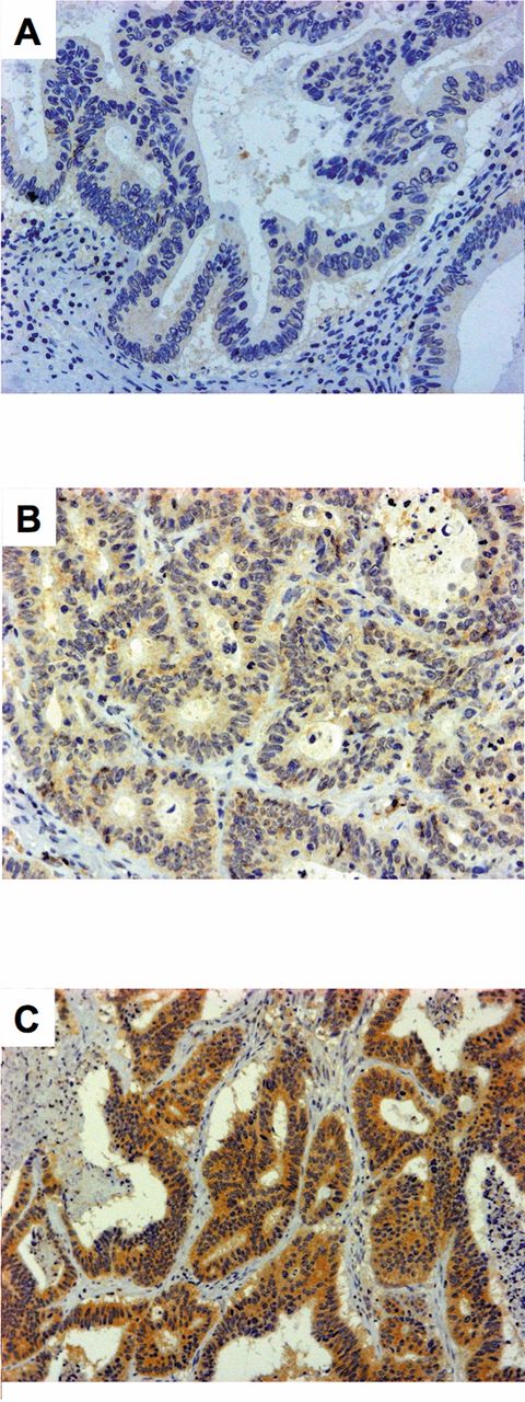
Immunohistochemical staining of herpesvirus entry mediator (HVEM) in human colorectal cancer tissues. Representative case of tumors with weak (A), intermediate (B) and strong (C) HVEM expression. Original magnification, ×200.

Figure 2.
  • Download figure
  • Open in new tab
  • Download powerpoint
Figure 2.

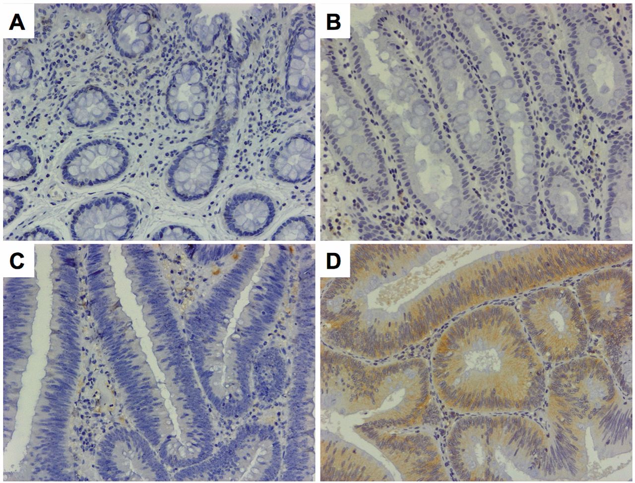
Immunohistochemical staining of herpesvirus entry mediator (HVEM) in normal colon epithelium, hyperplastic polyp and adenoma. Representative case of normal colonic epithelium with low HVEM (A), hyperplastic polyp with low HVEM (B), adenoma with low HVEM (C) and adenoma with high HVEM (D). Original magnification, ×200.

Results

HVEM expression in human normal and neoplastic colon tissues. To investigate the involvement of HVEM in CRC, we first examined HVEM expression in various colonic tissues including normal epithelium, and benign and malignant lesions by immunohistochemical analysis.

Among 10 samples of normal colonic epithelium examined, 3 had no staining for HVEM expression and 7 had a weak staining with score 1 at the protein level (Figure 2A). Out of the 10 hyperplastic polyps examined for HVEM expression, 5 had no staining, three weak, and 2 had an intermediate staining with score 1 (Figure 2B). Therefore, all normal epithelium and hyperplastic polyps were classified as having low HVEM expression (Table I). In 50 adenomas, 16% (8/50) showed no staining, 28% (14/50) weak, 42% (21/50) intermediate and 14% (7/50) strong staining for HVEM (Figure 2C and D). Furthermore, 54% (27/50) were defined as having score 1, 10% (5/50) with score 2, 8% (4/50) with score 3 and 28% (14/50) with score 4. As a result, 76% were classified as having low and 24% as having high HVEM expression (Table I).

In the present study, a total of 234 patients with CRC were evaluated for tumor HVEM expression. HVEM expression was identified in both the cytoplasm and plasma membrane of cancer cells (Figure 1). Among 234 CRCs, 5.1% had no staining, 26.1% weak, 49.6% intermediate and 19.2% strong staining for HVEM. In addition, 13.7% were defined with score 1, 16.2% with score 2, 20.0% with score 3 and 50.0% with score 4. As a result, 49.1% of patients with CRC were classified as having low HVEM expression and 50.9% as having high HVEM expression (Table I).

Correlation of tumor HVEM expression with clinicopathological factors. We then examined the correlation between tumor HVEM expression and various clinicopathological factors (Table II). As a result, tumors with high HVEM expression had more advanced tumor status and pathological stage. In contrast, there were no significant correlations of HVEM expression with other factors, including nodal status, metastatic status, age, gender, tumor location, histopathological grade.

View this table:
  • View inline
  • View popup
  • Download powerpoint
Table I.

Herpesvirus entry mediator (HVEM) expression in human colonic tissues.

View this table:
  • View inline
  • View popup
  • Download powerpoint
Table II.

Relationship between clinicopathological characteristics and herpesvirus entry mediator (HVEM) status in colorectal cancer.

Prognostic value of HVEM expression in human CRC. Among the 234 patients with CRC, the patients with high HVEM expression had significantly poorer survival than those with low HVEM expression (Figure 3). Furthermore, the univariate analysis showed that HVEM status, as well as age, histopathological grading, tumor status, nodal status, and metastatic status, were significant prognostic factors (Table III). In addition, the multivariate analysis indicated that HVEM status, as well as age, nodal status and metastatic status, were significant independent prognostic factors (Table III).

Figure 3.
  • Download figure
  • Open in new tab
  • Download powerpoint
Figure 3.

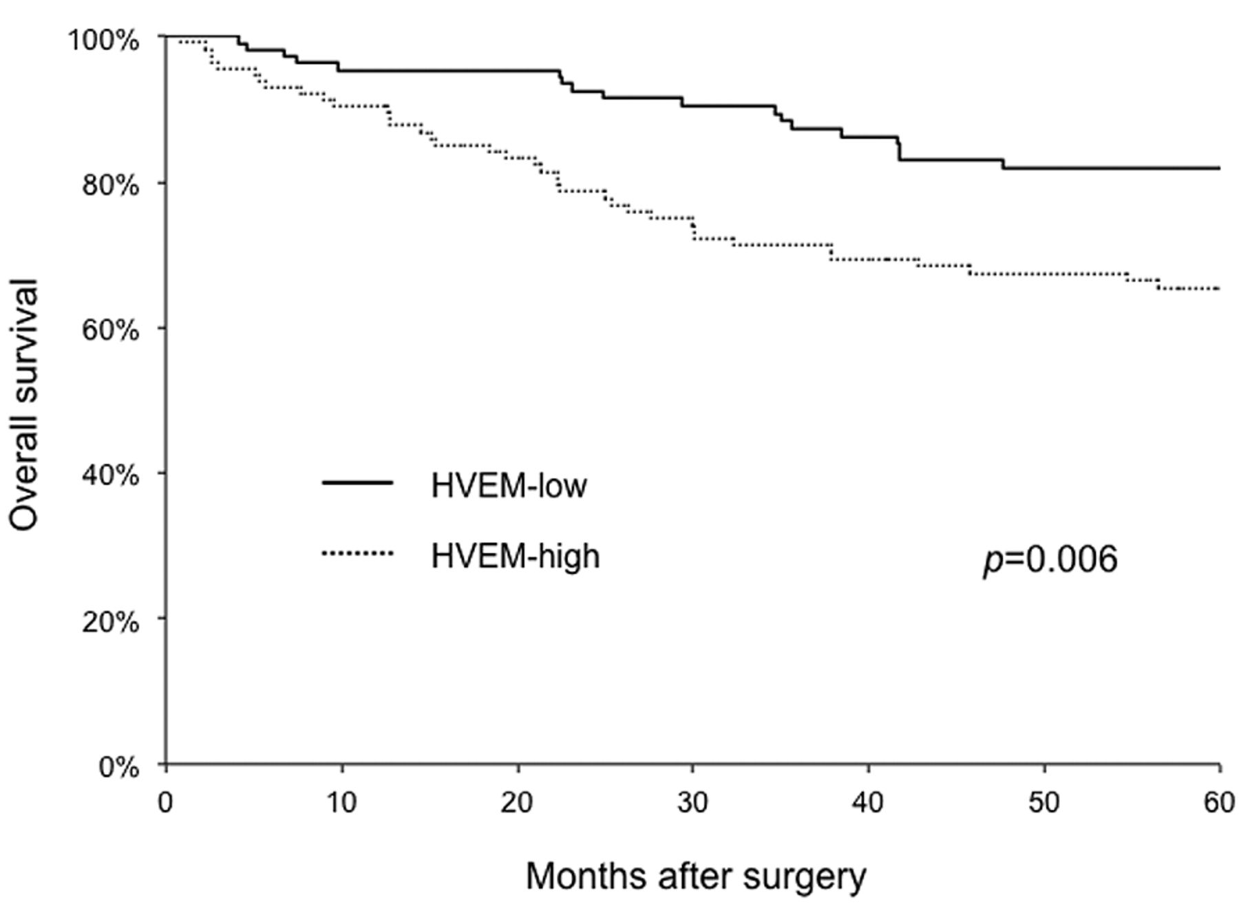
Overall survival of 234 patients with colorectal cancer in relation to their tumor herpesvirus entry mediator (HVEM) status. Patients with high HVEM expression had a significantly poorer prognosis than those with low expression.

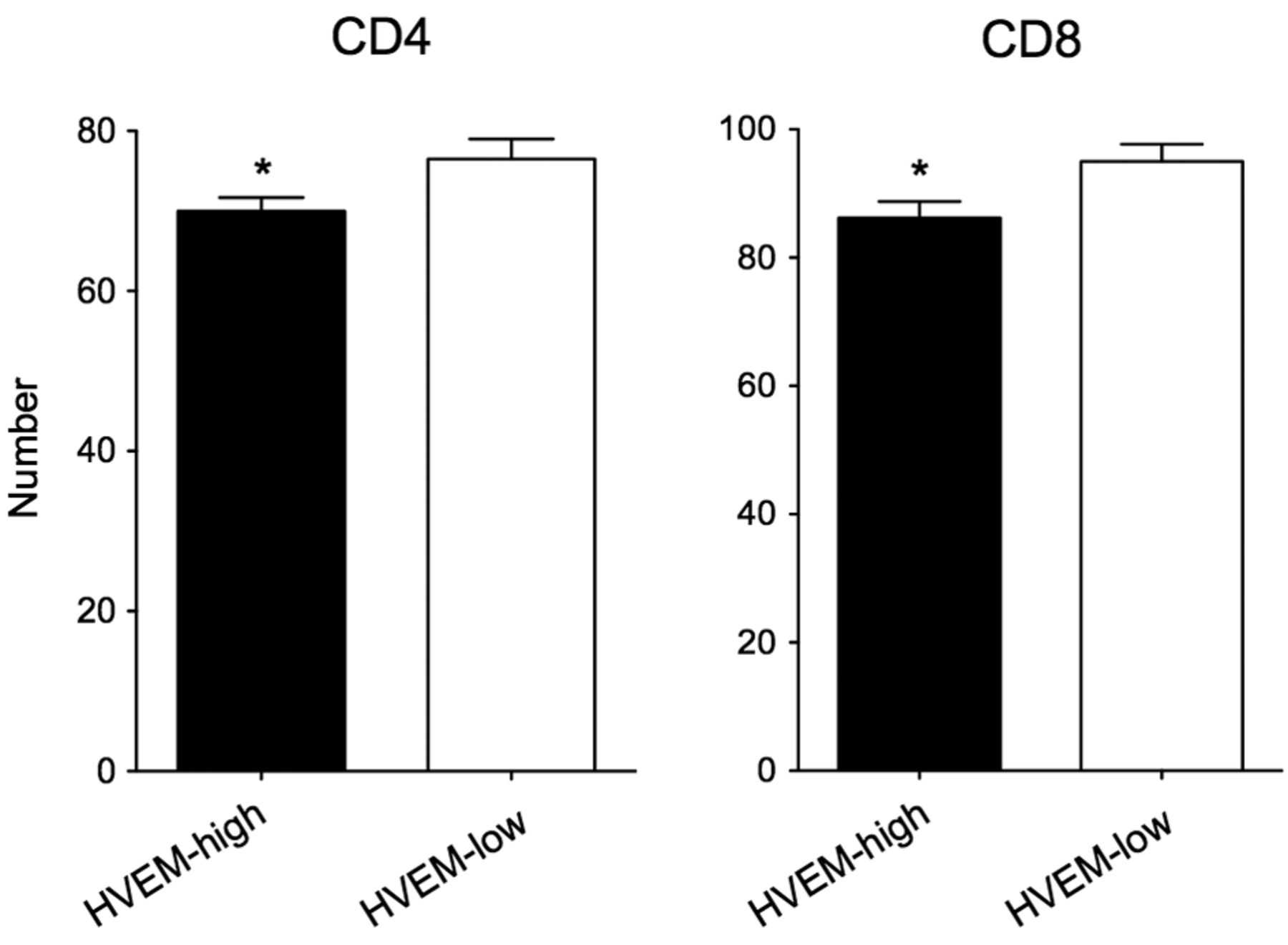
Inverse correlation between tumor HVEM expression and the presence of TILs. Finally, we investigated the correlation between tumor HVEM expression and the presence of TILs by immunohistochemical analysis. As a result, we found a significant inverse correlation between tumor HVEM expression and TILs, including both CD4+ and CD8+ T-cells (Figure 4). These data suggested that tumor HVEM expression might be involved in tumor immune evasion in human CRC.

Discussion

Tumors evade host immune response through several mechanisms. One of the critical mechanisms is the inhibition of antitumor T-cell activation by expression of immunosuppressive proteins on the tumor surface (23, 24). The B7/cytotoxic T-lymphocyte antigen-4 (CTLA4) pathway and the programmed cell death 1 ligand 1 (PD-L1)/programmed cell death protein 1 (PD1) pathway are known to be major negative regulatory T-cell pathways (25, 26). Recent studies have shown that other molecules and pathways might also play critical roles in tumor immunity (5, 27). Among them, HVEM has been suggested to play a potentially important role in several human malignancies (9, 17-19). However, there is searce knowledge about the clinical importance of HVEM in actual human cancer. Furthermore, to our knowledge, the clinical involvement of HVEM in human CRC tissues has not been addressed. Based on a series of our previous studies and recent reports, we hypothesized that HVEM may be functionally important in human CRC (9, 17-19, 27-29). To clarify this possibility, we first investigated the potential role of HVEM expression in various colorectal lesions. Immunohistochemical analysis demonstrated that intense HVEM expression was not observed in normal colonic epithelium and hyperplastic polyps. In contrast, 24% of adenomas and more than half of all CRCs had high HVEM expression. These data suggested that HVEM expression might be involved in the development and progression of CRC. It is well-known that CRC develops via an adenoma to carcinoma sequence with the accumulation of a number of genetic and epigenetic mutations (30). Although at present it is unknown whether tumor HVEM expression is the cause or consequence of tumor progression in CRC, HVEM may play a role during the adenoma–carcinoma sequence.

View this table:
  • View inline
  • View popup
  • Download powerpoint
Table III.

Univariate and multivariate prognostic analysis of 234 patients with colorectal cancer.

Next, we evaluated the clinicopathological significance of tumor HVEM expression in CRC. We found that tumors with high HVEM expression had more advanced tumor status. In contrast, there were no significant correlations of tumor HVEM expression with nodal and metastatic status. These data suggested that HVEM might be involved in tumor progression rather than metastasis in CRC. Furthermore, the univariate prognostic analysis indicated that not only age, histopathological grading, tumor status, nodal status, and metastatic status, but also HVEM status had a significant prognostic value for patients with CRC. More importantly, multivariate analysis revealed that tumor HVEM expression was defined as an independent prognostic factor. Taken together, these data show that HVEM status may play a critical role in CRC independently of conventional TNM factors. We recently reported similar data on prognostic value in esophageal cancer (18). Therefore, the importance of HVEM may be a universal phenomenon in a variety of human malignant diseases. Further studies in other gastrointestinal and nongastrointestinal tumors are required to confirm this interpretation.

Figure 4.
  • Download figure
  • Open in new tab
  • Download powerpoint
Figure 4.

The number of tumor-infiltrating CD4+ and CD8+ lymphocytes were significantly lower in tumors with high herpesvirus entry mediator (HVEM) expression compared with those with low expression. *p<0.05.

Finally, we investigated the underlying mechanism of involvement of tumor HVEM expression in CRC. We found that there was an inverse correlation between HVEM expression and the presence of TILs in CRC. The data suggested that HVEM might play a critical role in tumor immune evasion in human CRC. This is also consistent with our previous findings observed in esophageal cancer (18). Although TILs are thought to play a role in the inhibition of tumor progression and have prognostic significance in several human tumor types, including CRC, the precise underlying mechanisms are largely unknown (18, 21, 31, 32). Our data may highlight a potentially important role of HVEM in tumor immunity. Since other mechanisms, including cell proliferation and the cell cycle, may be involved in the role of HVEM in CRC, extensive basic studies are warranted in order to understand fundamental cancer biology and develop new therapeutic strategies.

Blocking negative regulatory pathways in T-cell activation has attracted much attention for cancer therapy. A randomized clinical trial has demonstrated that immunotherapy using antibody to CTLA4 improved overall survival in metastatic melanoma (25). Furthermore, both recent clinical trials and several basic studies have also suggested that targeting PD-L1/PD1 pathway holds promise for cancer treatment (26, 28, 29, 33). We and others have also reported that HVEM blockade enhanced tumor-reactive T-cell activation and inhibited tumor growth (18, 34). In fact, we have previously used a murine colon cancer model to investigate the in vivo efficacy of HVEM blocking and found that HVEM blockade exerted a significant antitumor effect (18). These data suggested that HVEM might be a novel target for cancer therapy against human CRC. However, HVEM blockade-alone may not be sufficient enough to induce significant clinical response especially for refractory tumors such as recurrent and metastatic CRC that acquired resistance to current conventional chemotherapy. Therefore, the combination of HVEM blockade with other anticancer strategies, including recent chemotherapy, anti-angiogeneic and molecular-targeted therapies, that are currently used as the standard treatment for patients with CRC, may be desirable (3, 35).

In conclusion, we have demonstrated as far as we are aware of for the first time that HVEM may play a critical role in tumor progression and immune evasion in CRC. Our data also revealed that HVEM may be a novel prognostic marker and potential therapeutic target for human CRC.

Acknowledgements

This work was supported by the following grants: Grants-in-Aid for Scientific Research from the Ministry of Education, Culture, Sports, Science and Technology of Japan; Research Grant from Daiwa Securities Health Foundation; Research Grant from The Japanese Society of Gastroenterology; a research grant from Takeda Science Foundation; and Research Grant from Nakayama Cancer Research Institute (M. Sho).

  • Received November 20, 2014.
  • Revision received November 28, 2014.
  • Accepted December 4, 2014.
  • Copyright© 2015 International Institute of Anticancer Research (Dr. John G. Delinassios), All rights reserved

References

  1. ↵
    1. Jemal A,
    2. Bray F,
    3. Center MM,
    4. Ferlay J,
    5. Ward E,
    6. Forman D
    : Global cancer statistics. CA Cancer J Clin 61: 69-90, 2011.
    OpenUrlCrossRefPubMed
  2. ↵
    1. Weeks JC,
    2. Catalano PJ,
    3. Cronin A,
    4. Finkelman MD,
    5. Mack JW,
    6. Keating NL,
    7. Schrag D
    : Patients' expectations about effects of chemotherapy for advanced cancer. N Engl J Med 367: 1616-1625, 2012.
    OpenUrlCrossRefPubMed
  3. ↵
    1. Van Cutsem E,
    2. Borras JM,
    3. Castells A,
    4. Ciardiello F,
    5. Ducreux M,
    6. Haq A,
    7. Schmoll HJ,
    8. Tabernero J
    : Improving outcomes in colorectal cancer: Where do we go from here? Eur J Cancer 49: 2476-2485, 2013.
    OpenUrlCrossRefPubMed
  4. ↵
    1. Cunningham D,
    2. Atkin W,
    3. Lenz HJ,
    4. Lynch HT,
    5. Minsky B,
    6. Nordlinger B,
    7. Starling N
    : Colorectal cancer. Lancet 375: 1030-1047, 2010.
    OpenUrlCrossRefPubMed
  5. ↵
    1. Pardoll DM
    : The blockade of immune checkpoints in cancer immunotherapy. Nat Rev Cancer 12: 252-264, 2012.
    OpenUrlCrossRefPubMed
  6. ↵
    1. Pasero C,
    2. Olive D
    : Interfering with coinhibitory molecules: BTLA/HVEM as new targets to enhance anti-tumor immunity. Immunol Lett 151: 71-75, 2013.
    OpenUrlPubMed
  7. ↵
    1. Montgomery RI,
    2. Warner MS,
    3. Lum BJ,
    4. Spear PG
    : Herpes simplex virus-1 entry into cells mediated by a novel member of the TNF/NGF receptor family. Cell 87: 427-436, 1996.
    OpenUrlCrossRefPubMed
    1. Kwon BS,
    2. Tan KB,
    3. Ni J,
    4. Oh KO,
    5. Lee ZH,
    6. Kim KK,
    7. Kim YJ,
    8. Wang S,
    9. Gentz R,
    10. Yu GL,
    11. Harrop J,
    12. Lyn SD,
    13. Silverman C,
    14. Porter TG,
    15. Truneh A,
    16. Young PR
    : A newly identified member of the tumor necrosis factor receptor superfamily with a wide tissue distribution and involvement in lymphocyte activation. J Biol Chem 272: 14272-14276, 1997.
    OpenUrlAbstract/FREE Full Text
  8. ↵
    1. Pasero C,
    2. Speiser DE,
    3. Derre L,
    4. Olive D
    : The HVEM network: new directions in targeting novel costimulatory/co-inhibitory molecules for cancer therapy. Curr Opin Pharmacol 12: 478-485, 2012.
    OpenUrlCrossRefPubMed
    1. Sarrias MR,
    2. Whitbeck JC,
    3. Rooney I,
    4. Ware CF,
    5. Eisenberg RJ,
    6. Cohen GH,
    7. Lambris JD
    : The three HveA receptor ligands, gD, LT-alpha and LIGHT bind to distinct sites on HveA. Mol Immunol 37: 665-673, 2000.
    OpenUrlCrossRefPubMed
  9. ↵
    1. Cai G,
    2. Anumanthan A,
    3. Brown JA,
    4. Greenfield EA,
    5. Zhu B,
    6. Freeman GJ
    : CD160 inhibits activation of human CD4+ T_cells through interaction with herpesvirus entry mediator. Nat Immunol 9: 176-185, 2008.
    OpenUrlCrossRefPubMed
  10. ↵
    1. Wang Y,
    2. Subudhi SK,
    3. Anders RA,
    4. Lo J,
    5. Sun Y,
    6. Blink S,
    7. Wang Y,
    8. Wang J,
    9. Liu X,
    10. Mink K,
    11. Degrandi D,
    12. Pfeffer K,
    13. Fu YX
    : The role of herpesvirus entry mediator as a negative regulator of T-cell-mediated responses. J Clin Invest 115: 711-717, 2005.
    OpenUrlCrossRefPubMed
    1. del Rio ML,
    2. Lucas CL,
    3. Buhler L,
    4. Rayat G,
    5. Rodriguez-Barbosa JI
    : HVEM/LIGHT/BTLA/CD160 co-signaling pathways as targets for immune regulation. J Leukoc Biol 87: 223-235, 2010.
    OpenUrlAbstract/FREE Full Text
    1. Sakoda Y,
    2. Park JJ,
    3. Zhao Y,
    4. Kuramasu A,
    5. Geng D,
    6. Liu Y,
    7. Davila E,
    8. Tamada K
    : Dichotomous regulation of GVHD through bidirectional functions of the BTLA-HVEM pathway. Blood 117: 2506-2514, 2011.
    OpenUrlAbstract/FREE Full Text
    1. Steinberg MW,
    2. Cheung TC,
    3. Ware CF
    : The signaling networks of the herpesvirus entry mediator (TNFRSF14) in immune regulation. Immunol Rev 244: 169-187, 2011.
    OpenUrlCrossRefPubMed
  11. ↵
    1. Steinberg MW,
    2. Turovskaya O,
    3. Shaikh RB,
    4. Kim G,
    5. McCole DF,
    6. Pfeffer K,
    7. Murphy KM,
    8. Ware CF,
    9. Kronenberg M
    : A crucial role for HVEM and BTLA in preventing intestinal inflammation. The Journal of experimental medicine 205: 1463-1476, 2008.
    OpenUrlAbstract/FREE Full Text
  12. ↵
    1. M'Hidi H,
    2. Thibult ML,
    3. Chetaille B,
    4. Rey F,
    5. Bouadallah R,
    6. Nicollas R,
    7. Olive D,
    8. Xerri L
    : High expression of the inhibitory receptor BTLA in T-follicular helper cells and in B-cell small lymphocytic lymphoma/chronic lymphocytic leukemia. Am J Clin Pathol 132: 589-596, 2009.
    OpenUrlAbstract/FREE Full Text
  13. ↵
    1. Migita K,
    2. Sho M,
    3. Shimada K,
    4. Yasuda S,
    5. Yamato I,
    6. Takayama T,
    7. Matsumoto S,
    8. Wakatsuki K,
    9. Hotta K,
    10. Tanaka T,
    11. Ito M,
    12. Konishi N,
    13. Nakajima Y
    : Significant involvement of herpesvirus entry mediator in human esophageal squamous cell carcinoma. Cancer 120: 808-817, 2014.
    OpenUrlPubMed
  14. ↵
    1. Zhai Y,
    2. Guo R,
    3. Hsu TL,
    4. Yu GL,
    5. Ni J,
    6. Kwon BS,
    7. Jiang GW,
    8. Lu J,
    9. Tan J,
    10. Ugustus M,
    11. Carter K,
    12. Rojas L,
    13. Zhu F,
    14. Lincoln C,
    15. Endress G,
    16. Xing L,
    17. Wang S,
    18. Oh KO,
    19. Gentz R,
    20. Ruben S,
    21. Lippman ME,
    22. Hsieh SL,
    23. Yang D
    : LIGHT, a novel ligand for lymphotoxin beta receptor and TR2/HVEM induces apoptosis and suppresses in vivo tumor formation via gene transfer. The Journal of clinical investigation 102: 1142-1151, 1998.
    OpenUrlCrossRefPubMed
  15. ↵
    1. Sobin L,
    2. Gospodarowicz M,
    3. Wittekind C
    : UICC-TNM Classification of Malignant Tumors, Seventh Edition. New York: Wiley-Blackwell, 2010.
  16. ↵
    1. Enomoto K,
    2. Sho M,
    3. Wakatsuki K,
    4. Takayama T,
    5. Matsumoto S,
    6. Nakamura S,
    7. Akahori T,
    8. Tanaka T,
    9. Migita K,
    10. Ito M,
    11. Nakajima Y
    : Prognostic importance of tumour-infiltrating memory T-cells in oesophageal squamous cell carcinoma. Clin Exp Immunol 168: 186-191, 2012.
    OpenUrlPubMed
  17. ↵
    1. Hotta K,
    2. Sho M,
    3. Fujimoto K,
    4. Shimada K,
    5. Yamato I,
    6. Anai S,
    7. Konishi N,
    8. Hirao Y,
    9. Nonomura K,
    10. Nakajima Y
    : Prognostic significance of CD45RO+ memory T-cells in renal cell carcinoma. British journal of cancer 105: 1191-1196, 2011.
    OpenUrlCrossRefPubMed
  18. ↵
    1. Dong H,
    2. Strome SE,
    3. Salomao DR,
    4. Tamura H,
    5. Hirano F,
    6. Flies DB,
    7. Roche PC,
    8. Lu J,
    9. Zhu G,
    10. Tamada K,
    11. Lennon VA,
    12. Celis E,
    13. Chen L
    : Tumor-associated B7-H1 promotes T-cell apoptosis: a potential mechanism of immune evasion. Nat Med 8: 793-800, 2002.
    OpenUrlCrossRefPubMed
  19. ↵
    1. Iwai Y,
    2. Ishida M,
    3. Tanaka Y,
    4. Okazaki T,
    5. Honjo T,
    6. Minato N
    : Involvement of PD-L1 on tumor cells in the escape from host immune system and tumor immunotherapy by PD-L1 blockade. Proc Natl Acad Sci USA 99: 12293-12297, 2002.
    OpenUrlAbstract/FREE Full Text
  20. ↵
    1. Hodi FS,
    2. O'Day SJ,
    3. McDermott DF,
    4. Weber RW,
    5. Sosman JA,
    6. Haanen JB,
    7. Gonzalez R,
    8. Robert C,
    9. Schadendorf D,
    10. Hassel JC,
    11. Akerley W,
    12. van den Eertwegh AJ,
    13. Lutzky J,
    14. Lorigan P,
    15. Vaubel JM,
    16. Linette GP,
    17. Hogg D,
    18. Ottensmeier CH,
    19. Lebbe C,
    20. Peschel C,
    21. Quirt I,
    22. Clark JI,
    23. Wolchok JD,
    24. Weber JS,
    25. Tian J,
    26. Yellin MJ,
    27. Nichol GM,
    28. Hoos A,
    29. Urba WJ
    : Improved survival with ipilimumab in patients with metastatic melanoma. N Engl J Med 363: 711-723, 2010.
    OpenUrlCrossRefPubMed
  21. ↵
    1. Topalian SL,
    2. Hodi FS,
    3. Brahmer JR,
    4. Gettinger SN,
    5. Smith DC,
    6. McDermott DF,
    7. Powderly JD,
    8. Carvajal RD,
    9. Sosman JA,
    10. Atkins MB,
    11. Leming PD,
    12. Spigel DR,
    13. Antonia SJ,
    14. Horn L,
    15. Drake CG,
    16. Pardoll DM,
    17. Chen L,
    18. Sharfman WH,
    19. Anders RA,
    20. Taube JM,
    21. McMiller TL,
    22. Xu H,
    23. Korman AJ,
    24. Jure-Kunkel M,
    25. Agrawal S,
    26. McDonald D,
    27. Kollia GD,
    28. Gupta A,
    29. Wigginton JM,
    30. Sznol M
    : Safety, activity and immune correlates of anti-PD-1 antibody in cancer. N Engl J Med 366: 2443-2454, 2012.
    OpenUrlCrossRefPubMed
  22. ↵
    1. Yamato I,
    2. Sho M,
    3. Nomi T,
    4. Akahori T,
    5. Shimada K,
    6. Hotta K,
    7. Kanehiro H,
    8. Konishi N,
    9. Yagita H,
    10. Nakajima Y
    : Clinical importance of B7-H3 expression in human pancreatic cancer. Br J Cancer 101: 1709-1716, 2009.
    OpenUrlCrossRefPubMed
  23. ↵
    1. Nomi T,
    2. Sho M,
    3. Akahori T,
    4. Hamada K,
    5. Kubo A,
    6. Kanehiro H,
    7. Nakamura S,
    8. Enomoto K,
    9. Yagita H,
    10. Azuma M,
    11. Nakajima Y
    : Clinical significance and therapeutic potential of the programmed death-1 ligand/programmed death-1 pathway in human pancreatic cancer. Clin Cancer Res 13: 2151-2157, 2007.
    OpenUrlAbstract/FREE Full Text
  24. ↵
    1. Ohigashi Y,
    2. Sho M,
    3. Yamada Y,
    4. Tsurui Y,
    5. Hamada K,
    6. Ikeda N,
    7. Mizuno T,
    8. Yoriki R,
    9. Kashizuka H,
    10. Yane K,
    11. Tsushima F,
    12. Otsuki N,
    13. Yagita H,
    14. Azuma M,
    15. Nakajima Y
    : Clinical significance of programmed death-1 ligand-1 and programmed death-1 ligand-2 expression in human esophageal cancer. Clin Cancer Res 11: 2947-2953, 2005.
    OpenUrlAbstract/FREE Full Text
  25. ↵
    1. Brenner H,
    2. Kloor M,
    3. Pox CP
    : Colorectal cancer. Lancet 383: 1490-1502, 2014.
    OpenUrlCrossRefPubMed
  26. ↵
    1. Ropponen KM,
    2. Eskelinen MJ,
    3. Lipponen PK,
    4. Alhava E,
    5. Kosma VM
    : Prognostic value of tumour-infiltrating lymphocytes (TILs) in colorectal cancer. J Pathol 182: 318-324, 1997.
    OpenUrlCrossRefPubMed
  27. ↵
    1. Zhang L,
    2. Conejo-Garcia JR,
    3. Katsaros D,
    4. Gimotty PA,
    5. Massobrio M,
    6. Regnani G,
    7. Makrigiannakis A,
    8. Gray H,
    9. Schlienger K,
    10. Liebman MN,
    11. Rubin SC,
    12. Coukos G
    : Intratumoral T-cells, recurrence and survival in epithelial ovarian cancer. N Engl J Med 348: 203-213, 2003.
    OpenUrlCrossRefPubMed
  28. ↵
    1. Brahmer JR,
    2. Tykodi SS,
    3. Chow LQ,
    4. Hwu WJ,
    5. Topalian SL,
    6. Hwu P,
    7. Drake CG,
    8. Camacho LH,
    9. Kauh J,
    10. Odunsi K,
    11. Pitot HC,
    12. Hamid O,
    13. Bhatia S,
    14. Martins R,
    15. Eaton K,
    16. Chen S,
    17. Salay TM,
    18. Alaparthy S,
    19. Grosso JF,
    20. Korman AJ,
    21. Parker SM,
    22. Agrawal S,
    23. Goldberg SM,
    24. Pardoll DM,
    25. Gupta A,
    26. Wigginton JM
    : Safety and activity of anti-PD-L1 antibody in patients with advanced cancer. N Engl J Med 366: 2455-2465, 2012.
    OpenUrlCrossRefPubMed
  29. ↵
    1. Lasaro MO,
    2. Sazanovich M,
    3. Giles-Davis W,
    4. Mrass P,
    5. Bunte RM,
    6. Sewell DA,
    7. Hussain SF,
    8. Fu YX,
    9. Weninger W,
    10. Paterson Y,
    11. Ertl HC
    : Active immunotherapy combined with blockade of a co-inhibitory pathway achieves regression of large tumor masses in cancer-prone mice. Mol Ther 19: 1727-1736, 2011.
    OpenUrlCrossRefPubMed
  30. ↵
    1. Yasuda S,
    2. Sho M,
    3. Yamato I,
    4. Yoshiji H,
    5. Wakatsuki K,
    6. Nishiwada S,
    7. Yagita H,
    8. Nakajima Y
    : Simultaneous blockade of programmed death 1 and vascular endothelial growth factor receptor 2 (VEGFR2) induces synergistic anti-tumour effect in vivo. Clin Exp Immunol 172: 500-506, 2013.
    OpenUrlPubMed
PreviousNext
Back to top

In this issue

Anticancer Research: 35 (3)
Anticancer Research
Vol. 35, Issue 3
March 2015
  • Table of Contents
  • Table of Contents (PDF)
  • Index by author
  • Back Matter (PDF)
  • Ed Board (PDF)
  • Front Matter (PDF)
Print
Download PDF
Article Alerts
Sign In to Email Alerts with your Email Address
Email Article

Thank you for your interest in spreading the word on Anticancer Research.

NOTE: We only request your email address so that the person you are recommending the page to knows that you wanted them to see it, and that it is not junk mail. We do not capture any email address.

Enter multiple addresses on separate lines or separate them with commas.
HVEM Expression Contributes to Tumor Progression and Prognosis in Human Colorectal Cancer
(Your Name) has sent you a message from Anticancer Research
(Your Name) thought you would like to see the Anticancer Research web site.
CAPTCHA
This question is for testing whether or not you are a human visitor and to prevent automated spam submissions.
13 + 4 =
Solve this simple math problem and enter the result. E.g. for 1+3, enter 4.
Citation Tools
HVEM Expression Contributes to Tumor Progression and Prognosis in Human Colorectal Cancer
TAKASHI INOUE, MASAYUKI SHO, SATOSHI YASUDA, SATOSHI NISHIWADA, SHINJI NAKAMURA, TAKESHI UEDA, NAOTO NISHIGORI, KEIJIRO KAWASAKI, SHINSAKU OBARA, TAKAYUKI NAKAMOTO, FUMIKAZU KOYAMA, HISAO FUJII, YOSHIYUKI NAKAJIMA
Anticancer Research Mar 2015, 35 (3) 1361-1367;

Citation Manager Formats

  • BibTeX
  • Bookends
  • EasyBib
  • EndNote (tagged)
  • EndNote 8 (xml)
  • Medlars
  • Mendeley
  • Papers
  • RefWorks Tagged
  • Ref Manager
  • RIS
  • Zotero
Reprints and Permissions
Share
HVEM Expression Contributes to Tumor Progression and Prognosis in Human Colorectal Cancer
TAKASHI INOUE, MASAYUKI SHO, SATOSHI YASUDA, SATOSHI NISHIWADA, SHINJI NAKAMURA, TAKESHI UEDA, NAOTO NISHIGORI, KEIJIRO KAWASAKI, SHINSAKU OBARA, TAKAYUKI NAKAMOTO, FUMIKAZU KOYAMA, HISAO FUJII, YOSHIYUKI NAKAJIMA
Anticancer Research Mar 2015, 35 (3) 1361-1367;
Reddit logo Twitter logo Facebook logo Mendeley logo
  • Tweet Widget
  • Facebook Like
  • Google Plus One

Jump to section

  • Article
    • Abstract
    • Materials and Methods
    • Results
    • Discussion
    • Acknowledgements
    • References
  • Figures & Data
  • Info & Metrics
  • PDF

Related Articles

  • No related articles found.
  • PubMed
  • Google Scholar

Cited By...

  • TNFRSF14 (HVEM) is a novel immune checkpoint for cancer immunotherapy in humanized mice
  • The Role of Hvem and its Interaction with Btla and Cd160 in b-Cell Lymphoma Progression
  • Prostate Cancer Cells Express More Androgen Receptor (AR) Following Androgen Deprivation, Improving Recognition by AR-Specific T Cells
  • Google Scholar

More in this TOC Section

  • TIG1 Inhibits the mTOR Signaling Pathway in Malignant Melanoma Through the VAC14 Protein
  • Novel α-Trifluoromethyl Chalcone Exerts Antitumor Effects Against Prostate Cancer Cells
  • Differential Effects of Anti-PD-1/PD-L1 Checkpoint Inhibitors on Adhesion Molecules and Cytokine Secretion by THP-1 Monocytes
Show more Experimental Studies

Similar Articles

Keywords

  • Colorectal cancer
  • HVEM
  • prognosis
  • surgery
  • tumor immunity
Anticancer Research

© 2023 Anticancer Research

Powered by HighWire