


Abstract
Background/Aim: 3-Phosphoinositide-dependent protein kinase-1 (PDK1) mediates the cellular effects of various growth factors. Increased PDK1 expression is present in various cancers, suggesting that PDK1 may be a critical oncogene in cancer progression. However, only limited data exist on PDK1 expression in ovarian serous cancer. Materials and Methods: We used tissue microarrays to analyze PDK1 expression in 253 primary ovarian serous carcinoma samples. Results: A statistically significant negative correlation between PDK1 expression and tumor grade was found. In the high-grade group of ovarian serous carcinomas (n=189), there was a statistically significant difference in overall survival between cases with positive and negative PDK1 expression (p=0.035); positive cases showed longer overall survival. Multivariate analysis confirmed that slight PDK1 expression was an independent indicator for prolonged overall survival (HR=0.51, 95% CI=0.28-0.92, p=0.025). Conclusion: PDK1 appears to be a prognostic marker and a possible therapeutic target in ovarian serous carcinoma.
- 3-phosphoinositide-dependent protein kinase 1
- serous carcinoma of the ovary
- immunohistochemistry
- intracellular signaling
Epithelial ovarian cancer is one of the most common causes of cancer-related death among women in western countries (1). Ovarian serous carcinoma is the most prevalent type of epithelial ovarian cancer and comprises of two different tumor entities: low-grade serous carcinomas and high-grade serous carcinomas. These entities have distinct pathogeneses, morphologies, genetic alterations, and clinical behaviours (2-7). Low-grade serous carcinomas arise from serous borderline tumors and show V-Ki-ras2 Kirsten rat sarcoma viral oncogene homolog (KRAS) and proto-oncogene B-Raf (BRAF) mutations, but few chromosomal abnormalities. In contrast, increasing evidence suggests that at least some high-grade tumors develop in the fallopian tube (3, 6-8). They show mutations of TP53 in virtually all tumors and inactivation of BRCA1 and BRCA 2 in about 50% of them (9). Another characteristic feature of these tumors is the high level of chromosomal alterations.
3-Phosphoinositide-dependent protein kinase-1 (PDK1) mediates the cellular effects of various growth factors. PDK1 activates multiple proteins including protein kinase B (Akt), some protein kinase C isoforms, Rheb-Target-of-rapamycin (TOR)-S6-kinase (S6K), and serum/glucocorticoid regulated kinase 1 (SGK). Increased PDK1 expression is seen in various malignant diseases, such as acute myeloid leukaemia, head and neck cancers, pancreatic cancer, and colorectal cancer (10-12). These findings suggest that PDK1 may be a critical oncogene in cancer progression.
Reduced expression of PDK1 in heterozygous phosphatase and tensin homolog (PTEN)+/− mice markedly protects these animals from developing a wide range of tumors (13). These results suggest that PDK1 may be a key mediator of neoplasia and validate PDK1 as a promising anticancer target for the prevention of tumor formation. Targeting PDK1 with oligonucleotides blocks the proliferation of U-87 glioblastoma cells by promoting apoptosis (14). However, only limited data are available about PDK1 expression in ovarian serous cancer. Ahmed et al. (15) showed the absence of PDK1 expression in normal ovaries, weak expression in benign tumors, and elevated expression in low- to high-grade ovarian carcinomas. This finding provides persuasive evidence for its oncogenic role in ovarian cancer progression.
Therefore, using tissue microarrays, we analyzed PDK1 expression in 253 primary ovarian serous carcinoma samples, aiming to examine the expression of PDK1 in ovarian carcinoma and to determine any correlation with clinicopathological features.
Materials and Methods
Study population. Ovarian serous carcinomas from 253 patients were retrieved from the archive of the Institute of Pathology, Charité Universitätsmedizin Berlin, Germany. Ninety-one patients had been included into the Tumorbank Ovarian Cancer (TOC) Network. The baseline patients characteristics are outlined in Table I. Histology has been reviewed by an experienced gynecologic pathologist (SDE). Classification as low- and high-grade carcinomas was performed according to the current WHO criteria (16).
Immunohistochemistry. Immunohistochemical staining was performed on tissue microarrays. For this, representative tumor areas of ovarian carcinoma samples were marked on hematoxylin and eosin–stained sections. Subsequently, two tissue cylinders 1 or 1.5 mm in diameter were punched from each tumor-bearing donor block and transferred to a paraffin block. Immunohistochemical staining was performed as previously described (17) using the pPDK1 (pPDK1, S241, 1:50) antibody from Cell Signalling Technology Inc. (Beverly, MA, USA). The overall intensity (negative, weak, moderate, and strong staining) and the percentage of stained tumor cells were scored visually for each tumor. Tumors in which more than 50% of the cells were positive, irrespective of staining intensity were scored as positive.
In a second step, we applied a semi-quantitative scoring system, which links the staining intensity to the percentage of positive cells in the following manner: the intensity was classified as: 0=negative staining, 1=weak staining, 2=moderate staining, and 3=strong staining. The percent of positive cells was classified as: 1: 0-25%; 2: 26-50%; 3: 51-75%; 4: >75%, the final score was achieved by multiplying the intensity and percent of positive cells. The cut-off was set to 1.
Statistical evaluation. Statistical analyses were conducted using SPSS 19 statistical software (SPSS Inc., Chicago, IL, USA). The association between PDK1 expression and age, stage of disease, and histological grades were determined by Pearson-correlation test. Cox regression method was used for multivariate analysis. Association analysis between PDK1 expression and survival was performed using the Kaplan-Meier method and the log-rank test. All tests were two-tailed, and the results were considered significant when p<0.05.
Results
PDK1 expression in low- and high-grade carcinomas of the ovary. In total, we examined 253 primary ovarian serous carcinomas for the expression of PDK1. Out of these, 40 (15.8%) showed a strong expression, 60 (23.7%) moderate expression, and 97 (38.3%) low expression of PDK1 in at least 50% of tumor cells. Forty-one tumors (16.2%) were negative for PDK1. We found a negative correlation between the percentage of tumor cells and tumor grade on one hand and staining intensity for PDK1, and tumor grade on the other hand (R=−0,116; p=0.75 and R=−0,114; p=0.78, respectively), although the difference between HGSC and LGSC was not statistically significant. Representative examples of staining intensities are depicted in Figure 1. We also observed that the TNM stage was negatively correlated with PDK1 positivity (R=−0,013; p=0.851).
Characteristics of the study group.
Clinical outcome. With univariate analysis, we found that the expression of PDK1 protein had a statistically significant impact on overall survival (OS) in the high-grade tumor group (Figure 2, Table II) but not for the whole group of LGSC and HGSC. Additionally, the FIGO stage and the residual tumor were significantly associated with OS in univariate analysis (Table II).
Under multivariate analysis, cases with PDK1 positivity showed longer OS. Residual tumor (macroscopic tumor-free cases) also showed a statistically significant impact on OS in multivariate analysis (Table II). PDK1 expression did not have a statistically significant impact on progression- free survival (PFS) either in univariate or multivariate analysis (Table II). There were no significant differences found in the results after applying a semi-quantitative score (data not shown).
Discussion
Serous carcinomas of the ovary, especially high-grade carcinomas, are very aggressive tumors with a dismal prognosis. Classical treatment options are limited and often have short-lasting results, resulting in short survival (1). Therefore, a better understanding over the specific molecular events associated with carcinogenesis and progression is critical for developing new treatments to improve survival rates of these patients.
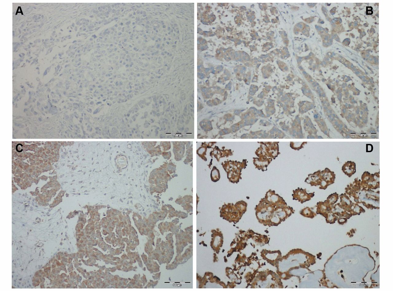
Immunohistochemical expression of PDK1 (3-phosphoinositide-dependent protein kinase-1) in serous ovarian carcinoma. Tumor cells with negative (A) and tumor cells with weak immunostaining (B). (C) Tumor cells with moderate immunostaining. (D) Tumor cells with strong immunostaining.

Kaplan-Meier survival curves of patients with serous ovarian carcinoma with positivity of pPDK1 in >50% of tumor cells. (A) Low-grade serous carcinoma. No significant difference was found in survival that was dependent on PDK1 expression (p=0.644). (B) High-grade serous carcinoma. A significant difference was found in survival that was dependent on PDK1 expression (p=0.035).
Survival analysis.
High- and low-grade primary ovarian serous carcinomas are two different tumor entities with different molecular events associated with carcinogenesis (18). In the current study, we examined PDK1 expression in a large cohort of primary ovarian serous carcinomas using immunohistochemical analysis. Our data revealed that PDK1 positivity was different between high- and low-grade tumors and was more frequently expressed in low-grade than in high-grade serous carcinomas. PDK1, a 63-kDa serine/threonine protein kinase consisting of an N-terminal kinase domain and a C-terminal pleckstrin homology domain (PH), is a constitutively active kinase that phosphorylates different substrates in cells (19, 20). Thus, PDK1 can activate kinases involved in the regulation of various physiological processes relevant to metabolism, growth proliferation, and survival (21). PDK1 was originally identified as a viable target for cancer by Bayascas and colleagues in 2005 (13), who generated transgenic mice hypomorphic for PDK1. Accumulating studies have since implicated PDK1 in various human cancers, such as acute myeloid leukaemia, head and neck cancer, pancreatic cancer, colorectal cancer, and oesophageal squamous cell cancer (10-12). Thus far, only one study has examined the expression of PDK1 in a small number of serous ovarian tumors (15). The specific group showed a gradual increase in the expression of PDK1 with increasing grade of ovarian tumors, with the high-grade tumors demonstrating high expression PDK1. Interestingly, using a much larger cohort of serous low- and high-grade carcinomas, we did not find any statistically significant difference in PDK1 expression between the two disease entities, but we observed a trend towards higher expression in the low-grade group.
PDK1 is required for anchorage-independent and xenograft growth of breast cancer cells harbouring either PI3KCA or KRAS mutations. A KRAS/PI3K/PDK1 axis may also exist in pancreatic cancer (10). Another study reported that PDK1 is an important effector of BRAF signaling (22). KRAS and BRAF mutations are well-known specific genetic alterations in low-grade serous carcinoma. Therefore, the higher percentage of PDK1-positive low-grade serous ovarian carcinomas may be due to the activation of PDK1 through oncogenic KRAS and BRAF. This may explain the higher percentage of PDK1-positive low-grade serous ovarian carcinomas in comparison to high-grade serous ovarian carcinomas.
Array comparative genomic hybridization studies have identified the PI3K/Akt/mammalian target of rapamycin (mTOR) pathway as the most frequently altered pathway in ovarian carcinomas (23). Over 35% of high-grade serous ovarian carcinomas show alterations in this pathway, including phosphatidylinositol-4,5-bisphosphate 3-kinase (PIK3CA) amplification or mutation, PTEN deletion, or amplification of AKT1 and AKT2 (24). In the current study, we found PDK1 expression in ~80% of high-grade and ~90% of low-grade tumors, and the expression ranged from weak to strong. This finding confirms the results of our previous study (13) in breast cancer, and agrees with the findings from other studies, showing PDK1-dependent and - independent Akt activation (23) respectively, and PDK1 activation are independent of the PIK3CA pathway.
Studies in serous ovarian carcinomas showed that the expression levels of PIK3CA and phosphorylated Akt (pAkt) are associated with decreased survival (26). However, in the current study, we found a significant correlation between pPDK1 expression and prolonged OS in high-grade, but not low-grade, serous ovarian carcinomas in our cohort. This finding suggests that the high-grade tumor group may be more heterogeneous than previously assumed. PDK1 signaling may be different in certain oncogenic contexts and may confer a growth advantage to tumor cells. Indeed, certain studies have shown that PDK1 is not linked to PIK3 signaling and instead goes through an Akt-independent pathway for cell survival in certain cancer cell lines (27). This finding could also explain the different survival outcomes in our patient group. However, this is not clear in the present study, and should be the subject of further investigations. Finally, previous studies have shown that loss of PDK1 expression sensitizes cells to chemotherapeutics and alters proliferation (14, 28). Thus, considering the broad PDK1 expression in serous ovarian cancer, PDK1 could also be a potential therapeutic target for the specific cancer type.
Acknowledgements
We would like to thank Kerstin Petri and Kerstin Witkowski for their excellent technical assistance.
- Received July 24, 2015.
- Revision received September 1, 2015.
- Accepted September 3, 2015.
- Copyright© 2015 International Institute of Anticancer Research (Dr. John G. Delinassios), All rights reserved


