
Main menu

  • Home
  • Current Issue
  • Archive
  • Info for
    • Authors
    • Subscribers
    • Advertisers
    • Editorial Board
  • Other Publications
    • In Vivo
    • Cancer Genomics & Proteomics
    • Cancer Diagnosis & Prognosis
  • More
    • IIAR
    • Conferences
    • 2008 Nobel Laureates
  • About Us
    • General Policy
    • Contact
  • Other Publications
    • Anticancer Research
    • In Vivo
    • Cancer Genomics & Proteomics

User menu

  • Register
  • Subscribe
  • My alerts
  • Log in
  • My Cart

Search

  • Advanced search
Anticancer Research
  • Other Publications
    • Anticancer Research
    • In Vivo
    • Cancer Genomics & Proteomics
  • Register
  • Subscribe
  • My alerts
  • Log in
  • My Cart
Anticancer Research

Advanced Search

  • Home
  • Current Issue
  • Archive
  • Info for
    • Authors
    • Subscribers
    • Advertisers
    • Editorial Board
  • Other Publications
    • In Vivo
    • Cancer Genomics & Proteomics
    • Cancer Diagnosis & Prognosis
  • More
    • IIAR
    • Conferences
    • 2008 Nobel Laureates
  • About Us
    • General Policy
    • Contact
  • Visit us on Facebook
  • Follow us on Linkedin
Research ArticleExperimental Studies

Recruitment of Cancer-Associated Fibroblasts and Blood Vessels by Orthotopic Liver Tumors Imaged in Red Fluorescent Protein (RFP) Transgenic Nude Mice

ATSUSHI SUETSUGU, YUKIHIKO HIROSHIMA, TAKURO MATSUMOTO, KOSUKE HASAGAWA, MIKI NAKAMURA, MASAHITO SHIMIZU, SHIGETOYO SAJI, HISATAKA MORIWAKI, MICHAEL BOUVET and ROBERT M. HOFFMAN
Anticancer Research November 2015, 35 (11) 5821-5825;
ATSUSHI SUETSUGU
1Gifu University Graduate School of Medicine, Gifu, Japan
2AntiCancer, Inc. San Diego, CA, U.S.A
3Department of Surgery, University of California, San Diego, CA, U.S.A.
  • Find this author on Google Scholar
  • Find this author on PubMed
  • Search for this author on this site
YUKIHIKO HIROSHIMA
2AntiCancer, Inc. San Diego, CA, U.S.A
3Department of Surgery, University of California, San Diego, CA, U.S.A.
  • Find this author on Google Scholar
  • Find this author on PubMed
  • Search for this author on this site
TAKURO MATSUMOTO
1Gifu University Graduate School of Medicine, Gifu, Japan
  • Find this author on Google Scholar
  • Find this author on PubMed
  • Search for this author on this site
KOSUKE HASAGAWA
1Gifu University Graduate School of Medicine, Gifu, Japan
  • Find this author on Google Scholar
  • Find this author on PubMed
  • Search for this author on this site
MIKI NAKAMURA
1Gifu University Graduate School of Medicine, Gifu, Japan
  • Find this author on Google Scholar
  • Find this author on PubMed
  • Search for this author on this site
MASAHITO SHIMIZU
1Gifu University Graduate School of Medicine, Gifu, Japan
  • Find this author on Google Scholar
  • Find this author on PubMed
  • Search for this author on this site
SHIGETOYO SAJI
1Gifu University Graduate School of Medicine, Gifu, Japan
  • Find this author on Google Scholar
  • Find this author on PubMed
  • Search for this author on this site
HISATAKA MORIWAKI
1Gifu University Graduate School of Medicine, Gifu, Japan
  • Find this author on Google Scholar
  • Find this author on PubMed
  • Search for this author on this site
MICHAEL BOUVET
3Department of Surgery, University of California, San Diego, CA, U.S.A.
  • Find this author on Google Scholar
  • Find this author on PubMed
  • Search for this author on this site
ROBERT M. HOFFMAN
2AntiCancer, Inc. San Diego, CA, U.S.A
3Department of Surgery, University of California, San Diego, CA, U.S.A.
  • Find this author on Google Scholar
  • Find this author on PubMed
  • Search for this author on this site
  • For correspondence: all@anticancer.com
  • Article
  • Figures & Data
  • Info & Metrics
  • PDF
Loading

Abstract

Background/Aim: The tumor microenvironment (TME) is critical for tumor growth and progression. We report here an imageable model of the TME of orthotopic liver cancer. Materials and Methods: The transgenic red fluorescent protein (RFP)-expressing nude mouse was used as the host. The RFP nude mouse expresses RFP in all organs. Non-colored Huh-7 human hepatoma cells were injected in the spleen of RFP nude mice to establish an orthotopic liver cancer model. TME formation resulting from the orthotopic liver tumor was observed using the Olympus OV100 small animal fluorescence imaging system. Results: Non-colored liver cancer cells formed tumor colonies in the liver 28 days after cell transplantation to the spleen. RFP-expressing host cells and blood vessels were recruited by the liver tumors as visualized by fluorescence imaging. A desmin- and sirus-red-positive area increased around and within the liver tumor over time. Conclusion: These results indicate cancer-associated fibroblasts (CAFs) were recruited by the liver tumors suggesting that CAFs, along with the angiogenic tumor blood vessels, were necessary for liver-tumor growth and could serve as visible therapeutic targets.

  • Tumor microenvironment
  • cancer-associated fibroblasts
  • blood vessels
  • liver metastasis
  • RFP
  • fluorescence
  • color-coded imaging

Our laboratory previously developed a color-coded model of the tumor microenvironment (TME) using red fluorescent protein (RFP), cyan fluorescent protein (CFP), and green fluorescent protein (GFP) transgenic nude mice as hosts for cancer cells expressing a different color fluorescent protein (1-11).

Our color-coded TME imaging technology was used by Egeblad et al. (12) to show that stromal cells had higher motility in the periphery of the tumor. Our color-coded model was also used by Egeblad to demonstrate macrophages, fibroblasts, dendritic cells, and lymphocytes in the TME (12).

We have also demonstrated, using color-coded imaging, that tumors acquired fluorescent protein-expressing stroma over time, during growth in fluorescent protein-expressing nude mice, including during passage. The acquired stroma grew along with the tumor. The bright stroma enabled non-invasive imaging of a pancreatic-cancer patient-derived orthotopic xenograft (PDOX) mouse model (7-9). We previously showed that CAFs are recruited by metastasis, which stimulate their growth (6, 8, 11). We also discovered that stromal cells are necessary for metastasis (13).

We previously used a GFP-expressing transgenic nude mouse as the host which expresses GFP in all organs, but only dimly in the parenchymal cells of the liver, to study the role of the TME in liver metastasis of colon cancer. Non-colored HCT-116 human colon cancer cells were injected in the spleen of GFP nude mice, which led to the formation of experimental liver metastasis. TME formation resulting from the liver metastasis was observed using the Olympus OV100 small animal fluorescence imaging system. HCT-116 cells formed tumor colonies in the liver by 28 days after cell transplantation to the spleen. GFP-expressing host cells were recruited by the metastatic tumors as visualized by fluorescence imaging. A desmin-positive area increased around and within the liver metastasis over time, suggesting cancer-associated fibroblasts (CAFs) were recruited by the liver metastasis (6).

The present study images the recruitment of blood vessels and cancer-associated fibroblasts by orthotopic liver tumors growing in transgenic RFP nude mice.

Figure 1.
  • Download figure
  • Open in new tab
  • Download powerpoint
Figure 1.

A: Schematic representation of experimental protocol. Non-colored Huh-7 human hepatoma cells (2.0×10⁶/50 μl) were injected in the spleen of RFP nude mice to establish an orthotopic liver tumor model. B: Imaging of an orthotopic liver tumor on day 28 after cancer-cell injection. RFP fluorescence was observed in the orthotopic liver tumor. The liver tumor was observed using the Olympus OV100 small animal fluorescence imaging system. Upper panels are low magnification (left panel: bright-field; central panel: fluorescence; right panel: merge). Yellow arrows indicate non-colored tumor (Bar=10 mm). Lower panels are high magnification (left panel: bright-field; central panel: fluorescence; right panel: merge). Green arrows indicate non-color tumor (Bar=1 mm).

Materials and Methods

Cell culture. Huh-7 human liver cancer cells were maintained in RPMI 1640 medium supplemented with 10% FCS (14) as well as with penicillin and streptomycin (Gibco BRL, Grand Island, NY, USA). The Huh-7 cell line was cultured at 37°C in a 5% incubator.

Transgenic red fluorescent protein-expressing nude mice. Transgenic red fluorescent protein (RFP) nude mice (AntiCancer, Inc., San Diego, CA, USA), expressing RFP (DsRed.T3) under the control of a chicken β-actin promoter and cytomegalovirus enhancer, were used in this study. All tissues from this transgenic line, with the exception of erythrocytes, were red under blue excitation light.

Orthotopic liver-cancer model. Non-colored Huh-7 cells were first harvested by trypsinization and washed three times with cold serum-free medium and then resuspended with serum-free RPMI 1640 medium. RFP nude mice were anesthetized with a ketamine mixture (0.02 ml solution of 20 mg/kg ketamine, 15.2 mg/kg xylazine, and 0.48 mg/kg acepromazine maleate) injected into the peritoneal cavity. Non-colored Huh-7 cancer cells (2.0×10⁶) were injected in the spleen of RFP nude mice during open laparotomy in order for tumors in the liver to form (Figure 1).

Figure 2.
  • Download figure
  • Open in new tab
  • Download powerpoint
Figure 2.

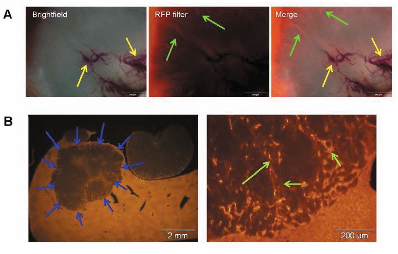
Fluorescence imaging of liver sections. A: High magnification image of the orthotopic Huh-7 liver tumors. The orthotopic liver tumor was observed using the Olympus OV100 small animal fluorescence imaging system. Yellow arrows indicate tumor vessels. Green arrows indicate RFP stroma. (Bar=500 μm). B: Image of Huh-7 liver tumor section. Blue arrows indicate Huh-7 tumor. Green arrows indicate RFP stroma recruited by the liver tumor. (left panel: Bar=2 mm); (right panel: Bar=200 μm).

Figure 3.
  • Download figure
  • Open in new tab
  • Download powerpoint
Figure 3.

Hematoxylin and eosin, sirus-red and desmin staining. Liver sections from RFP nude mice with orthotopic Huh-7 liver tumors were stained with hematoxylin and eosin (H&E), sirus-red and desmin. There were significantly more sirus-red and desmin positive areas in the tumor than in the non-tumor part of the liver. The liver tumor contained RFP-, and desmin-expressing cells, sirus-red-stained cells as well as suggesting that cancer-associated fibroblast (CAFs) have a role in liver tumor development. Left panel: H&E stain; central panel: sirus-red stain; right panel: desmin stain. Non-colored Huh-7 liver tumor sections (original magnification, ×200). Red arrows indicate sirus red-positive area. Blue arrows indicate desmin-positive area. (Bar=100 μm).

In vivo imaging. For in vivo imaging based on fluorescent proteins (16-19), the Olympus OV100 Small Animal Imaging System (Olympus Corp., Tokyo, Japan) was used. The Olympus OV100, which contains an MT-20 light source (Olympus Biosystems, Planegg, Germany) and DP70 CCD camera (Olympus), were used for cellular imaging in live mice. The optics of the OV100 fluorescence imaging system have been specially developed for macro-imaging as well as micro-imaging with high light-gathering capacity. High-resolution images were captured directly on a PC (Fujitsu Siemens, Munich, Germany). Images were processed for contrast and brightness and analyzed with the use of Paint Shop Pro 8 and CellR (Olympus Biosystems) (20).

Histological and immunohistochemical analysis. The host livers were fixed with 10% buffered formalin, sectioned at a thickness of 4 μm, and stained with hematoxylin and eosin. Desmin was stained with anti-desmin antibody (Lab Vision, Fremont, CA, USA) using an avidin–biotin–peroxidase complex technique (Vector, Burlingame, CA). Collagen deposition was assessed by Sirius-red staining.

Results and Discussion

Imaging recruitment of blood vessels and cancer-associated fibroblasts by a non-colored orthotopic liver tumor. Huh-7 human liver cancer cells were injected in the spleen of RFP nude mice. Low-magnification imaging showed the contrast between the non-colored liver tumor and RFP-expressing liver (Figure 1). High-magnification fluorescence imaging showed extensive RFP fluorescence in the tumor due to recruitment of RFP-expressing blood vessels and fibroblast-like cells (Figure 2). Thus, host RFP cells were imaged extensively accumulating in the liver tumor.

Immunohistochemical experiments showed that expression of desmin was prominent in the liver tumor, but not in the host liver, suggesting that cancer-associated fibroblasts (CAFs) were recruited and grew in the tumor (Figure 3). The tumor also was stained with sirus-red, which marks collagen, possibly produced by the CAFs. CAFs have an important role in tumor progression (21). CAFs have a high rate of proliferation and differential expression of extracellular matrix (ECM) components, such as collagen, and growth factors compared to normal fibroblasts (21-23). CAFs promote tumor growth by inducing angiogenesis, recruiting bone marrow-derived endothelial-progenitor cells, and remodeling the ECM (24-28). CAFs can confer resistance to anti-angiogenic therapy (21, 29). CAFs also mediate tumor-enhancing inflammation mediated by NF-kB (21).

However, the formation of the TME is still largely unknown. The present report demonstrates a color-coded imaging model in which the development of the TME can be visualized in an orthotopic liver cancer model. RFP transgenic nude mice are useful to image stromal development in the tumor, since bright RFP-expressing stromal cells are recruited by the liver tumors. A desmin- and sirus-red-positive area increased around the liver tumors over time, suggesting CAFs were recruited by the liver tumors and have an important role in tumor progression. Stromal cells essential for the liver tumors to develop can be imaged as visual therapeutic targets. RFP-expressing blood vessels are also recruited by the liver tumor, enabling visual therapeutic targeting of angiogenesis as well.

Acknowledgements

This study was supported in part by National Cancer Institute grant CA132971.

Footnotes

  • Dedication

    This paper is dedicated to the memory of A.R. Moossa, M.D.

  • Received July 17, 2015.
  • Revision received September 1, 2015.
  • Accepted September 3, 2015.
  • Copyright© 2015 International Institute of Anticancer Research (Dr. John G. Delinassios), All rights reserved

References

  1. ↵
    1. Yang M,
    2. Li L,
    3. Jiang P,
    4. Moossa AR,
    5. Penman S,
    6. Hoffman RM
    : Dual-color fluorescence imaging distinguishes tumor cells from induced host angiogenic vessels and stromal cells. Proc Natl Acad Sci USA 100: 14259-14262, 2003.
    OpenUrlAbstract/FREE Full Text
    1. Yang M,
    2. Reynoso J,
    3. Jiang P,
    4. Li L,
    5. Moossa AR,
    6. Hoffman RM
    : Transgenic nude mouse with ubiquitous green fluorescent protein expression as a host for human tumors. Cancer Res 64: 8651-8656, 2004.
    OpenUrlAbstract/FREE Full Text
    1. Yang M,
    2. Reynoso J,
    3. Bouvet M,
    4. Hoffman RM
    : A transgenic red fluorescent protein-expressing nude mouse for color-coded imaging of the tumor microenvironment. J Cell Biochem 106: 279-284, 2009.
    OpenUrlCrossRefPubMed
    1. Amoh Y,
    2. Li L,
    3. Yang M,
    4. Jiang P,
    5. Moossa AR,
    6. Katsuoka K,
    7. Hoffman RM
    : Hair follicle-derived blood vessels vascularize tumors in skin and are inhibited by doxorubicin. Cancer Res 65: 2337-2343, 2005.
    OpenUrlAbstract/FREE Full Text
    1. Amoh Y,
    2. Yang M,
    3. Li L,
    4. Reynoso J,
    5. Bouvet M,
    6. Moossa AR,
    7. Katsuoka K,
    8. Hoffman RM
    : Nestin-linked green fluorescent protein transgenic nude mouse for imaging human tumor angiogenesis. Cancer Res 65: 5352-5357, 2005.
    OpenUrlAbstract/FREE Full Text
  2. ↵
    1. Suetsugu A,
    2. Osawa Y,
    3. Nagaki M,
    4. Saji S,
    5. Moriwaki H,
    6. Bouvet M,
    7. Hoffman RM
    : Imaging the recruitment of cancer-associated fibroblasts by liver-metastatic colon cancer. J Cell Biochem 112: 949-953, 2011.
    OpenUrlPubMed
  3. ↵
    1. Suetsugu A,
    2. Katz M,
    3. Fleming J,
    4. Moriwaki H,
    5. Bouvet M,
    6. Saji S,
    7. Hoffman RM
    : Multi-color palette of fluorescent proteins for imaging the tumor microenvironment of orthotopic tumorgraft mouse models of clinical pancreatic cancer specimens. J Cell Biochem 113: 2290-2295, 2012.
    OpenUrlCrossRefPubMed
  4. ↵
    1. Suetsugu A,
    2. Katz M,
    3. Fleming J,
    4. Truty M,
    5. Thomas R,
    6. Saji S,
    7. Moriwaki H,
    8. Bouvet M,
    9. Hoffman RM
    : Imageable fluorescent metastasis resulting in transgenic GFP mice orthotopically implanted with human-patient primary pancreatic cancer specimens. Anticancer Res 32: 1175-1180, 2012.
    OpenUrlAbstract/FREE Full Text
  5. ↵
    1. Suetsugu A,
    2. Katz M,
    3. Fleming J,
    4. Truty M,
    5. Thomas R,
    6. Saji S,
    7. Moriwaki H,
    8. Bouvet M,
    9. Hoffman RM
    : Non-invasive fluorescent-protein imaging of orthotopic pancreatic-cancer-patient tumorgraft progression in nude mice. Anticancer Res 32: 3063-3068, 2012.
    OpenUrlAbstract/FREE Full Text
    1. Suetsugu A,
    2. Hassanein MK,
    3. Reynoso J,
    4. Osawa Y,
    5. Nagaki M,
    6. Moriwaki H,
    7. Saji S,
    8. Bouvet M,
    9. Hoffman RM
    : The cyan fluorescent protein nude mouse as a host for multicolor-coded imaging models of primary and metastatic tumor microenvironments. Anticancer Res 32: 31-38, 2012.
    OpenUrlAbstract/FREE Full Text
  6. ↵
    1. Suetsugu A,
    2. Snyder CS,
    3. Moriwaki H,
    4. Saji S,
    5. Bouvet M,
    6. Hoffman RM
    : Imaging the interaction of pancreatic cancer and stellate cells in the tumor microenvironment during metastasis. Anticancer Res 35: 2545-2551, 2015.
    OpenUrlAbstract/FREE Full Text
  7. ↵
    1. Egeblad M,
    2. Ewald AJ,
    3. Askautrud HA,
    4. Truitt ML,
    5. Welm BE,
    6. Bainbridge E,
    7. Peeters G,
    8. Krummel MF,
    9. Werb Z
    : Visualizing stromal cell dynamics in different tumor microenvironments by spinning disk confocal microscopy. Dis Model Mech 1: 155-167, 2008.
    OpenUrlAbstract/FREE Full Text
  8. ↵
    1. Bouvet M,
    2. Tsuji K,
    3. Yang M,
    4. Jiang P,
    5. Moossa AR,
    6. Hoffman RM
    : In vivo color-coded imaging of the interaction of colon cancer cells and splenocytes in the formation of liver metastases. Cancer Res 66: 11293-11297, 2006.
    OpenUrlAbstract/FREE Full Text
  9. ↵
    1. Suetsugu A,
    2. Osawa Y,
    3. Nagaki M,
    4. Moriwaki H,
    5. Saji S,
    6. Bouvet M,
    7. Hoffman RM
    : Simultaneous color-coded imaging to distinguish cancer “stem-like” and non-stem cells in the same tumor. J Cell Biochem 111: 1035-1041, 2010.
    OpenUrlCrossRefPubMed
    1. Hoffman RM
    : The multiple uses of fluorescent proteins to visualize cancer in vivo. Nat Rev Cancer 5: 796-806, 2005.
    OpenUrlCrossRefPubMed
  10. ↵
    1. Hoffman RM,
    2. Yang M
    : Subcellular imaging in the live mouse. Nat Protoc 1: 775-782, 2006.
    OpenUrlCrossRefPubMed
    1. Hoffman RM,
    2. Yang M
    : Color-coded fluorescence imaging of tumor–host interactions. Nat Protoc 1: 928-935, 2006.
    OpenUrlCrossRefPubMed
    1. Hoffman RM,
    2. Yang M
    : Whole-body imaging with fluorescent proteins. Nat Protoc 1: 1429-1438, 2006.
    OpenUrlCrossRefPubMed
  11. ↵
    1. Hoffman RM
    : Application of GFP imaging in cancer. Laboratory Investigation 95: 432-452, 2015.
    OpenUrlPubMed
  12. ↵
    1. Yamauchi K,
    2. Yang M,
    3. Jiang P,
    4. Xu M,
    5. Yamamoto N,
    6. Tsuchiya H,
    7. Tomita K,
    8. Moossa AR,
    9. Bouvet M,
    10. Hoffman RM
    : Development of real-time subcellular dynamic multicolor imaging of cancer-cell trafficking in live mice with a variable-magnification whole-mouse imaging system. Cancer Res 66: 4208-4214, 2006.
    OpenUrlAbstract/FREE Full Text
  13. ↵
    1. Erez N,
    2. Truitt M,
    3. Olson P,
    4. Arron ST,
    5. Hanahan D
    : Cancer-associated fibroblasts are activated in incipient neoplasia to orchestrate tumor-promoting inflammation in an NF-kappaB-dependent manner. Cancer Cell 17: 135-147, 2010.
    OpenUrlCrossRefPubMed
    1. Bhowmick NA,
    2. Neilson EG,
    3. Moses HL
    : Stromal fibroblasts in cancer initiation and progression. Nature 432: 332-337, 2004.
    OpenUrlCrossRefPubMed
  14. ↵
    1. Kalluri R,
    2. Zeisberg M
    : Fibroblasts in cancer. Nat Rev Cancer 6: 392-401, 2006.
    OpenUrlCrossRefPubMed
  15. ↵
    1. Egeblad M,
    2. Nakasone ES,
    3. Werb Z
    : Tumors as organs: Complex tissues that interface with the entire organism. Dev Cell 18: 884-901, 2010.
    OpenUrlCrossRefPubMed
    1. Olumi AF,
    2. Grossfeld GD,
    3. Hayward SW,
    4. Carroll PR,
    5. Tlsty TD,
    6. Cunha GR
    : Carcinoma-associated fibroblasts direct tumor progression of initiated human prostatic epithelium. Cancer Res 59: 5002-5011, 1999.
    OpenUrlAbstract/FREE Full Text
    1. Allinen M,
    2. Beroukhim R,
    3. Cai L,
    4. Brennan C,
    5. Lahti-Domenici J,
    6. Huang H,
    7. Porter D,
    8. Hu M,
    9. Chin L,
    10. Richardson A,
    11. Schnitt S,
    12. Sellers WR,
    13. Polyak K
    : Molecular characterization of the tumor microenvironment in breast cancer. Cancer Cell 6: 17-32, 2004.
    OpenUrlCrossRefPubMed
    1. Orimo A,
    2. Gupta PB,
    3. Sgroi DC,
    4. Arenzana-Seisdedos F,
    5. Delaunay T,
    6. Naeem R,
    7. Carey VJ,
    8. Richardson AL,
    9. Weinberg RA
    : Stromal fibroblasts present in invasive human breast carcinomas promote tumor growth and angiogenesis through elevated SDF-1/CXCL12 secretion. Cell 121: 335-348, 2005.
    OpenUrlCrossRefPubMed
  16. ↵
    1. Pietras K,
    2. Pahler J,
    3. Bergers G,
    4. Hanahan D
    : Functions of paracrine PDGF signaling in the proangiogenic tumor stroma revealed by pharmacological targeting. PLoS Med 5: e19, 2008.
    OpenUrlCrossRefPubMed
  17. ↵
    1. Crawford Y,
    2. Kasman I,
    3. Yu L,
    4. Zhong C,
    5. Wu X,
    6. Modrusan Z,
    7. Kaminker J,
    8. Ferrara N
    : PDGF-C mediates the angiogenic and tumorigenic properties of fibroblasts associated with tumors refractory to anti-VEGF treatment. Cancer Cell 15: 21-34, 2009.
    OpenUrlCrossRefPubMed
PreviousNext
Back to top

In this issue

Anticancer Research: 35 (11)
Anticancer Research
Vol. 35, Issue 11
November 2015
  • Table of Contents
  • Table of Contents (PDF)
  • Index by author
  • Back Matter (PDF)
  • Ed Board (PDF)
  • Front Matter (PDF)
Print
Download PDF
Article Alerts
Sign In to Email Alerts with your Email Address
Email Article

Thank you for your interest in spreading the word on Anticancer Research.

NOTE: We only request your email address so that the person you are recommending the page to knows that you wanted them to see it, and that it is not junk mail. We do not capture any email address.

Enter multiple addresses on separate lines or separate them with commas.
Recruitment of Cancer-Associated Fibroblasts and Blood Vessels by Orthotopic Liver Tumors Imaged in Red Fluorescent Protein (RFP) Transgenic Nude Mice
(Your Name) has sent you a message from Anticancer Research
(Your Name) thought you would like to see the Anticancer Research web site.
CAPTCHA
This question is for testing whether or not you are a human visitor and to prevent automated spam submissions.
3 + 5 =
Solve this simple math problem and enter the result. E.g. for 1+3, enter 4.
Citation Tools
Recruitment of Cancer-Associated Fibroblasts and Blood Vessels by Orthotopic Liver Tumors Imaged in Red Fluorescent Protein (RFP) Transgenic Nude Mice
ATSUSHI SUETSUGU, YUKIHIKO HIROSHIMA, TAKURO MATSUMOTO, KOSUKE HASAGAWA, MIKI NAKAMURA, MASAHITO SHIMIZU, SHIGETOYO SAJI, HISATAKA MORIWAKI, MICHAEL BOUVET, ROBERT M. HOFFMAN
Anticancer Research Nov 2015, 35 (11) 5821-5825;

Citation Manager Formats

  • BibTeX
  • Bookends
  • EasyBib
  • EndNote (tagged)
  • EndNote 8 (xml)
  • Medlars
  • Mendeley
  • Papers
  • RefWorks Tagged
  • Ref Manager
  • RIS
  • Zotero
Reprints and Permissions
Share
Recruitment of Cancer-Associated Fibroblasts and Blood Vessels by Orthotopic Liver Tumors Imaged in Red Fluorescent Protein (RFP) Transgenic Nude Mice
ATSUSHI SUETSUGU, YUKIHIKO HIROSHIMA, TAKURO MATSUMOTO, KOSUKE HASAGAWA, MIKI NAKAMURA, MASAHITO SHIMIZU, SHIGETOYO SAJI, HISATAKA MORIWAKI, MICHAEL BOUVET, ROBERT M. HOFFMAN
Anticancer Research Nov 2015, 35 (11) 5821-5825;
Reddit logo Twitter logo Facebook logo Mendeley logo
  • Tweet Widget
  • Facebook Like
  • Google Plus One

Jump to section

  • Article
    • Abstract
    • Materials and Methods
    • Results and Discussion
    • Acknowledgements
    • Footnotes
    • References
  • Figures & Data
  • Info & Metrics
  • PDF

Related Articles

  • No related articles found.
  • PubMed
  • Google Scholar

Cited By...

  • Sutureless Surgical Orthotopic Implantation Technique of Primary and Metastatic Cancer in the Liver of Mouse Models
  • Differential Organ-targeting and Cellular Characteristics of Metastatic Human Pancreatic Cancer Cell Lines in Mouse Models
  • Google Scholar

More in this TOC Section

  • Docosahexaenoic Acid Potentiates the Anticancer Effect of the Menadione/Ascorbate Redox Couple by Increasing Mitochondrial Superoxide and Accelerating ATP Depletion
  • Streptonigrin Mitigates Lung Cancer-induced Cachexia by Suppressing TCF4/TWIST1-induced PTHLH Expression
  • Atezolizumab Retains Cellular Binding to Programmed Death Ligand 1 Following Aerosolization via Mesh Nebulizer
Show more Experimental Studies

Similar Articles

Keywords

  • tumor microenvironment
  • cancer-associated fibroblasts
  • blood vessels
  • liver metastasis
  • RFP
  • Fluorescence
  • color-coded imaging
Anticancer Research

© 2023 Anticancer Research

Powered by HighWire