


Abstract
Background: Sixteen 3-styryl-2H-chromenes were subjected to quantitative structure–activity relationship analysis based on their cytotoxicity, tumor selectivity and anti-HIV activity, in order to find their new biological activities. Materials and Methods: Cytotoxicity against four human oral squamous cell carcinoma (OSCC) cell lines, three mesenchymal and two epithelial normal oral cells was determined by the 3-(4,5-dimethylthiazol-2-yl)-2,5-diphenyltetrazolium bromide method. Tumor-selectivity (TS) was evaluated by the ratio of the mean CC50 (50% cytotoxic concentration) against normal human oral cells to that against OSCC cell lines. Anti-HIV activity was evaluated by the ratio of CC50 to EC50 (50% cytoprotective concentration from HIV infection). Potency-selectivity expression (PSE) was determined by the ratio of TS/CC50 against OSCC. Physicochemical, structural and quantum-chemical parameters were calculated based on the conformations optimized by the LowModeMD method. Results: All 3-styryl-2H-chromene derivatives showed relatively high tumor selectivity. Especially, the compound that has a methoxy group at 7-position of the chromene ring and chlorine at 4’-position of phenyl group in styryl moiety [12] showed the highest TS and PSE values, exceeding those of resveratrol, doxorubicin and 5-FU. All compounds showed no anti-HIV activity. Among 330 chemical descriptors, 8, 74 and 16 descriptors significantly correlated to the cytotoxicity of normal and tumor cells, and tumor-specificity, respectively. Conclusion. Multivariate statistics with chemical descriptors for molecular shape and flatness may be useful for the evaluation of tumor-specificity of 3-styryl-2H-chromenes.
Benzopyran (called chromene by IUPAC nomenclature) is a polycyclic organic compound that results from the fusion of a benzene ring to a heterocyclic pyran ring. There exist two isomers of benzopyran that vary by the orientation of the fusion of the two rings compared to the oxygen, resulting in 1-benzopyran (chromene) and 2-benzopyran (isochromene). It has been reported that [(2S)-methyl-2-methyl-8-(3’’-methylbut-2’’-enyl)-2-(4’-methylpent-3’-enyl)-2H-chromene-6-carboxylate], having electron-donating groups as substituents on the aromatic ring, showed potent trypanocidal activity (1). 4H-chromenes have strong cytotoxicity against a panel of human cancer cell lines involving pathways that include microtubule depolarization and tumor vasculature disruption (2). A chromene analog, Crolibulin™ (EPC2407) is currently in Phase I/II clinical trials for the treatment of advanced solid tumors (3). Substituted (E)-3-styryl-2H-chromenes demonstrated profound cytotoxic activity against MCF-7 cell line, via its electron-donation via [4 + 2] Diels-Alder cycloaddition (4). Also, (E)-3-styryl-2H-chromene showed potent anti-human rhinovirus activity by interfering with the early stages of virus infection probably at the adsorption and/or uncoating level (5). However, the cytotoxicity of these compounds against malignant and non-malignant cells have not been evaluated at the same time.
In order to further explore novel biological activities of 3-styryl-2H-chromenes, we recently synthesized a series of sixteen 3-styryl-2H-chromene derivatives (Figure 1). In the present study, we investigated their cytotoxicity and anti-HIV activity and then performed the quantitative structure–activity relationship (QSAR) analysis.

Structure of sixteen 3-styryl-2H-chromenes.
For the cytotoxicity assay, both human normal oral cells (gingival fibroblast, HGF; pulp cells, periodontal ligament fibroblast, HPLF; pulp cell, HPC) and human oral squamous cell carcinoma (OSCC) cell lines (Ca9-22, HSC-2, HSC-3, HSC-4) were used as target cells. The anti-tumor potential was evaluated by the tumor-selectivity index (TS), calculated by dividing the mean 50% cytotoxic concentration (CC50) against normal oral cells by that against OSCC cell lines. We have already confirmed that the TS value determined by this method reflects the anti-tumor potential of test samples, although these normal oral cells and OSCC cell lines are classified as different types of cells (mesenchymal or epithelial) (6). As a second stage of confirmation of tumor-specificity, the effects on human oral keratinocyte (HOK) and primary human gingival epithelial cells (HGEP) together with epithelial OSCC were investigated.
For the anti-HIV assay, mock- and HIV-infected-human T-cell lymphotropic virus-I (HTLV-I) carrying human T-cell line MT4 was used. The selectivity index (SI) was calculated by dividing the CC50 by the 50% cytoprotective concentration from HIV infection (EC50) (7).
Materials and Methods
Materials. The following chemicals and reagents were obtained from the indicated companies: Dulbecco's modified Eagle's medium (DMEM), from GIBCO BRL, Grand Island, NY, USA; fetal bovine serum (FBS), 3-(4,5-dimethylthiazol-2-yl)-2,5-diphenyltetrazolium bromide (MTT), doxorubicin, azidothymidine and 2’,3’-dideoxycytidine from Sigma-Aldrich Inc., St. Louis, MO, USA; resveratrol, dimethyl sulfoxide (DMSO), dextran sulfate (molecular mass, 5 kDa) from Wako Pure Chem. Ind., Osaka, Japan; 5-fluorouracil (5-FU) from Kyowa, Tokyo, Japan; curdlan sulfate (molecular mass, 79 kDa) from Ajinomoto Co. Ltd., Tokyo, Japan. Culture plastic dishes and plates (96-well) were purchased from Becton Dickinson (Franklin Lakes, NJ, USA).
Synthesis of test compounds. (E)-3-Styryl-2H-chromene [1], (E)-3-(4-methoxystyryl)-2H-chromene [2], (E)-3-(4-fluorostyryl)-2H-chromene [3], (E)-3-(4-chlorostyryl)-2H-chromene [4], (E)-6-methoxy-3-styryl-2H-chromene [5], (E)-6-methoxy-3-(4-methoxystyryl)-2H-chromene [6], (E)-3-(4-fluorostyryl)-6-methoxy-2H-chromene [7], (E)-3-(4-chlorostyryl)-6-methoxy-2H-chromene [8], (E)-7-methoxy-3-styryl-2H-chromene [9], (E)-7-methoxy-3-(4-methoxystyryl)-2H-chromene [10], (E)-3-(4-fluorostyryl)-7-methoxy-2H-chromene [11], (E)-3-(4-chlorostyryl)-7-methoxy-2H-chromene [12], (E)-6-chloro-3-styryl-2H-chromene [13], (E)-6-chloro-3-(4-methoxystyryl)-2H-chromene [14], (E)-6-chloro-3-(4-fluorostyryl)-2H-chromene [15] and (E)-6-chloro-3-(4-chlorostyryl)-2H-chromene [16] (Figure 1) were synthesized by Horner-Wadsworth-Emmons reaction of the appropriate 2H-chromene-3-carbaldehydes with selected diethyl benzylphosphonate derivatives, according to previous methods (5). All compounds were dissolved in DMSO at 80 mM and stored at −20°C before use.
Cell culture. HGF, HPLF and HPC cells, established from the first premolar tooth extracted from the lower jaw of a 12-year-old girl (8), and OSCC cell lines (Ca9-22, HSC-2, HSC-3, HSC-4), purchased from Riken Cell Bank, Tsukuba, Japan were cultured at 37°C in DMEM supplemented with 10% heat-inactivated FBS, 100 units/ml, penicillin G and 100 μg/ml streptomycin sulfate under a humidified 5% CO2 atmosphere. HOK cells (purchased from COSMO BIO Co. Ltd., Tokyo) were cultured in keratinocyte growth supplement (OKGS, Cat, No. 2652). HGEP cells (purchased from CELLnTEC Advanced Cell Systems AG, Bern, Switzerland) was growing in CnT-PR medium. Cells were then harvested by treatment with 0.25% trypsin-0.025% EDTA-2Na in PBS(−) and either subcultured or used for experiments. Cells were then harvested by treatment with 0.25% trypsin-0.025% EDTA-2Na in PBS(−) and either subcultured or used for experiments.
Assay for cytotoxic activity. Cells were inoculated at 2.5×10³ cells/0.1 ml in a 96-microwell plate (Becton Dickinson Labware, Franklin Lakes, NJ, USA). After 48 h, the medium was removed by suction with an aspirator and replaced with 0.1 ml of fresh medium containing different concentrations of single test compounds. Control cells were treated with the same amounts of DMSO present in each diluent solution. Cells were incubated for 48 h and the relative viable cell number was then determined by the MTT method. In brief, the treated cells were incubated for another three hours in fresh culture medium containing 0.2 mg/ml MTT. Cells were then lysed with 0.1 ml of DMSO and the absorbance at 540 nm of the cell lysate was determined using a microplate reader (Biochromatic Labsystem, Helsinki, Finland). The CC50 was determined from the dose–response curve and the mean value of CC50 for each cell type was calculated from triplicate assays.
Cytotoxic activity of sixteen 3-styryl-2H-chromenes. Each value represents the mean of triplicate determinations.
Calculation of TS. The tumor-selectivity index (TS) was calculated by the following equation: TS=mean CC50 against normal cells (D)/mean CC50 against tumor cells (B) [(D/B) in Table I]. Since Ca9-22 cells were derived from gingival tissue (9), the relative sensitivity of Ca9-22 and HGF was also compared [(C/A) in Table I].
When HOK cells were used, TS was calculated by the following equation: TS=CC50 against HOK (E)/mean CC50 against tumor cells (B) [(E/B) in Table II]. When HGEP cells were used, TS is calculated by the following equation: TS=CC50 against HGEP (F)/mean CC50 against tumor cells (B) [(F/B) in Table II].
Calculation of PSE. When HGF, HPLF and HPC cells were used, the potency-selectivity expression (PSE) value of each compound was calculated by the following equation: PSE=(TS/meanCC50 against tumor cells (B) ×100 (10) [(D/B2) in Table I]. When HOK and HGEP cells were used, PSE was calculated by the following equation: PSE=E/B2 or F/B2, respectively (Table II).
Assay for HIV activity. HTLV-I-carrying human T-cell line MT4 cells, highly sensitive to human immunodeficiency virus-1 (HIV-1), were infected with HIV-1IIIB at a multiplicity of infection (m.o.i.) of 0.01. HIV- and mock-infected (control) MT-4 cells were incubated for five days with different concentrations of samples and the relative viable cell number was determined by the MTT assay. The CC50 and EC50 were determined from the dose–response curve for mock-infected and HIV-infected cells, respectively (7). All data represent the mean values of triplicate measurements. The anti-HIV activity was evaluated by SI (SI=CC50/EC50).
Estimation of CC50 values. Original data contain the sign of inequality such as ”>”. For the convenience of analysis, these values were changed into forms suitable for arithmetic calculation. Since “>400” is equal to “from 400 to ∞”, we calculated the harmonic mean as follows: 1/[average(1/400,1/∞)]=800. Since the CC50 values had a distribution pattern close to a logarithmic normal distribution, we used the pCC50 (i.e., the −log CC50) for the comparison of the cytotoxicity between the compounds. The mean pCC50 values for normal cells and tumor cell lines were defined as N and T, respectively (11).
Calculation of the representative value for tumor selectivity. Tumor selectivity is defined by the balance between pCC50 values for normal (N) and tumor (T) cells. The difference (T–N) was used as a tumor-selectivity index in the following analyses.
Cytotoxic activity of compounds [10, 12, 14] and anticancer drugs against human normal epithelial cells (HOK and HGEP). Each value represents the mean of triplicate determinations.
Calculation of chemical descriptors. Each chemical structure was optimized by the LowModeMD method (12), a suitable search method for minimum energy conformers of flexible molecules, with Merck Molecular Force Field (MMFF94x) in Molecular Operating Environment (MOE) 2013.08 (Chemical Computing Group Inc., Quebec, Canada). The descriptors used were: (a) std_dim2, std_dim3 (standard dimension 2 or 3: the square root of the second largest eigenvalue of the covariance matrix of the atomic coordinates) that depend on the structure connectivity and conformation; (b) E_tor (torsion potential energy) and E_oop (out-of-plane potential energy) that use the MOE potential energy model to calculate energetic quantities from stored 3D conformations; (c) vsurf_A (amphiphilic moment), vsurf_D4 (hydrophobic volume 4), vsurf_D5 (hydrophobic volume 5) and vsurf_R (surface rugosity) that are similar to the VolSurf descriptors (13) and depend on the structure connectivity and conformation; (d) BCUT_SMR_1 (using atomic contribution to molar refractivity1) and BCUT_SLOGP_1 (using atomic contribution to logP1) that are adjacency and distance matrix descriptors and calculated from the distance and adjacency matrices of the heavy atoms; (e) chi1v (atomic valence connectivity index), KierFlex (Kier molecular flexibility index), KierA1 (First alpha modified shape index) and KierA3 (Third alpha modified shape index) that are the Kier and Hall chi connectivity indices (14); (f) SMR_VSA7 (sum of vi such that Ri >0.56) (The Subdivided Surface Areas are descriptors based on an approximate accessible van der Waals surface area calculation for each atom, vi along with some other atomic property, pi. The vi is calculated using a connection table approximation. Ri denotes the contribution to Molar Refractivity for atom i as calculated in the SMR descriptor) (15); (g) Weight (molecular weight).
Statistical treatment. The relation among cytotoxicity, tumor specificity index, and chemical descriptors was investigated using simple regression analyses by JMP Pro version 10.0.2 (SAS Institute Inc., Cary, NC, USA). The significance level was set at p<0.05.
Results
Cytotoxicity. Among sixteen 3-styryl-2H-chromene derivatives, compound [12] showed the highest cytotoxicity against four human OSCC cell lines (mean CC50=4.7±2.9 μM) followed by [14] (4.8±1.7 μM), [6] (5.0±3.7 μM) and [10] (8.4±1.6 μM) (Table I). Since compounds [12], [10], [14] and [6] showed much lower cytotoxicity against three human oral normal cells (mean CC50=280.4±207.1, 332.1±64.4, 189.9±183.7 and 128.2±160.2 μM, respectively), they showed the highest tumor-selectivity [TS (D/B)=59.9 [12], 39.7 [10], 39.3 [14] and 25.4 [6], respectively]. It should be noted that their tumor-selectivity was higher than that of resveratrol (TS=2.4), doxorubicin (TS=12.6) and 5-FU (TS=11.3) (Table I).
When tumor-selectivity was calculated by different equation using Ca9-22 and HGF cells, both derived from gingival tissues ([TS(C/A)], compounds [12], [10], [14] and [6] again showed very high tumor-selectivity (115.4, 34.0, 24.3 and 6.9, respectively). Their tumor-selectivity exceeded that of resveratrol, doxorubicin and 5-FU (1.3, 6.3 and 3.2, respectively) (Table I).
In order to identify compounds which have both good potencies and are selectively toxic to neoplasms, the potency-selectivity expression (PSE) values of the compounds were calculated. This property is the product of the reciprocal of the average CC50 value and the average TS figure multiplied by 100 (10). When HGF/HPLF/HPC was used as normal cells, doxorubicin showed the highest value (2588.5), followed by [12] (1277.5) > [14] (811.4) > [6] (504.6) > [10] (474.2) >> 5-FU (12.8) > resveratrol (PSE=2.2) (D/B2 in Table I).
Confirmation of tumor-specificity using epithelial normal oral cells together with OSCC cell lines. When HOK cells were used as normal cells, compounds [10, 12, 14] showed much lower cytotoxicity (CC50=>400, >400 and 333 μM, respectively) (E), giving higher tumor-specificity (TS=>47.6, >85.1 and 69.4, respectively) (E/B) and potency-selectivity expression (PSE=>567, >1811 and 1446, respectively)(E/B2 ×100) (Table II). On the other hand, doxorubicin and 5-FU showed unexpectedly higher cytotoxicity (CC50=0.55 and 412 μM, respectively) (E), resulting in unexpectedly lower tumor-specificity (TS=1.1 and 4.7, respectively) (E/B) and potency-selectivity expression (PSE=222 and 5.3, respectively) (E/B2 ×100) (Table II).
Similarly, compounds [10, 12, 14] showed much lower cytotoxicity against HGEP cells (CC50=>400, >400 and 89.1 μM, respectively) than doxorubicin and 5-FU (CC50=0.42 and 46.9 μM, respectively) (F), thus producing much higher tumor-specificity (TS=>46.7, >85.1 and 18.6, respectively) (F/B) and potency-selectivity expression (PSE=>567, >1811 and 388, respectively) (F/B2) than doxorubicin and 5-FU (TS=0.86 and 0.53, respectively; PSE=176 and 0.60, respectively) (Table II).
It should be noted that among compounds [10, 12, 14], [12] again showed the highest TS and PSE values when tumor-specificity and potency-selectivity expression were determined with epithelial malignant (Ca9-22, HSC-2, HSC-3, HSC-4) and non-malignant cells (HOK, HGEP).
Anti-HIV activity. In contrast to popular anti-HIV agents (dextran sulfate, curdlan sulfate, azidothymidine, 2’,3’-dideoxycytidine) (SI=1935, 6028, 10403, 1916), none of 3-styryl-2H-chromene 1-16 protected the cells from the cytopathic effect of HIV infection (SI<1) (Table III). Based on these data, the following QASR analysis was focused on the cytotoxicity of 3-styryl-2H-chromenes.
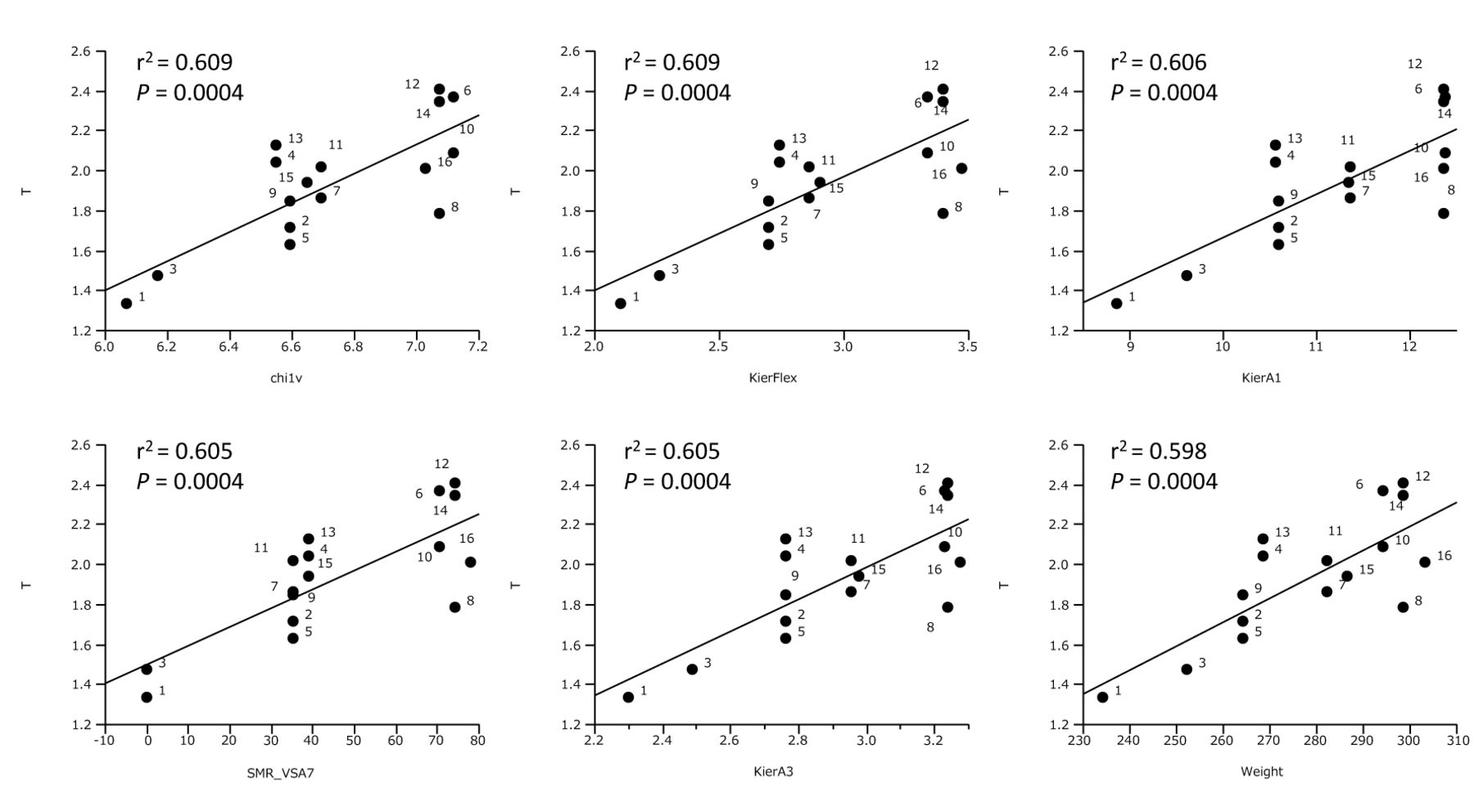
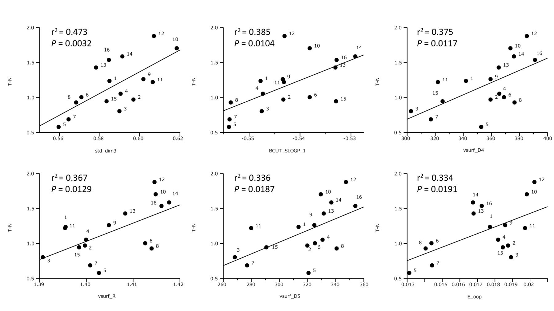
Computational analysis. All calculations of QSAR analysis of 3-styryl-2H-chromenes were done with MOE, using 330 descriptors including Hamiltonian (AM1. PM3 and MNDO). QSAR analysis of cytotoxicity against normal cells demonstrated that eight descriptors showed correlation with N [mean pCC50 (i.e., −log CC50) for normal cells]. Scatter plots of the top six descriptors [std_dim2 (r2=0.447, p=0.0046), E_tor (r2=0.410. p=0.075), E_oop (r2=0.339, p=0.0181), std_dim3 (r2=0.329, p=0.0202), vsurf_A (r2=0.292, p=0.0306) and BCUT_SMR_1 (r2=0.278, p=0.0357)] are shown (Figure 2). QSAR analysis of cytotoxicity against tumor cells demonstrated that seventy four descriptors showed correlation with T (mean pCC50 for tumor cells). Scatter plots of the top six descriptors [chi1v (r2=0.609, p=0.0004), KierFlex (r2=0.609, p=0.0004), KierA1 (r2=0.606, p=0.0004), SMR_VSA7 (r2=0.605, p=0.0004), KierA3 (r2=0.605, p=0.0004) and Weight (r2=0.598, p=0.0004)] are shown (Figure 3). QSAR analysis of selective cytotoxicity demonstrated that sixteen descriptors showed correlation with T-N. Scatter plots of the top six descriptors [std_dim3 (r2=0.473, p=0.0032), BCUT_SLOGP_1 (r2=0.385, p=0.0104), vsurf_D4 (r2=0.375, p=0.0117), vsurf_R (r2=0.367, p=0.0129), vsurf_D5 (r2=0.336, p=0.0187) and E_oop (r2=0.334, p=0.0191)] are shown (Figure 4).
We found that the introduction of methoxy group at R1 of chromene ring significantly [5, 6, 7, 8] increased the cytotoxicxity against normal cells (N) (p=0.0054), but reduced tumor-specificity (N-T) (p=0.0134). On the other hand, the introduction of methoxy group at R2 of chromene ring [9, 10, 11, 12] significantly increased the N-T (p=0.0293) (Figure 5).
Anti-HIV activity of sixteen 3-styryl-2H-chromenes and chemotherapeutic agents. Each value represents the mean of triplicate determinations.
We searched the combination sets that are useful to estimate the cytotoxicity and tumor-selectivity by using multiple regression analysis with leave-one-out cross verification method. We could construct the good estimation model for T-N, using two descriptors (vsurf_R and E_oop) (Figure 6), producing the following equation:

Discussion
The present study demonstrated for the first time that sixteen 3-styryl-2H-chromenes showed moderate to potent tumor-specificity and no detectable anti-HIV activity (Table I). Especially, compound that have a methoxy group at 7-position of the chromene ring and chlorine at 4’-position of phenyl group in styryl moiety [12] showed the highest tumor-selectivity [TS=59.9 (D/B); 115.4 (C/A)], exceeding that of resveratrol [TS=2.4 (D/B); 1.3 (C/A)], doxorubicin [TS=12.6 D/B); 6.3(C/A)] and 5-FU [TS=11.3 (D/B); 3.2 (C/A)] (Table I). Compound [12] showed the highest PSE value (1277.5) among sixteen 3-styryl-2H-chromenes, exceeding that of 5-FU (12.8) and resveratrol (2.2) (Table I). Since HGF, HPLF and HPC cells are mesenchymal cells, we investigated whether similar results with HOK and HGEP, human epithelial normal oral cells. The experiments showed this was the case. All three selected 3-styryl-2H-chromenes [10, 12, 14] showed much higher tumor specificity and potency-selectivity expression than doxorubicin and 5-FU. Compound [12] again showed the highest TS and PSE values, whereas doxorubicin and 5-FU showed essentially no tumor specificity. These results suggest the necessity of investigation about the safety of doxorubicin for treatment of oral cancer patients.

Determination of coefficient between chemical descriptors and cytotoxicity of 3-styryl-2H-chromenes against normal cells (defined as N). The mean (pCC50 i.e., the −log CC50) values for normal cells were defined as N.

Determination of coefficient between chemical descriptors and cytotoxicity of 3-styryl-2H-chromenes against tumor cells (defined as T). The mean (pCC50 i.e., the −log CC50) values for tumor cell lines were defined as T.

Determination of coefficient between chemical descriptors and tumor specificity of 3-styryl-2H-chromenes (defined as T–N).

Effect of introduction of methoxy group on R1 or R2 of chromene ring on N and N-T.
We found that addition of chlorine at 4’-position of phenyl group in styryl moiety of [1] increased the cytotoxicity (mean CC50 (B)=96.3→12.6 μM), tumor-specificity [TS(D/B)=4.2→12.8] and potency-selectivity expression [PSE(D/B2)=4.3→101.6) (compare 1 and 4). Similarly, addition of chlorine at 4’-position of phenyl group in styryl moiety of [9] increased the cytotoxicity (mean CC50 (B)=14.8→4.7 μM), tumor-specificity [TS(D/B)=18.6→59.9] and potency-selectivity expression [PSE(D/B2)=125.5→ 1277.5) (compare 9 and 12). However, chlorine at 4’-position of phenyl group in styryl moiety of [5] and [13] did not increase the cytotoxicity, tumor-specificity nor potency-selectivity expression (compare 5 and 8, 13 and 16) (Table I). These results suggest the importance of 3-dimentional structure in determining these parameters. It is known that the presence of halogens influences the molecular conformation and the biological activity by its steric and/or electronic effects. Conti and Desideri also reported on the inhibition of anti-picornavirus activity with chlorine-substituted 3-styrylchromene (5). The present study demonstrated that the introduction of methoxy group at R1 of chromene ring significantly increased the cytotoxicity against normal cells, but reduced the tumor-specificity, whereas the introduction of methoxy group at R2 of chromene ring significantly increased the tumor-specificity (Figure 5).

Multiple regression models for the estimation of T-N. T-N=32.1(±4.39)vsurf_R+121(±17)E_oop−46.1(±6.2), n=16, R2=0.870, Q2=0.821, s=0.145.
QSAR analysis demonstrated that (i) cytotoxicity against normal cells correlated well with six descriptors (std_dim2, E_tor, E_oop, std_dim3, vsurf_A and BCUT_SMR_1) that reflect structure connectivity and conformation, torsion potential energy, out-of -plane potential energy, amphiphilic moment and molar refractivity (Figure 2), (ii) cytotoxicity against tumor cells correlated well with six (chi1v, KierFlex, KierA1, SMR_VSA7, KierA3 and Weight) that reflect atomic valence connectivity, flexibility, molecular shape, surface area and molecular weight (Figure 3) and (iii) tumor-selectivity correlated well with six descriptors (std_dim3, BCUT_SLOGP_1, vsurf_D4, vsurf_R, vsurf_D5 and E_oop) that reflect structure connectivity and conformation, hydrophobicity, surface rugosity and out-of-plane potential energy (Figure 4). By searching various sets of combinations, we could construct the good estimation model for T-N, using two descriptors (vsurf_R and E_oop).
We have found that all sixteen 3-styryl-2H-chromenes did not protect the cells from cytophathic effect of HIV infection. The lack of anti-HIV activity is not in agreement with a previous study where 4H-chromen-4-one and 2H-chromene derivatives inhibited the replication of picornavirus in vitro with high therapeutic indexes (250 and 36, respectively) (5). Recently, their group newly synthesized (E)-6-chloro-3-(3-phenylprop-1-en-1-yl)-2H-chromene (16) and [2-(2H-chromen-3-yl)vinyl]pyridines and 3-[2-(pyridinyl)vinyl]-4H-chromen-4-ones (17), that inhibited the infection of human rhinoviruse (HRV) 1B and 14 replications, probably by acting at the uncoating level as a capsid-binder. The discrepancy between our and their results may be due to the different assay systems of anti-viral activity (whether using both mock-infected and infected cells at the same time or not).
We have recently found that (E)-3-(4-hydroxystyryl)-6-methoxy-4H-chromen-4-one induced mitochondrial vacuolization, and inhibited the autophagy [expression of The microtubule-associated protein 1 light chain 3 (LC3)], and finally induced apoptosis [limited degradation of Poly (ADP-ribose) polymerase (PARP)] in HSC-2 cells (Sakagami et al., submitted). It remains to be investigated whether (E)-3-(4-chlorostyryl)-7-methoxy-2H-chromene [12] shows similar actions.
In conclusion, the present study suggests that multivariate statistics with chemical descriptors for molecular shape and flatness may be useful for evaluation of tumor-specificity of 3-styryl-2H-chromenes.
Acknowledgements
This work was partially supported by KAKENHI from the Japan Society for the Promotion of Science (JSPS) (15K08111). The annual license of the statistical software, JMP Pro, was supported by the grant-in-aid of the oncology specialists promotion program by the Ministry of Education, Culture, Sports, Science and Technology, Japan.
Footnotes
Conflicts of Interest
The Authors wish to confirm that there are no known conflicts of interest associated with this publication and there has been no significant financial support for this work that could have influenced its outcome.
- Received June 22, 2015.
- Revision received July 16, 2015.
- Accepted July 20, 2015.
- Copyright© 2015 International Institute of Anticancer Research (Dr. John G. Delinassios), All rights reserved
In this issue


Jump to section
Related Articles
Cited By...
- Quantitative Structure-Cytotoxicity Relationship of 2-Arylazolylchromones and 2-Triazolylchromones
- Quantitative Structure-Cytotoxicity Relationship of 3-(N-Cyclicamino)chromone Derivatives
- Quantitative Structure-Cytotoxicity Relationship of Pyrano[4,3-b]chromones
- Quantitative Structure-Cytotoxicity Relationship of 2-(N-cyclicamino)chromone Derivatives
- Quantitative Structure-Cytotoxicity Relationship of Furo[2,3-b]chromones
- In Vitro Anti-tumor Activity of Azulene Amide Derivatives
- Quantitative Structure-Cytotoxicity Relationship of Cinnamic Acid Phenetyl Esters
- Quantitative Structure-Cytotoxicity Relationship of 2-Azolylchromones
- Search for New Type of Anticancer Drugs with High Tumor Specificity and Less Keratinocyte Toxicity
- Enhancement of Cytotoxicity of Three Apoptosis-inducing Agents Against Human Oral Squamous Cell Carcinoma Cell Line by Benzoxazinotropone