


Abstract
Background/Aim: The expression of the CD44 variant exon 9 (CD44v9) was investigated in order to elucidate its significance for cancer stem cells in circulating human colorectal cancer cells (CTCs). Materials and Methods: After peripheral blood was drawn from patients with colorectal cancer, CTCs were collected. Using the reverse transcription-polymerase chain reaction method, we examined the relationship between expression of CD44v9 mRNA and prognosis. Results: In 60 out of 150 patients with colorectal cancer, expression of CD44v9 mRNA was positive in CTCs. In patients with stage III disease, the 5-year survival rate was 89% for patients with negative CD44v9 expression, whereas it was 52.4% in patients with positive expression (p<0.05). In patients with stage IV unresectable cancer, the 2-year survival rate was 70.1% in cases with CD44v9-negative expression and 33.3% in cases of positive expression (p<0.05). Conclusion: CD44v9 mRNA in the CTCs of colorectal cancer is useful as a factor predicting recurrence, prognosis, and treatment efficacy.
Abbreviations: CD; Cluster of differentiation, RT-PCR; reverse transcription-polymerase chain reaction, CTC; circulating tumor cells, CD44v9; CD44 variant exon9, xCT: cystine glutamate exchanger.
Colorectal cancer is highly prevalent compared to other malignant tumors, and recurrence/metastasis frequently occur in the form of hematogenous metastasis to liver and lungs (1-4). In other words, effective counter-measures against hematogenous metastasis will improve the survival rate of patients with colorectal cancer. In general, hematogenous metastasis of colorectal cancer proceeds in the following sequence: dissociation of cancer cells from the primary lesion, entry into capillary vessels, spread to the whole body via the portal and greater circulatory systems, adhesion to vascular endothelial cells in the target organ, extravasation, and invasion and growth (5). Thanks to the recent elucidation of the metastatic mechanism, treatments have become available in clinical settings that target several molecules expressed on the cell surface and angiogenic growth factor, and prognoses have improved (6-8). According to recent perception, not all cancer cells are able to grow or resist drugs, only certain cancer cells, namely cancer stem cells, have such capabilities (9).
It is generally understood that the role of stem cells in healthy tissue is to continuously retain the capacity to produce the constitutive cells of the tissue (10). Likewise, cancer stem cells are considered to retain the capability of self-duplication and self-differentiation, in addition to drug resistance and immune evasion. In 1994, Dick et al. first identified cancer stem cells in malignant tumors when they found leukemia-developing cells in human acute myeloid leukemia by using a molecular marker. Subsequently, stem cells were found in such solid tumors as breast cancer, brain tumor and colorectal cancer (11-18). For colorectal cancer in particular, leucine-rich repeat-containing G-protein-coupled receptor 5, CD133, and CD44 were identified as the markers for cancer stem cells (11, 19, 20). The CD44 gene is positioned on the short arm of chromosome 11 and has a transmembrane structure (21, 22). The gene consists of multiple exons inserted, by an alternative splicing mechanism, into the extracellular domain near the transmembrane domain. It have also been reported that this is associated with growth, invasion, and metastasis of cancer cells. In stomach cancer and colorectal cancer with hematogenous metastasis in particular, CD44 variant exon 6 and CD44 variant exon 9 (CD44v9) are frequently expressed in the primary lesion (23-26). According to recent reports, CD44v8 through 10 bind to the xCT proteins that constitute the cystine/glutamic acid transporter on cell membranes to inhibit accumulation of active oxygen in cancer cells that can therefore escape activation of oxidative stress (27). Furthermore, CD44v9 is an important factor for cancer stem cells in colorectal cancer (28).
Accordingly, we investigated the relationship between expression of CD44v9 in the blood and the recurrence and survival rates for colorectal cancer, considering that cancer cells travel through blood vessels in hematogenous metastasis, and that CD44v9 plays an important role in cancer stem cells.
Materials and Methods
Patients and sample collection. Blood (20 ml) was drawn by vein puncture from 150 patients with sporadic colorectal cancer and from 15 healthy volunteers at the First Department of Surgery, University of Fukui, Japan between 2003 and 2011. According to the TNM classification (29), 24, 35, 55, and 36 tumors were Dukes' stage I, II, III, and IV respectively.
To avoid contamination with skin cells, 5 ml blood were discarded before the study samples were taken. Blood was processed with the OncoQuick density gradient system according to the manufacturer's instructions (Greiner Bio-One GmbH, Frickenhausen, Germany).
Tumor cells obtained by density gradient centrifugation were suspended in 400 μl phosphate-buffered saline. Negative control(no epithelial cells) of this system was used by 20ml blood of healthy volunteers.
RT-PCR. Total RNA was extracted from tumor cells using ISOGEN (Wako, Tokyo Japan), and reverse transcribed using using Prime Script RT reagent kit (Takara, Otsu Japan) (30). The primers for PCR to amplify CD44v9 gene-coding regions were as follows: The 5’ primer, CD44v9-AX, was the published human CD44v9 sequence (31): TTCTCTACATCACATGAAGGC. The 3’ primer, CD44v9-BX, was GCTTGATGTCAGAGTAGAAGT. Thirty cycles of denaturation (94°C, 1 min), annealing (55°C, 1.0 min), and extension (72°C, 2 min) were carried out in a thermal cycler (PTC-100, Programmable Thermal Controller; NJ Research Inc., MA, USA). The amplified products were purified using QIAquick PCR Purification kit (Qiagen, Hilden, Germany). The products were used for a second round of PCR amplification for 30 cycles using two primers: the 5’ primer, CD44v9-CX, ATGAAGGCTTGGAAGAA encompassed the published human regular CD44v9 sequence; the 3’ primer, CD44v9-DX, was GTAGAAGTTGTTG. Thirty cycles of denaturation (94°C, 1 min), annealing (50°C, 1.5 min), and extension (72°C, 2 min) were carried out in a thermal cycler (PTC-100, Programmable Thermal Controller; NJ Research Inc.). All PCR product, were resolved by electrophoresis in 1.2% agarose gel. The sequencing was performed on PCR products that showed bands in RT-PCR analysis. Sequence analysis showed the presence of the CD44v9 gene.
Semi-quantitative detection of mRNA. Ethidium bromide staining of the gels identified a band of the CD44v9 gene. To ensure reproducibility, all PCR amplifications were performed in duplicate. Densitometric analysis of photographed gels was used for band quantification (32). Positive case; the band was confirmed, Negative case; the band was not confirmed.
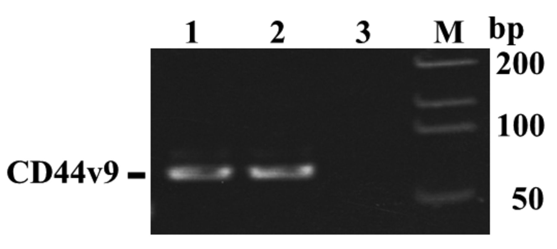
CD44 variant exon 9 (CD44v9) mRNA expression in the CTCs of patients with colorectal cancer. CD44v9 mRNA expression was shown in patients with colorectal cancer, although at different levels. The expression of CD44v9 mRNA was observed in 60 (40%) out of the 150 samples of CTCs from patients with primary colorectal cancer. Lane 1 and Lane2; Positive expression of CD44v9 mRNA, Lane 3; Negaitive expression of CD44v9 mRNA,, M; DNA size marker.
Statistical considerations. Survival time was calculated using the Kaplan–Meier method, and the log-rank test was used to compare the curves of the survival times using Stat Mate IV (ATMS Co., Ltd., Japan). Other characteristics of the two treatment arms were compared using the chi-square test using Stat Mate IV (ATMS Co., Ltd.). The Cox proportional hazards model was used in multivariate regression analyses of survival date (IBM SPSS Statistics for Windows, Version21.0, IBM Corporation, Armonk, NY, USA). Differences were considered significant at p-values less than 0.05.
Results
Expression of CD44v9 mRNA in the CTCs of patients with colorectal cancer. Figure 1 shows representative cases of CD44v9 mRNA expression, as demonstrated by the nested PCR technique. Out of 150 colorectal cancer cases, 60 (40%) had the expression of CD44v9 mRNA. The expression of CD44v9 mRNA was not detected in fifteen healthy volunteers.
Relationship between expression of CD44v9 mRNA and clinicopathological findings. The correlations between expression of CD44v9 mRNA and lymph node metastasis, peritoneal dissemination, hematogenous metastasis and TNM stage were significantly high, although no signicant difference in expression of CD44v9 mRNA was noted regarding gender, age, location, histological type, serosal invasion, lymphatic invasion, and venous invasion (Table I).
CD44v9 mRNA expression in the CTCs of patients with different stages of colorectal cancer. The expression of CD44v9 mRNA was found in the CTCs of 17 out of 59 patients with stage I-II colorectal cancer (28.8%); 20 out of 55 patients with stage III (36.4%); and 23 out of 36 patients with stage IV disease (63.9%), indicating that the number of cases with CD44v9 mRNA expression increases with progression of tumor stage (Table I).
Correlation between clinicopathological features and CD44v9 mRNA expression.
Expression of CD44v9 mRNA and recurrence rate in stage I-III colorectal cancer. The recurrence rate in stage III colorectal cancer was five out of 35 cases (14.3%) when expression of CD44v9 mRNA was negative in CTCs, and significantly higher, eight out of 20 cases (40%), when expression was positive (Table II). In Stages I and II, no significant difference was observed in the recurrence rate between cases with negative and those with positive expression of CD44v9 mRNA.
Correlation between CD44v9 expression and recurrence rate in stage III colorectal cancer.
Expression of CD44v9 mRNA in CTCs and survival rate in stage III colorectal cancer. In stage III colorectal cancer, the 5-year survival rate was 89.5% in cases with negative expression, and 52.4% in positive cases, showing that survival rate was significantly poorer in cases of positive CD44v9 mRNA expression (Figure 2). In stages I-II, no significant difference was observed in survival rate between cases with negative and those with positive CD44v9 mRNA expression.
Relationship between CD44v9 mRNA expression in CTCs and survival rate in cases with unresectable stage IV colorectal cancer. The 2-year survival rate in patients with unresectable stage IV colorectal cancer was 70.1% in cases of negative CD44v9 mRNA expression, and significantly lower at 33.3% in cases of positive CD44v9 mRNA expression (Figure 3).
CD44v9 mRNA as a new prognostic factor in colorectal cancer. Univariate analysis using the Cox proportional hazard model showed significant differences in CD44v9 mRNA expression, serosal invasion, venous invasion, lymph node metastasis, peritoneal metastasis, and hematogenous metastasis. Multivariate analysis showed significant differences in CD44v9 mRNA expression, seroral invasion, lymph node metastasis, and hematogenous metastasis. The hazard ratio for CD44v9 mRNA expression was 4.445 (95% confidence interval=1.689-11.693) (Table III).
Discussion
Hematogenous metastasis is considered a prognosis-determining factor in colorectal cancer. In actual clinical settings, stage classification is conducted with regard to tumor depth, lymph node metastasis, and distant metastasis (1-4). Depending on stage, chemotherapy, etc. is carried out. However, for patients with stages I-III disease, recurrence occurs in some cases but not in others, even within the same stage (4). Therefore, important factors may exist other than those currently considered.
Pathological findings and CD44v9 mRNA as prognostic factor for patients with colorectal cancer. The Cox proportional hazards model was used in multivariate regression analyses of survival data.

Relationship between CD44v9 mRNA expression and survival rate in patients with stage III colorectal cancer. The 5-year survival rate was 89.5% in patients with CD44v9 mRNA-negative cancer but significantly poorer, 52.4% in those with CD44v9 mRNA-positive cancer. Life-table analysis was performed using the Kaplan–Meier technique. The outcomes of different groups of patients were compared by the log-rank test (p<0.05).
Recently the concept of intratumoral heterogeneity has undergone substantial change. At the 2006 American Association for Cancer Research Congress, cancer stem cells were defined as the “cells existing in the tumor with self-renewal capacity and the ability to produce various lineages of tumor-forming cancer cells” (33). Furthermore, cancer stem cells may be drug-resistant and have immune evasion capability, possibly causing treatment refractoriness and recurrence (28, 33). In 2007, stem cells of colorectal cancer were reported and CD44 was extracted as one of their markers (11). CD44 is a transmembrane protein, and a variant exon insertion may sometimes be found outside the cell membrane. A CD44 molecule with several variant exon insertions has been identified in hematopoietic cells and various healthy tissues, as well as in epithelial cancer cells (34). In malignant tumors, the level of malignancy, including metastasis, has been reported to be closely related to the type of variant exon insertions in malignant tumors, and its importance has been reported (23-26, 35-37). In our recent study on colorectal cancer variants, we found that cells with CD44v9 expression have the characteristics of cancer stem cells, such as drug resistance, survival capability, and tumor-forming ability, reconfirming the importance of the expression of CD44v9 (28). The spread of cancer cells to distant organs in hematogenous metastasis is considered to occur via blood, and the involvement of CTCs has been confirmed (5). Previous studies on CTCs in the blood referred to the measurement of carcinoembryonic antigen (CEA) mRNA and cytokeratin mRNA, as well as expression of epithelial cell adhesion molecule (38-42). However, these studies may have included cancer stem cells, non-cancer stem cells, and other various types of cancer cells, since the presence of cancer cells in the blood did not match the recurrence rate. Therefore, our study on the expression of CD44v9 mRNA, a marker for cancer stem cells in CTCs, is significant in view of recurrence and treatment resistance of cancer. According to our findings, prognosis is poorer in cases of unresectable colorectal cancer with CD44v9 mRNA expression in CTCs compared to cases with no such expression. This is probably because of the acquisition of drug resistance and immune evasion capability, possibly involving cancer stem cells. The same theory may be applicable to stage III cases associated with a high prevalence of recurrence, as the recurrence rate was significantly higher and prognosis poorer in cases with CD44v9 mRNA expression compared to the cases without expression. According to our results, cancer stem cells are likely to be involved when CD44v9 mRNA is expressed in CTCs, and the prognoses for these cases will be poor owing to a high recurrence rate and drug resistance.

Relationship between CD44v9 mRNA expression and survival rate in patients with unresectable stage IV colorectal cancer. The 2-year survival rate was 70.1% in patients with CD44v9 mRNA-negative cancer but significantly poorer, 33.3% in those with CD44v9 mRNA-positive cancer. Life-table analysis was performed using the Kaplan–Meier technique. The outcomes of different groups of patients were compared by the log-rank test (p<0.05).
Expression of the CD44v9 mRNA marker for human colorectal cancer in CTCs is considered to be a good indicator for recurrence rate, drug resistance, and subsequent prognosis.
Acknowledgements
The technical assistance of Ms M. Saitoh with this research was appreciated.
This work was supported in part by the Organization for Life Science Advancement Programs and Translational Research Program.
Footnotes
-
Conflicts of Interest
The Authors do not have any significant financial interest in any company (or its competitor) making any products discussed in the article. The Authors report no conflicts of interest.
- Received August 20, 2014.
- Revision received September 25, 2014.
- Accepted September 30, 2014.
- Copyright© 2015 International Institute of Anticancer Research (Dr. John G. Delinassios), All rights reserved


