


Abstract
Background: Compounds containing ethylenediamine (−NCH2CH2N-) moiety are known to exhibit antimicrobial, -fungal, -bacterial, -tuberculosis and -cancer activities. Materials and Methods: In the present study, we evaluated the in vitro cytotoxic activity of N,N’-bis(2-hydroxybenzyl)- (6), N,N’-bis(5-bromo-2-hydroxybenzyl)- (7) and N,N’-bis(5-chloro-2-hydroxybenzyl) (8)- ethylenediamine dihydrochlorides; and N,N’-bis(2-hydroxybenzyl)- (9), N,N’-bis(5-bromo-2-hydroxybenzyl)- (10) and N,N’-bis(5-chloro-2-hydroxybenzyl) (11)- ethylenediamine toward human lung (A549), breast (MDA-MB-231) and prostate (PC3) cancer cell lines after 24-h treatment using crystal violet dye binding assay. Effects on the cell cycle the using flow cytometry, and mitochondrial membrane potential using rhodamine-123 florescent dye were also evaluated. Results: Compounds 7 and 8 exhibit cytotoxic activity, causing cell arrest at different phases of the cell cycle and loss of mitochondrial membrane potential in the above cancer cell lines. Conclusion: These findings clearly demonstrate, to our knowledge for the first time, that ethylenediamine dihydrochloride salts–compounds 7 and 8–exhibit concentration-dependent cytotoxic activity towards A549, MDA-MB-231 and PC3 cancer cell lines, which may serve as a basis for future work on novel therapeutic agents.
- N,N’-Bis(2-hydroxybenzyl)ethylenediamine derivatives
- cytotoxic activity
- cell cycle phase
- mitochondrial transmembrane potential
- MMP
Metal complexes containing an ethylenediamine (−NCH2CH2N) backbone have attracted significant interest as potential anticancer agents due to their rich redox chemistry and relative ease of manipulation (1-3). They have been found to induce cytotoxic activity in various types of cancer cell lines (2, 4-7). However, recent studies have ascribed the activities of some ethylenediamine-metal complexes to the presence of their ligands, prompting the search for the development of novel ethylenediamine-type ligands as chemotherapeutic agents (8, 9). Examples of ethylenediamine-type ligands that have been reported to show anticancer activity are protein farnesyltransferase (FTase) inhibitors (1), (S,S)-ethylendiamine-N,N-di-2-(3-cyclohexyl) propanoic acid esters (2), and N-solanesyl-N,N’-bis(3,4-dimethoxybenzyl)ethylenediamine (SDB-ethylenediamine) (3) (Figure 1) (9-12).
In addition to their anticancer activity, ethylenediamine-type ligands have been reported to have anti-microbial, -fungal, - bacterial, -tuberculosis, -malarial, -leishmanial and -histaminic activities (13-17). For example, N-geranyl-N’-(2-adamantyl) ethane-1,2-diamine (SQ109; 4), a new drug candidate based on the 1,2-ethylenediamine pharmacophore of ethambutol, is a potential anti-tuberculosis drug that has entered phase I/II clinical trials (Figure 1) (18-20). Ethylenediamine ligands have found application in iron-chelating therapy, as radiopharmaceutical for tumor imaging etc. such as N,N’-di(o-hydroxybenzyl) ethylenediamine-N,N’-diacetic acid (HBED, 5, Figure 1) (21-22). Recently, our group reported that N,N’-bis(2-hydroxy-5-bromo benzyl)ethylenediamine (10) and N,N’-bis(2-hydroxy-5-chlorobenzyl) ethylenediamine (11) (Table I) showed significant antimicrobial and antifungal activities against Staphylococcus aureus, Pseudomonas aeruginosa and Salmonella enterica, respectively (23).
The result of our previous investigation (23) prompted us to evaluate the cytotoxic activity of N,N’-bis(2-hydroxybenzyl)ethylenediamine derivatives 6-11 in human cancer cell (Table I). The active compounds cytotoxicity on cell-cycle distribution and mitochondrial membrane potential (MMP) were also evaluated.
Materials and Methods
Materials. F12K medium, RPMI medium, penicillin-streptomycin antibiotic solution (100×), fetal bovine serum (FBS), Trypsin-EDTA solution (1×), phosphate buffer (PBS), 50% glutaraldehyde, RNase, IGPAL CA-630, crystal violet, tetramethyl rhodamine methyl ester (rhodamine-123; TMRM) florescent dye and propidium iodide were obtained from Sigma-Aldrich Company (St. Louis, MO, USA). Monobasic and dibasic potassium phosphate, EDTA, D-glucose, Triton X-100 and ethanol were obtained from Thomas Scientific Company (Swedesboro, NJ, USA).
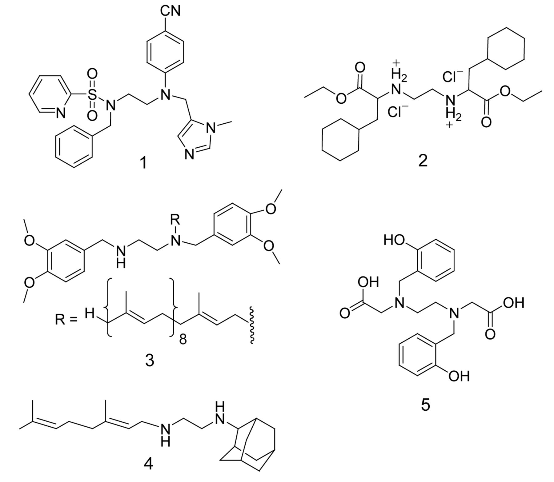
Chemical structure of ligands containing ethylenediamine pharmacophore. 1:protein farnesyltransferase (FTase) inhibitors; 2:(S,S)-ethylendiamine-N,N-di-2-(3-cyclohexyl) propanoic acid esters; 3:N-solanesyl-N,N’-bis(3,4-dimethoxybenzyl)ethylenediamine (SDB-ethylene-diamine); 4:N-geranyl-N’-(2-adamantyl)ethane-1,2-diamine (SQ109); 5:N,N’-di(o-hydroxybenzyl)ethylenediamine-N,N’-diacetic acid (HBED).
Cell line maintenance. Human lung (A549), breast (MDA-MB-231) and prostate (PC3) cancerous cell lines were obtained from the American Type Culture Collection (ATCC, Rockville, MD, USA) and cultured under the supplied guidelines. The cells were maintained in F12K (A549, PC3) or RPMI (MDA-MB-231) medium containing 100 units of penicillin/ml, 100 μg of streptomycin/ml, 2 mM L-glutamine and 10% FBS in T-75 cm2 flasks at 37°C in a 5% CO2 incubator.
Treatment of cells.
Cell viability assay: The cells were plated at a density of 5×10⁴ cells per well in polystyrene, flat-bottom 24-well microtiter plates (Corning Costar, Rochester, NY, USA) in complete medium containing 10% FBS and allowed to stabilize overnight in a CO2 incubator at 37°C The cells were then treated with compounds 6-11 at different concentrations (0, 10, 25, 50, 75, and 100 μM) in a final volume of 1 ml per well in triplicate wells for each treatment for 24-h at 37°C in a 5% CO2 incubator. All studies were repeated at least thrice.
Cell-cycle analysis: The cells at a density of 0.65×10⁶ cells per T-25 cm2 flask (Corning Costar) in complete medium were plated and allowed to stabilize overnight in a CO2 incubator at 37°C. The cells were then treated with active compounds 7 and 8 at different concentrations (0, 10 and 20 μM) in a final volume of 5 ml per flask in triplicate flasks for 24-h at 37°C in an incubator with 5% CO2.
Evaluation of cell viability. At the end of the incubation period, cell viability was evaluated by crystal violet dye uptake assay according to our previously reported method (24). The lethal dose (LD50) of the compound (i.e. the dose of tested compound where 50% cell death was observed) was calculated according to the method of Ipsen and Feigl (25).
Cell-cycle analysis by flow cytometry. The active compound cytotoxic effect on different stages of cell cycle in cancer cell lines was studied using Accuri Flow Cytometer (BD Biosciences, San Jose, CA, USA). Cells at a density of 0.65×10⁶ cells per T-25 cm2 flask were plated overnight. The next day, cells were treated with 0, 10 and 20 μM of active compound in triplicate flasks for 24-h with 5% CO2 in an incubator at 37°C. At the end of incubation, the cells were trypsinized and centrifuged at 1055 ×g for 10 min at room temperature. The pellet was suspended in 1 ml of PBS and transferred to eppendorf tube. The cells were pelleted by centrifugation at 1055 ×g for 5 min. The cells were then stained with 500 μl of Vindelov's reagent containing 0.01 mg/ml of ribonunuclease A and 0.075 mg/ml of propidium iodide and 1 μl/ml of IGPAL CA-630 in PBS for overnight at 4°C in dark. The stained cells were run through Accuri Flow Cytometer at low speed (Becton Dickinson, San Jose, CA, USA). In each sample, a total of 50,000 events were analyzed separately. The Accuri C6 software was used for acquisition and analysis of the data and the percentage of cells in each phase.

Effect of compounds 7 and 8 on A549, MDA-MB-231 and PC3 cell viability. Data are represented as the mean ±SEM (error bars) for n=9.
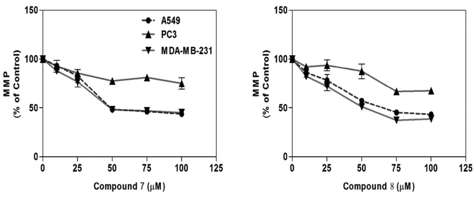
Measurement of MMP. MMP was determined using rhodamine-123 florescent dye assay in A549, MDA-MB-231 and PC3 cancer cells. At the end of incubation, cells were fixed with 400 μl of 0.25% aqueous glutaraldehyde containing rhodamine-123 to yield a final concentration of 1 μM for 30 min at room temperature. The supernatant was discarded, and the plates were washed with tap water and air-dried in the hood for 5 min. Finally, 500 μl of 0.1% Triton × 100 in dPBS was added per well and incubated at 37°C for 1-h. The plates were read with the excitation filter set at 485 nm and the emission filter at 538 nm on a microplate Tecan Fluorometer.
Statistical analysis. The viability (n=9) and cell-cycle analysis (n=3) results were presented as the mean ±standard deviation. All data for treated cells were presented as percentage values in comparison to the untreated control (100%). The data were analyzed for significance by one-way ANOVA, followed by Dunnett's multiple comparison test, using Graph Pad Prism Software, version 3.0 (Graph Pad Software, Inc., San Diego, CA, USA). Differences from the respective untreated control were considered statistically significant when *p<0.05.
Results
Cytotoxic activity of compounds 6-11 on cancer cell lines. In vitro cytotoxic activity of compounds 6-11 was evaluated at different concentrations (0, 10, 25, 50, 75 and 100 μM) in A549, MDA-MB-231 and PC3 cancer cell lines after 24-h drug treatment using crystal violet dye binding assay. The LD50 for all tested compounds were calculated according to the Ipsen method (25) and are shown in Table I. It was observed that compounds 7 and 8 caused concentration-dependent cell death (Figure 2), while compounds 6 and 9-11 did not cause cell death (inactive; LD50>100 μM) in these cell lines after 24-h treatment in comparison to the untreated control cells. The cytotoxicity of compound 7 was higher than compound 8 in these cell lines. Furthermore, compound 7 (LD50=17.5 μM) and 8 (LD50=21.0 μM) were more cytotoxic to MDA-MB 231 cell line in comparison to A549 and PC3 cell lines, based on their calculated LD50 value (Table I).
The (LD50) values (μM) for compounds 6-11 tested in lung (A549), breast (MDA-MB-231) and prostate (PC3) cancer cell lines after 24-h treatment.

Effect of compounds 7 and 8 on A549 (A), MDA-MB-231 (B) and PC3 (C) cell cycle distribution. Data are represented as mean and SEM (error bars) for n=3. *Statistically significant difference from the control (p<0.05) using Dunnett's multiple comparison test.

Effect of compounds 7 and 8 on mitochondrial membrane potential (MMP) of A549, MDA-MB-231 and PC3 cells. Data are represented as the mean ± SEM (error bars) for n=3.
Effect of compounds 7 and 8 on the different phases of cell cycle. The cytotoxic activity of compounds 7 and 8 on cell-cycle phases was evaluated by treating cells (A549, MDA-MB-231 and PC3) with compounds, followed by staining with propidium iodide. The percentage of cells in the G1/G0, S and G2/M phases of the cell cycle were analyzed using a flow cytometer. It was observed that treatment of A549, MDA-MB-231 and PC3 cells with compounds 7 and 8 at 10 and 20 μM after 24 h treatment resulted in significant cell-cycle arrest (p<0.05) in different-cell cycle phases. Interestingly, there was a marked increase in the percentage of cells in specific cell-cycle phases with respect to control cells (Figure 3). Results indicate that compound 7 caused significant accumulation of (i) A549 cells in the G2/M phase at 10 μM and at the S phase at 20 μM (Figure 3A), (ii) MDA-MB-231 cells in S phase at both 10 μM and 20 μM (Figure 3B), and (iii) PC3 cells in G2/M phase at 10 μM and G1/G0 phase at 20 μM (Figure 3C). However, compound 8 caused significant accumulation of (i) A549 cells in S phase at 20 μM (Figure 3A), (ii) MDA-MB-231 cells in S phase at both 10 μM and 20 μM (Figure 3B), and (iii) PC3 cells in G1/G0 phase at 20 μM (Figure 3C).
MMP. The effect of compounds 7 and 8 on MMP was evaluated at different concentrations (0, 10, 25, 50, 75 and 100 μM) in A549, MDA-MB-231 and PC3 cells after 24-h treatment using rhodamine-123 florescent dye. Results indicate that compounds 7 and 8 caused loss of MMP in A549, MDA-MB-231 and in PC3 cells in comparison to the untreated control cells (Figure 4).
Discussion
The in vitro cytotoxicity of N,N’-bis(2-hydroxylbenzyl) ethylenediamine derivatives 6-11 in different cancer cell lines, and the active compounds cytotoxicity on cell-cycle phases and MMP were evaluated for the first time. It was observed from the present investigation that compounds 7 and 8 displayed cytotoxicity towards A549, MDA-MB-231 and PC3 cancer cell lines after 24-h treatment (Table I). The observed cytotoxic activity was dose-dependent in the three cancer cell lines. However, compound 7 (containing a Br group) displayed higher cytotoxic activity in comparison to compound 8 (containing a Cl group) based on the calculated LD50 values. Another interesting observation is that compounds 10 and 11, the non-hydrochloride salts (free diamine) of the active compounds 7 and 8, were found to be inactive (LD50 >100 μM). This observation is consistent with previous finding that the ethylenediamine dihydrochloride salt (such as compound 2, Figure 1) is more toxic to cancer cells than its free diamine form (11). The absence of aromatic bromine or chlorine atom in the dihydrochloride 6 resulted in loss of cytotoxic activity (Table I). Based on the structure-activity relationship (SAR), we may associate the cytotoxic activity of these ethylenediamine dihydrochloride salts (7 and 8) to the presence of aromatic halogen (Cl or Br groups).
In recent years, cell-cycle regulation has been an effective strategy for controlling tumor growth (26, 27). The effect of toxic substances on different phases of the cell cycle can activate or inhibit growth and proliferation of cancer cells (28). In the present investigation, cell-cycle analysis indicated that compounds 7 and 8 caused accumulation of A549, MDA-MB-231 and PC3 cells in different (G1/G0, S and G2/M) cell-cycle phases (Figure 3). Compound 7 led to (i) S phase cell arrest in A549 at 20 μM and in MDA-MB-231 cells at both 10 and 20 μM, indicating that cells may have been undergoing inhibition of DNA synthesis (Figure 3A and B); (ii) G2/M phase cell arrest in A549 and PC3 at 10 μM, indicating that cells may have been undergoing DNA replication (Figure 3A and B); and (iii) G1/G0 phase cell arrest in the PC3 cell line at 20 μM, indicating that cells may have been undergoing apoptosis (Figures 3C). Compound 8 had similar effects.
Studies have shown that evaluation of the MMP in living cells is of critical importance in accessing the toxic effect of chemicals on mitochondrial function (29-32). In the present study, MMP measurement using rhodamine-123 indicated that compounds 7 and 8 led to loss of MMP in A549, PC3 and MDA-MD-231 cell lines at different concentrations after 24-h treatment (Figure 4). Rhodamine-123, a green-fluorescent dye, is selectively taken up by mitochondria and the amount taken up by cells is directly proportional to the MMP (29). Interestingly, for both compounds, the cancer cell lines exhibited similar sensitivity and resistance in the order of MDA-MB-231 (most sensitive) >A549 >PC3 (most resistant) cells (Figure 4). Based on this finding, we believe that the elicited cytotoxicity of compounds 7 and 8 may be associated with the loss of MMP in these cancer cells.
In conclusion, our studies demonstrated that the ethylenediamine dihydrochloride salts 7 and 8 exhibited cytotoxic effects, caused cell arrest in different cell-cycle phases and loss of MMP in human lung (A549), breast (MDA-MB-231) and prostate (PC3) cancer cell lines after 24-h treatment. Further studies are needed to achieve a better understanding over the cytotoxicity mechanism of these compounds.
Acknowledgements
The Authors gratefully acknowledge Florida A&M University TITLE III PROGRAM for their financial support.
Footnotes
-
This article is freely accessible online.
-
Conflicts of Interest
The Authors declare that they have no financial or non-financial competing interests.
- Received September 30, 2013.
- Revision received November 7, 2013.
- Accepted November 8, 2013.
- Copyright© 2014 International Institute of Anticancer Research (Dr. John G. Delinassios), All rights reserved


