


Abstract
Background: There exist substantial evidence that laryngeal cancer represents a unique entity among squamous head and neck carcinomas. Materials and Methods: Tumors from 289 patients with squamous cell laryngeal cancer were assessed for protein (immunohistochemistry) and mRNA (qRT-PCR) expression of Notch pathway components (Notch1 to 4 receptors and Jagged1 ligand) on tissue microarrays. Results: In univariate analysis, enhanced nuclear Jagged1 expression conferred a longer disease-free survival (DFS) (p=0.013) and overall survival (OS) (p=0.004), in contrast to the unfavorable prognostic value of Notch3 for both DFS (p=0.009) and OS (p=0.024). In multivariate analysis, overexpression of either Notch or cytoplasmic Jagged1 conferred an unfavorable effect on DFS (Hazard Ratio=1.88, 95% Confidence Interval=1.03-3.43, p=0.04). Conclusion: Our study indicates a consistent unfavorable effect of Notch3 and cytoplasmic Jagged1 protein expression, a favorable impact of nuclear Jagged 1 localization, and a differential prognostic value of Notch2 expression according to the presence of cytoplasmic Jagged 1.
- Laryngeal cancer
- immunohistochemistry
- mRNA expression
- Notch
- Jagged1
- multivariate analysis
- prognostic value
The survival rates of patients with laryngeal cancer have remained unchanged over the past 30 years (1). Although the larynx is anatomically part of the head and neck area and is covered by squamous epithelium, there is substantial evidence that it represents a unique entity (2). Ideally, research efforts should distinctly focus on laryngeal carcinogenesis and overt cancer characteristics, instead of extrapolating knowledge from biology of squamous cell head and neck cancer.
Among regulatory pathways that control cell proliferation, differentiation, survival and apoptosis, Notch signaling is considered to be fundamental (3). The first human trials of Notch inhibitors have recently been reported (4, 5), a fact that emphasizes that much research still lies ahead.
In the current translational study, we utilized tissue microarrays (TMAs) from 289 laryngeal tumors to assess the immunohistochemical (IHC) and mRNA expression of Notch pathway components, including the transmembrane receptors Notch1 to 4 and their associated ligand, Jagged1. The selection was based on a previous study by our group exploring prognostic gene expression models in early laryngeal cancer (6). The aims of the study were to evaluate the expression profiles of these biomolecules, their association with clinicopathological characteristics, and their prognostic value in patient outcome. To the best of our knowledge, this is the largest study of expression of Notch signaling pathway components performed on tumors from patients with laryngeal cancer.
Materials and Methods
Patients with localized stage I-III squamous cell laryngeal cancer, managed between 1985 and 2008 at the Otolaryngology Department of the Aristotle University of Thessaloniki with potentially curative surgery with/without radical external beam irradiation, were retrospectively identified. All patients gave their informed consent prior to their inclusion in the study for the provision of biological material for research purposes. The present study was approved by the Bioethics Committee of the Aristotle University of Thessaloniki, School of Medicine (A9926/28-05-2008).
Histological evaluation. An experienced pathologist (M.B.) reviewed representative hematoxylin and eosin (H&E)-stained slides from tissue blocks for adequacy of material and calculation of the percentage of tumor cells in each case. Manual dissection was performed on routinely deparaffinized sections in order to increase tumor cell content in the extracted molecular templates (>50% in all samples). A REMARK flow chart is shown in Figure 1.
TMA construction and IHC. The methodology of TMA construction and IHC staining has already been described by Pentheroudakis et al. (7). The procedure, including the antibodies and conditions used for IHC, is fully presented in the Online Supplement.
RNA extraction and quantitative real-time qPCR methodology. The methodology for RNA extraction and relative gene expression assessment with qPCR has been described elsewhere (7). A detailed description of the method, including the mRNA targets assessed in this study (Notch1, Notch2, Notch3 and Notch4), is available in the Online Supplement.
Processing of biomarker variables for statistical analysis. Protein and mRNA biomarker data were assessed as continuous and categorical variables with immunoreactive scores (IRS) and relative quantification (RQ) values, respectively. Literature search was not useful in indicating any specific cut-off values for the biomolecules included in this study. Thus, cut-off setting was based on the distribution of continuous values. For IRS, these were determined as follows: median for nuclear and cytoplasmic Notch1, Notch2 and Notch3; negative vs. positive expression for cytoplasmic and membranous Jagged1 (natural cut-offs); and, 75% for nuclear Jagged1. Cut-offs for mRNA RQ values for Notch family members were as follows: the 25th percentile for Notch1, the median for Notch2 and Notch3 and the 75th percentile for Notch4. Receiver-operating curve (ROC) analysis of the IRS for each protein with 3-year disease-free survival (DFS) rate as the indicator parameter did not yield any cut-off values with significant sensitivity and specificity.
In order to study the complex interactions and the possible underlying biological role of simultaneous protein expression, the combined biomolecule expression was studied by utilizing the following 2×2 combination IHC variables: nuclear Jagged1–cytoplasmic Jagged1; nuclear Notch1–cytoplasmic Notch1; Notch2–cytoplasmic Jagged1; Notch3–cytoplasmic Jagged1; and Notch3–nuclear Jagged1. In cases that log-rank analysis pointed to a significant expression pattern, binary expression patterns (e.g. any high vs. both low) were subsequently pursued.
Statistical analysis. Categorical data are displayed as frequencies and corresponding percentages, while continuous data as median and range. Correlations among all continuous data were assessed with the Pearson's correlation coefficient, while the comparison of categorical data between groups was performed by Fisher's exact tests. Associations between categorical and continuous data were examined using the Kruskall–Wallis test. The prognostic role of IHC and mRNA biomarkers was assessed based on the cut-offs defined above. Overall survival (OS) was measured from the date of diagnosis to the date of death or last contact. DFS was measured from the date of diagnosis to documented disease progression, death without prior documented progression or last contact. Time-to-event distributions were estimated using Kaplan–Meier curves and compared using the log-rank test. Unsupervised hierarchical clustering analysis was utilized for the study of Notch1-4 mRNA expression. Univariate Cox regression analyses were performed for DFS and OS to assess the prognostic significance of biomarkers. A backward selection procedure with a removal criterion of p>0.10 was utilized in the multivariate Cox regression analysis in order to identify significant factors among the examined biomarkers and basic clinicopathological characteristics (according to the categories given in Table I). For all comparisons, the level of significance was set at α=0.05. All results are presented according to reporting recommendations for prognostic studies of tumor markers (8). Analyses were performed with the use of SAS version 9.3 (SAS Institute Inc., Cary, NC, USA).

REMARK flow chart of the study. FFPE: Formalin-fixed paraffin-embedded; TMA: tissue microarray; IHC: immunohistochemistry.
Clinicopathological characteristics of patients (N=289).
Results
In total, data of 289 patients with laryngeal cancer were analyzed. Selected patient and tumor characteristics are given in Table I.
Expression of Notch pathway molecules. The descriptives of IHC and mRNA protein expression are given in the Online Supplement. Notch expression by IHC was common in examined cases (positive/tested cases for nuclear Notch1: 247/281=88%; cytoplasmic Notch1: 261/279=94%; Notch2: 270/286=94%; Notch3: 275/285=96%). In contrast to Notch positivity, Jagged1 expression by IHC was less common, especially with regard to cytoplasmic and membranous localization (positive/tested cases for nuclear Jagged1: 104/285=36%; cytoplasmic Jagged1: 27/179=15%; membranous Jagged1: 5/179=3%). Representative cases of Notch1-3 and Jagged1 IHC staining is shown in Figure 2.
Associations between Notch pathway components at the mRNA and protein levels (p-values).
Table II presents all paired correlations of protein vs. mRNA expression. Notch mRNA expression was not correlated with protein expression of Notch or Jagged1. Statistically significant associations were observed between protein and mRNA expression only when combined IHC variables were assessed.
Unsupervised hierarchical clustering of mRNA RQ values did not reveal any distinct profiles of the laryngeal carcinomas in the present series and was not further pursued. Association of clinicopathological characteristics with expression of Notch pathway molecules. All correlations of patients' clinicopathological characteristics with the protein and mRNA expression were examined. The most interesting findings were: a) decreased Jagged1 cytoplasmic IHC staining in tumors with advanced T stage (p=0.005); b) association of well-differentiated tumors with either concurrent overexpression of Notch2 protein and underexpression of cytoplasmic Jagged1 protein (p=0.01), or enhanced cytoplasmic Notch1 protein localization (p=0.07); c) tumors of advanced T stage were characterized by high levels of Notch2 mRNA expression (p=0.001).

Immunohistochemical expression of Notch1-3 and Jagged1 proteins in the laryngeal tumors of our study. A: Notch1-positive case: nuclear staining in tumor cells, indicative of the presence of a cleaved, activated form of the protein; B: Notch1-negative case: very mild focal cytoplasmic Notch1 expression; C: Notch2-positive tumor: cytoplasmic expression with a granular or dot-like pattern; D: Notch2-negative tumor; E: Notch3-positive case: moderate-to-strong protein expression; F: Notch3-negative tumor; G: Jagged1-positive case: strong cytoplasmic protein expression; H: Jagged1-negative tumor, in contrast to strong non-neoplastic epithelial expression. Bar: 10 μm (in green; bottom right of each figure).
Prognostic impact of Notch-related protein expression. Univariate analysis: The prognostic significance of clinicopathological characteristics is presented in the Online Supplement.
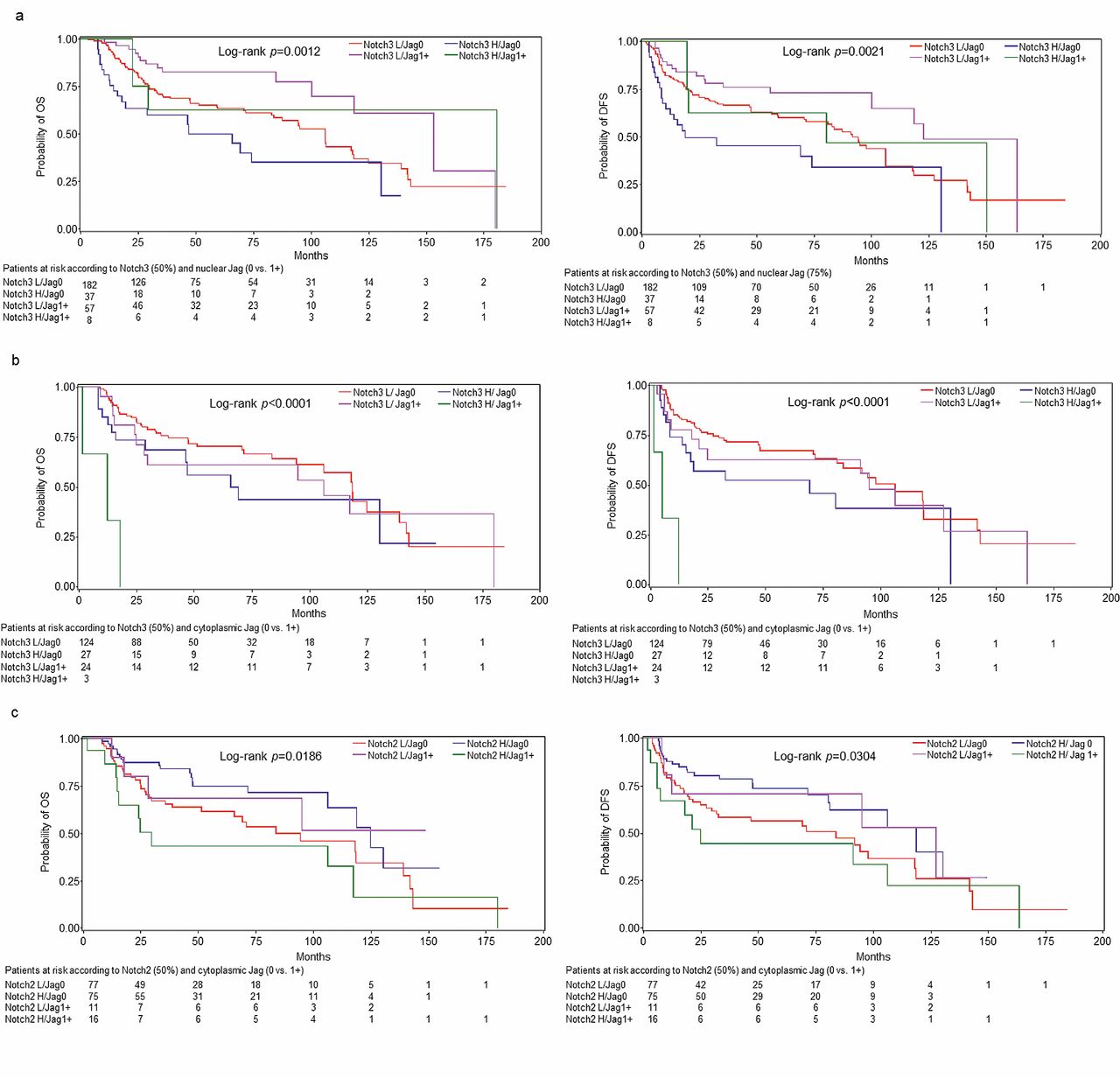
With regard to IHC, enhanced nuclear expression Jagged1 protein was linked to longer DFS (p=0.013) and OS (p=0.004), contrary to the unfavorable prognostic value of Notch3 protein expression for both DFS (p=0.009) and OS (p=0.024). Importantly, in log-rank (Figure 3a-c) and univariate Cox regression analysis (Table III), combined over- or underexpression of the receptors and the respective ligand appeared to bear even more significant prognostic information. Emphasis should be attributed on: a) the complete absence of any prognostic value of Notch1 at the protein level; b) the favorable prognostic effect of Notch2 only when paired with low cytoplasmic expression of Jagged1 protein; and c) the consistent prognostic impact of nuclear protein expression of Jagged1 (favorable when overexpressed) and Notch3 (unfavorable when overexpressed), when studied both alone and in combination.

Prognostic significance of Cartesian protein co-expression of Notch2 or 3 and Jagged1 (nuclear or cytoplasmic). a: Notch3 and nuclear Jagged1; b: Notch3 and cytoplasmic Jagged1; c: Notch2 and cytoplasmic Jagged1. H: High; L: low; OS: overall survival; DFS: disease-free survival; Jag: Jagged1. Cut-off values: Nuclear Jagged1-low vs. -high (75 percentile); cytoplasmic Jagged1-negative vs. positive (0 vs. 1+); Notch1-3 50%.
As for mRNA expression, only Notch1 had a favorable prognostic significance for DFS (p=0.039).
Multivariate analysis: The prognostic value of clinicopathological parameters is quoted in Table IV.
Cox multivariate analysis assigned no independent prognostic value to any mRNA marker. On the contrary, a remarkable finding was the independent prognostic significance of the combined protein expression study of the ligands with the respective receptor. Notably, when either protein expression of Notch3 or cytoplasmic Jagged1 was high, an independent unfavorable effect on DFS was revealed (HR=1.88, 95% CI=1.03-3.43, Wald's p=0.040). In contrast, when high protein expression of Notch2 was coupled with low cytoplasmic protein expression of Jagged1, a favorable independent effect emerged for both DFS (HR=0.48, 95% CI=0.29-0.81, Wald's p=0.006) and OS (HR=0.47, 95% CI=0.27-0.81, p=0.007). However, in the case of simultaneous protein overexpression of Notch2 and cytoplasmic Jagged1, a trend for a detrimental effect on OS was found (HR=2.24, 95% CI=0.93-5.44, Wald's p=0.074).
Univariate Cox regression analysis for 4-level immunohistochemistry combinations (overall and disease-free survival).
Discussion
Marked differences between oral carcinomas and cancer of the larynx have recently been reported, with regard to the frequency of gene hypermethylation and allelic loss (2). Head and neck carcinomas are still treated according to the one-size-fits-all concept, while the development of prognostic and predictive biomarkers in laryngeal cancer is scant.
Our study confirmed that laryngeal tumors, similarly to other types of head and neck cancers, are characterized by the predominant expression pattern of Notch proteins and an active Notch–Jagged axis. In particular, Notch2 and Notch3 proteins were ubiquitously expressed with strong intensity on IHC, as already described for tongue carcinomas (9). Contrary to reports of the fundamental presence of Notch1 protein in head and neck neoplasms (9, 10), Notch1 protein expression was less prominent in our laryngeal tumor set.
Jagged1 localization was much more frequent in the nucleus compared to the cytoplasm, contrary to reported evidence for oral squamous carcinomas (11). Our observation is, however, in line with the study of Ascano et al. (12) and could be explained by proteolytic processing and subsequent nuclear translocation of Jagged1 protein. Similar mechanisms have been described for other Notch ligands, such as Delta, Serrate and Jagged2 (13-15). As Bland et al. speculated (13), this could suggest a receptor-like function for Jagged1 protein and a resulting Notch–Jagged interaction between neighboring cells; ultimately, this finding could imply the presence of bi-directional signaling between Notch and Jagged1. The proposed theory is not novel, since it has already been portrayed for avian erythroblastosis oncogene B 4 (ERBB-4) receptors (16).
Multivariate Cox regression analysis (overall and disease-free survival).
Interestingly, nuclear protein expression of Jagged1 was not only found to exhibit an independent prognostic value in univariate analysis, but was also a favorable prognostic factor (both for DFS and OS) in multivariate analysis. In contrast, well-established evidence exists in favor of a tumor-promoting role of Jagged1 in head and neck cancer, both at the protein and mRNA level (9, 17, 18). Moreover, Jagged1 has been linked to the induction of epithelial-to-mesenchymal transition (19) and angiogenesis (20). This discrepancy should be interpreted under the view that the Notch pathway and its components function in a highly context-dependent fashion, behaving either as oncogenes or tumor suppressors in different tissues (21). Thus, in laryngeal cancer, nuclear Jagged1 protein might acquire a tumor-preventing role.
Notch1 protein expression is linked to advanced clinical stage and poor prognosis in tongue carcinoma (9, 22). However, our study did not reveal any prognostic value for Notch1 protein expression. Quite the opposite, Notch1 mRNA expression had a positive effect on DFS, in contrast to a recent report on tongue cancer (11).
Univariate and multivariate analyses indicated that Notch3 might be an important independent predictor of unfavorable outcome in laryngeal cancer, which is in agreement with its role in tongue carcinoma (9). In parallel, cytoplasmic Jagged1 protein expression conferred a worse DFS in multivariate analysis. Similar reports exist for head and neck cancer (9, 18), one of which especially denotes the tumor-promoting effect of cytoplasmic Jagged1 localization (11). Admittedly, our study reveals a discrepancy regarding the prognostic role of Jagged1 protein expression in laryngeal cancer, where localization acts as a regulator; the outcome is favorable when Jagged1 IHC localization is nuclear and dismal when cytoplasmic. This could truly reflect a distinct prognostic role for nuclear Jagged1, as described above, or could just represent a statistical artifact, as a result of the considerable number of cases without data regarding Jagged1.
The hypothesis that bi-directional Notch-Jagged1 signaling could drive carcinogenesis in laryngeal tumors served as the rationale for studying combined protein expression in our study. Importantly, the Notch2–cytoplasmic Jagged1 co-expression patterns acquired a significant prognostic value both in univariate and multivariate analyses, supporting a cross-talk effect and corresponding complex interactions. Such co-expression patterns are already known to confer a worse prognosis in head and neck cancer (23), with a Jagged–Notch activation loop between tumor cells being implicated. However, in our study the most stimulating finding is the differential prognostic effect of the Notch2–cytoplasmic Jagged1 pair, which depends on cytoplasmic Jagged1 expression. One possible scenario is the following: if cytoplasmic Jagged1 is present, then it binds with Notch2 and promotes the neoplastic phenotype as a tumor promoter; conversely, in cases where Jagged1 is not expressed in the cytoplasm, another ligand (not assessed in this study) might alternatively bind to Notch2 to exert a favorable ligand-dependent effect.
One restriction of our study is that only the expression status of the Notch pathway components was examined. However, although NOTCH gene mutations have not been reported in solid tumors until recently, there is growing evidence that NOTCH1 mutations are particularly frequent (up to 15%) in head and neck cancer, with only TP53 mutations out-numbering them (24). In contrast to hematological malignancies, in which NOTCH1 and -2 gain-of-function mutations have been described (25, 26), head and neck tumors seem to be driven by loss-of-function NOTCH1 mutations, thus indicating a tumor suppressor rather than an oncogenic role for the NOTCH1 gene in these tumor types (24, 27). However, in contrast to NOTCH1, genes encoding the Notch2-4 proteins and the Notch ligands are very rarely mutated in carcinomas arising in the head and neck area.
In conclusion, the expression of Notch pathway proteins seems to play a critical role in laryngeal cancer biology and behavior. The most appealing findings of the present study are: a) the unfavorable effect of protein expression of Notch3 and cytoplasmic Jagged1; b) the favorable impact of nuclear localization of Jagged1; and c) the differential prognostic value of Notch2 according to the presence of cytoplasmic Jagged1. As a following step, validation prospective studies in larger laryngeal cancer sets need to be performed, ideally combining protein expression with DNA sequence analysis data.
Footnotes
-
Conflicts of Interest
The Authors declare that they have no conflicts of interest.
-
Online Supplementary Material
(http://hecog.gr/en/activities/publications/96-publications2011-today/220-2013)
Additional File 1: TMA construction and immunohistochemistry.
Additional File 2: RNA extraction and qRT-PCR methodology.
Additional File 3: Descriptives of protein and mRNA expression.
Additional File 4: Associations between clinicopathological variables and Notch pathway components at the mRNA and protein levels (p-values).
Additional File 5: Prognostic significance of clinicopathological characteristics.
Additional File 6: Prognostic significance of mRNA expression on disease-free survival.
- Received July 12, 2014.
- Revision received August 27, 2014.
- Accepted September 2, 2014.
- Copyright© 2014 International Institute of Anticancer Research (Dr. John G. Delinassios), All rights reserved


