


Abstract
Background/Aim: This study investigated the effects of magnolol, a compound from Magnolia officinalis, on the behavior of LNCaP and PC3 human prostate cancer cells in vitro. Materials and Methods: In vitro cell culture approach with biochemical tests and Western blot analyses was used. Results: Magnolol, (80 μM, 6 hour exposure) was found to affect the expression of insulin-like growth factor-1 (IGF-1) and associated proteins. In both cell lines, protein expression of IGF-1 and insulin-like growth factor binding protein-5 (IGFBP-5) were significantly decreased, while protein expression of IGFBP-3 was significantly increased. Additionally, protein expression of insulin-like growth factor-1 receptor (IGF-1R) was significantly increased and the phosphorylated form of IGF-1 (p-IGF-1R) was significantly decreased in PC3 cells, while IGFBP-4 protein expression was significantly increased in LNCaP cells. Conclusion: This study has demonstrated for the first time that magnolol can alter the expression of IGF-1 and associated proteins in human prostate cancer cells in vitro and suggests that magnolol may have a potential role as a novel anti-prostate cancer agent.
In Canada and the United States, prostate cancer is the most commonly diagnosed form of cancer among men and the second most common cancer among men worldwide (1). This study continued previous investigation on the effects of magnolol, a compound found in the roots and bark of the magnolia tree Magnolia officinalis, by determining further pathways by which magnolol exposure may alter cellular proliferation. Specifically, this study investigated the effect of magnolol on insulin-like growth factor-1 (IGF-1), the insulin-like growth factor-1 receptor (IGF-1R) and the associated insulin-like growth factor binding proteins (IGFBPs) in PC3 and LNCaP human prostate cancer cells in vitro.
Insulin-like growth factors (IGFs) are involved in cellular growth, differentiation and apoptosis avoidance (2, 3). The cellular response to IGF is initiated through IGF binding to IGF-receptors (IGF-Rs). IGF action is regulated by IGFBPs, whether by increasing IGF's half-life or by promoting/inhibiting IGF's binding to IGF-Rs (4). The IGFBPs function by having a greater binding affinity for IGF than for IGF-Rs, by blocking the normal signaling interaction and allowing IGFBPs to functionally remove IGFs from the system in this way (3). IGFBPs also exhibit a range of IGF-independent activities: increased IGFBP-3 can result in reduced protein synthesis or induce apoptosis, while IGFBP-2 and IGFBP-5 can induce mitosis (5). Dramatic changes in IGFBP expression has also been associated with cancer, particularly in that IGFBP-2 and IGFBP-5 over-expression correlates with the change from androgen sensitivity to androgen independence in prostate cancer cells (6-8). The present study examines the effect of magnolol on IGF-1 and associated protein expression in two prostate cancer cell lines in vitro.
Materials and Methods
Materials. All antibodies used in this study were purchased from Santa Cruz Biotechnology, Inc. (Santa Cruz, CA, USA). The antibodies employed included: mouse polyclonal anti-actin, anti-IGF-1R, anti-IGFBP-2 and anti-IGFBP-3; goat polyclonal anti-IGF-1; and rabbit polyclonal anti-p-IGF-1R, anti-IGFBP-4 and anti-IGFBP-5. All other chemicals and materials were purchased from Sigma-Aldrich Canada (Oakville, ON, Canada), unless otherwise indicated.
Cell culture and treatment with magnolol. Human PC3 prostate adenocarcinoma cells (ATCC, Manasses, VA, USA) and human LNCaP prostate adenocarcinoma cells (ATCC) were cultured on 100 mm plastic tissue-culture dishes (Falcon, Mississauga, ON, Canada) in RPMI (Gibco, Burlington, ON, Canada) supplemented with 10% fetal bovine serum (FBS) and 1% antibiotic-antimycotic (Gibco) and were incubated at 37°C in 5% CO2. After incubation, the cells were exposed to 80 μM magnolol, dissolved in dimethyl sulfoxide (DMSO), for 6 hours. Control cells received only DMSO. Magnolol (80 μM) was previously determined to be the effective concentration via cytotoxicity studies (9). After treatment, the cells were removed by trypsin diluted in phosphate buffered saline (PBS) and re-suspended in RPMI with 10% FBS and centrifuged for 5 min at 500 × g. The media was then aspirated off leaving only a cell pellet and the cells were re-suspended and washed in cold PBS. The cells were again centrifuged for 5 min at 500 × g. After centrifugation, the PBS was aspirated off and the cell pellet was stored at −80°C until further analysis.
Immunoblot analysis. Cell pellets were re-suspended in 100 μl of 10 mM Tris-HCl buffer (pH 7.4) containing 0.5 mM PMSF and briefly sonicated. Cell lysates were then centrifuged at 9,300 × g for 20 min at 4°C. The supernatant was removed from the pellet and evaluated for protein content. Equal amounts of protein from this extract were mixed in a 3:1 ratio with standard Laemmli buffer consisting of 50 mM Tris-HCl (pH 6.8), 10% SDS, 0.1% bromophenol blue, 10% glycerol and 100 mM β-mercaptoethanol and boiled for 3 min. Electrophoresis through 10% SDS-PAGE gels was used to resolve proteins, which were then transferred onto nitrocellulose membranes (Bio-Rad, Mississauga, ON, Canada) by electroblotting. The membranes were then incubated in the presence of a 1% BSA (w/v) TBS-Tween (0.05% v/v) solution overnight at 4°C. Primary antibodies diluted to 1:200 (v/v) were then applied to membranes and incubated for 1 hour at room temperature. After incubation, the membranes were washed three times with TBS-Tween (0.05% v/v) for a total of 30 min, then incubated with alkaline phosphatase (AP)-conjugated secondary antibodies (1:1000) for 1.25 h at room temperature. After incubation, the membranes were again washed three times with TBS-Tween (0.05%) for a total of 30 min and rinsed quickly with distilled water and subsequently exposed to SigmaFast BCIP-NBT tablets (Sigma) dissolved in distilled water to visualize protein expression levels. Western blots were then photographed using the Infinity Capture Software (Lumenera Corp., Ottawa, ON, Canada) and densitometry was performed with the ImageJ Software (National Institute of Health, Bethesda, MD, USA).
Statistical analysis. Statistical analysis was conducted using the Graphpad Prism 4.03 for Windows (Graphpad Software, Inc. San Diego, CA, USA). The results of the immunoblot analysis were compared using a simple t-test and results were considered statistically significant at p≤0.1.
Results
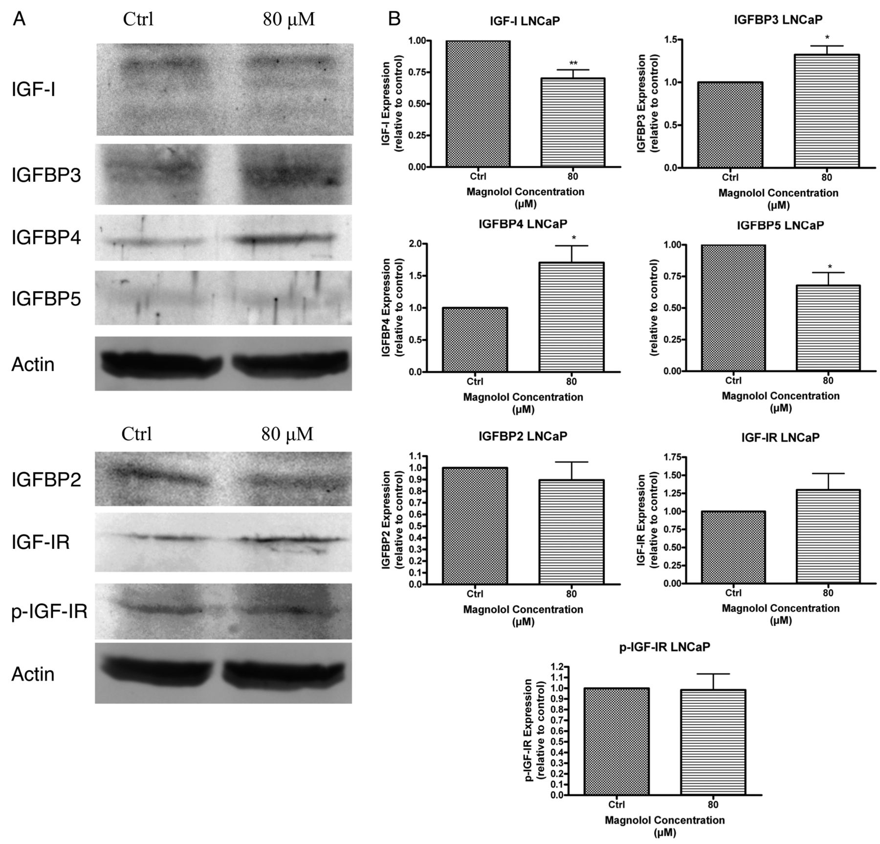
Magnolol affects the expression of IGF-1, IGF-1R and IGFBPs. Protein expression of IGF-1, IGF-1R and IGFBPs were altered in the PC3 (Figure 1) and LNCaP (Figure 2) human prostate cancer cells after 6 hours of exposure to 80 μM magnolol. This magnolol exposure resulted in a reduction to 56% of control IGF1 in PC3 cells. Similarly, in LNCaP cells, magnolol exposure resulted in a significant IGF-1 reduction to 70% of control. To determine if changes in IGF-1 expression in response to magnolol were affected by changes in the IGFBPs, protein expression of IGFBP-2, IGFBP-3, IGFBP-4 and IGFBP-5 was also examined. Protein expression of IGFBP-2 was not significantly affected in either LNCaP or PC3 cells. IGFBP-3 protein expression increased significantly in both cell lines: to 132% of control in LNCaP cells and to 142% of control in PC3 cells. IGFBP-4 protein expression was not significantly affected by magnolol exposure in PC3 cells, however, the expression was increased to 160% of control value without attaining significance. In LNCaP cells the IGFBP-4 protein expression was increased to 171% of control. The final IGFBP evaluated, IGFBP-5, showed a significant decrease in protein expression in both LNCaP cells and PC3 cells in response to magnolol treatment at 79% and 68% of control values, respectively. Finally, to determine if magnolol exposure would affect IGF signaling, the protein expression of IGF-1R and its phosphorylated form was examined. Neither IGF-1R nor p-IGF-1R were significantly affected by magnolol exposure in LNCaP cells, however, IGF-1R protein expression increased to 130% of control without attaining statistical significance. In PC3 cells the protein expression of IGF-1R increased to 132% of control, while the protein expression of p-IGF-1R decreased to 65% of control values.
Discussion
The IGFBPs are a complicated group of proteins with a variety of functions both dependent and independent of IGFs. In this study, the effects of magnolol, a compound found in the roots and bark of the magnolia tree Magnolia officinalis, have been examined on the expression of IGF-1, IGF-1R and several IGFBPs in LNCaP cells and PC3 cells in vitro. Magnolol exposure resulted in significantly reduced protein expression of IGF-1 in both cell lines after 6 hours at a concentration of 80 μM. Changes were also observed in IGF-1R and the IGFBPs at this concentration and time point.
IGF-1 functions to increase cellular uptake of amino acids and to promote carbohydrate metabolism through stimulation of glycogen and protein synthesis (3). Stimulation of the cell cycle can also result from elevated IGF-1 levels as IGF-1 can increase the synthesis and stimulation of cyclin D1 (2, 10), while IGF-I expression has been associated with increased protein expression of cyclin D1 and CDK4 in MCF-7 cells (11). The decrease in IGF-1 observed in the present study corresponds to a decrease in cyclin D1 expression in PC3 and DU145 cells exposed to magnolol in vitro as observed by McKeown et al. in a previous study (9). As alterations in the cell cycle were also observed in the aforementioned study (9), it is possible that decreased IGF-1 protein expression observed in the present study may be involved in the altered cell cycle response to magnolol previously described (9).
IGF-1 function is mediated by IGF-1R. IGF-R is activated via phosphorylation and subsequently activates the phosphatidylinositol 3-kinase (PI3K) and mitogen activated protein kinase (MAPK) pathways (12). As IGF-1R protein expression was increased by similar amounts in both LNCaP cells and PC3 cells, achieving statistical significance only in PC3 cells, this would suggest an increase in IGF-1 signaling and therefore increased cellular proliferation. Protein expression of the phosphorylated form, p-IGF-1R, was significantly decreased in PC3 cells, while the expression was unchanged in LNCaP cells. This suggests that, while the expression of IGF-1R is increasing, perhaps in a compensatory manner, in response to decreased IGF-1 expression, p-IGF-1R is either decreasing in protein expression or remains unchanged and, therefore, IGF-1 signaling is not increasing. This relationship of decreased IGF-1 and either decreased or unchanged p-IGF-1R could be one further explanation of changes to the PI3K and MAPK signal transduction pathways in response to magnolol previously observed (13-17).
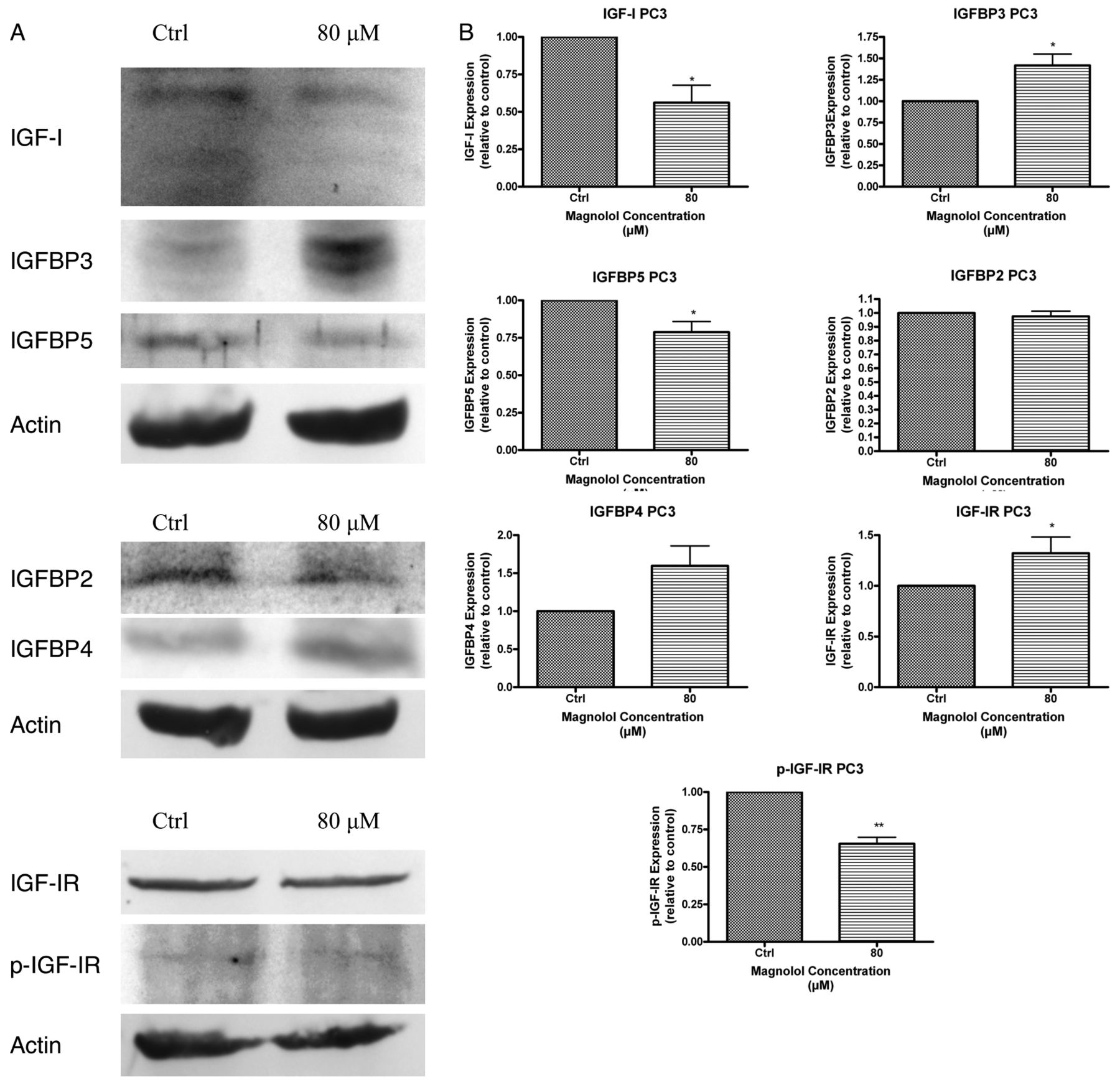
Magnolol affects expression of IGF-1 and associated proteins in PC3 human prostate cancer cells in vitro. Cells were treated with either dimethyl sulfoxide (DMSO) as control (Ctrl) or 80 μM magnolol for 6 h. Actin was used as a loading control. Each blot shown in Figure 1A is representative of results obtained from three separate experiments, each assayed at least in triplicate. These collective results are represented as changes in protein expression in the bar graphs in Figure 1B. (*) indicates statistical significance at p≤0.1 and (**) indicates statistical significance at p≤0.05.

Magnolol affects expression of IGF-1 and associated proteins in LNCaP human prostate cancer cells in vitro. Cells were treated with either dimethyl sulfoxide (DMSO) as control (Ctrl) or 80 μM magnolol for 6 h. Actin was used as a loading control. Each blot shown in Figure 2A is representative of results obtained from three separate experiments, each assayed as least in triplicate. These collective results are represented as changes in protein expression in the bar graphs in Figure 2B. (*) indicates statistical significance at p≤0.1 and (**) indicates statistical significance at p≤0.05.
IGFBP-2 is directly involved in growth stimulation and has been suggested to be implicated with progression from androgen sensitivity to androgen independence (7, 8, 18). In the present study, the protein expression of IGFBP-2 was not significantly altered from control values in either PC3 or LNCaP cells. This was unexpected since previously decrease in IGF-1 has been associated with decreased IGFBP-2 protein expression as IGFBP-2 functions to stabilize IGF-1 and, thereby, increase its half-life (19).
IGFBP-3 protein expression was significantly increased in both LNCaP and PC3 cells after 6 hours exposure to magnolol in vitro. IGFBP-3 is generally associated with the induction of anti-metastatic functions and acts as a tumor suppressor in mouse models (20). This increase in IGFBP-3 expression may be involved in other magnolol-mediated changes to metastasis observed previously (13, 17, 21).
In the present study, the most pronounced change to protein expression in both LNCaP and PC3 cells occurred in IGFBP-4, with IGFBP-4 increasing in both cell lines, although this increase was only statistically significant in LNCaP cells. IGFBP-4 acts by inhibiting IGF-1 action and, therefore, inhibiting cellular growth and proliferation, with the main role of IGFBP-4 in normal cells being the protection of cells from overstimulation by IGF (19). The increased expression of IGFBP-4 observed in the present study provides an additional and novel pathway by which alterations to cellular growth and proliferation previously described may occur (9). In vivo studies have shown that both an increase and a decrease in IGFBP-4 expression can delay tumor formation (22).
IGFBP-5 expression was significantly lower in both LNCaP cells and PC3 cells exposed to magnolol. Decreases in IGFBP-5 have previously been associated with decreases in anti-apoptotic activity and may be involved in the anti-apoptotic response to magnolol previously described (23, 24). Decreases in IGFBP-5 expression have also been associated with inhibition of IGF-1 activities involving DNA synthesis and cellular metabolism (19).
The progression from androgen sensitivity to androgen independence may be inhibited by decreased IGFBP-5 expression (6). By decreasing IGFBP-5 expression, there is a resultant loss of IGF-1 bioavailability, which in turn results in decreased progression through the cell cycle (2). This is supported by in vivo models where IGFBP-5 was found to have no effect on apoptosis but functioned by regulating the cell cycle in prostate cancer cells in a rat model (25).
Magnolol exposure appears to decrease IGF-1 expression through two mechanisms: protein expression of IGF-1 is directly decreased, as well as indirectly affected by the IGFBPs. By decreasing the availability of IGF-1 in these two ways, the anti-proliferative and anti-apoptotic effects of magnolol may be controlled by this system of IGF-1 and IGFBPs interacting both individually, as well as additively or synergistically. Further research studies examining the effect of magnolol on other aspects of cancer cell progression and control, such as metastasis and apoptosis, are warranted.
Acknowledgements
Support for this work was provided by a research grant from the Jeanne and J.-Louis Lévesque Foundation. B. McKeown is a recipient of a graduate student research assistantship from the Jeanne and J.-Louis Lévesque Foundation and R. Hurta is a recipient of the Lévesque Professorship in Nutrisciences and Health.
- Received August 28, 2014.
- Revision received September 11, 2014.
- Accepted September 18, 2014.
- Copyright© 2014 International Institute of Anticancer Research (Dr. John G. Delinassios), All rights reserved


